
Sometimes, diagnosing urinary bladder stones in dogs and cats is simple: one set of x-rays and the mineral-dense uroliths glow bright white on your viewing screen.
Other times, it’s not so straightforward… especially for small or radiolucent stones.
Here’s how radiographs and ultrasound can be used to help you find pesky, hard-to-view bladder stones.
Are bladder stones on your differential diagnosis list?
Bladder stones may be at the forefront of your mind if you see a dog or cat who’s…
Having blood in their urine.
Urinating more frequently, and in short streams.
Having urinary accidents in the home.
Straining or experiencing pain during urination.
Excessively grooming around their genitals.
Usually, a client will bring their pet into you for these concerns, and your physical exam will help to determine that there’s no urinary obstruction.
With urinary bladder stones, you may notice some discomfort on palpation of the caudal abdomen. On a cat or small dog, you may even feel stones or crepitus in the area of the bladder.
Some patients, on the other hand, may exhibit minimal symptoms and their physical exam may be normal (sometimes bladder stones are an incidental finding).
Either way, most pets will need some type of imaging to confirm that bladder stones are there. Radiographs are a great place to start…
Finding uroliths via radiographs
In addition to any other needed tests—such as a urine analysis or bloodwork—radiographs are often recommended for pets with urinary symptoms, in order to look for uroliths or other abnormalities.
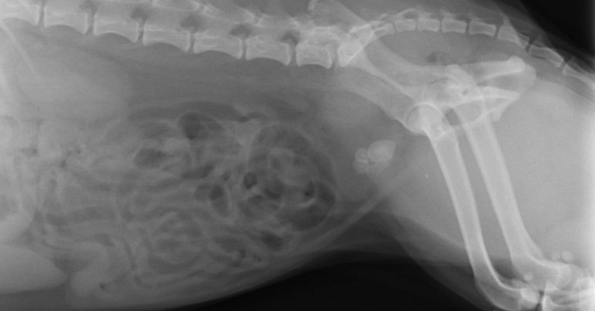
Typically, you’ll start with two simple views of the abdomen—a lateral and a VD.
Take a close look at the urinary bladder to look for radiopaque stones, which should show up as a white opacity relative to soft tissues thanks to their mineral composition.
Stones can range in size from small sand-like grains to more than two inches across. There may be just one or two stones present… or a small group… or even more than 100.
Remember to check the urethra for small stones that could be stuck—especially if the patient is straining or in pain during urination.
For better visualization of the entire urethra in male dogs, consider taking another lateral view with the hindlimbs pulled forward.
Also, check the kidneys and the areas of the ureters. While much less common in pets than in people, occasionally uroliths may be seen higher up in the urinary tract.
If you see stones now—you have your answer.
If you suspect urinary bladder stones but still don’t see them, a contrast study may allow better visualization.
For finding bladder stones, a double-contrast study is ideal.
This includes using both a positive contrast agent (soluble iodinated contrast medium) and a negative contrast agent (room air, or ideally carbon dioxide to reduce the risk of an air embolus) in the bladder together.
Anesthesia or sedation may be needed for the patient’s safety and comfort since the contrast agents are administered via a urinary catheter.
In addition to radiographs, an ultrasound is a useful tool…

Finding uroliths via ultrasound
An ultrasound study is another great option for finding bladder stones, especially radiolucent ones.
The fluid-filled bladder provides contrast for the ultrasound waves so that stones can be found (and often emphasized by acoustic shadowing).
Besides radiolucent stones, you may also see…
Bladder stones (radiopaque or radiolucent) that were too small to visualize radiographically (smaller than 1-3mm).
Other problematic issues in the bladder, such as ‘sludge’ buildup in cats with crystalluria.
Damage to the urinary bladder itself, such as inflammation.
The condition of the upper urinary tract—the kidneys and ureters.
Unexpected findings, such as tumors or anatomical abnormalities of the bladder.
Because of this, ultrasound imaging is a valuable tool for helping you diagnose and treat problems of the bladder, including urinary stones.
Follow-up
Depending on your findings, you may recommend a diet change for dissolvable stones, or a cystotomy to remove the stones.
For dissolution, follow-up imaging can help to track the patient’s progress and see whether or not the stone is dissolving.
When a cystotomy is recommended, remember to use imaging on the day of surgery…
Take pre-op radiographs to confirm the stones are still there, and that your urinary catheter is in place.
Include post-op views to confirm and document that all stones were successfully removed.
Since most stones are radiopaque, standard radiographs are a good option for follow-ups—and typically the imaging choice on the day of surgery.
But ultrasound can also be used in conjunction with other diagnostic tests to monitor the health of the urinary system long-term and to look for early signs of a problem such as a reoccurrence of stones.

Catching stones early, when they’re small, may allow less invasive treatment options such as voiding urohydropropulsion.
With the right combination of imaging modalities, you can help your clients stay on top of treating, monitoring, and preventing urinary bladder stones in their pets.
Written by: Dr. Tammy Powell, DVM
Disclaimer: This article is for general informational purposes only, and not intended as a guide to the medical treatment of any specific animal.
