


Fluoroscopy - the difference between II images & FPD

Understanding the Contrast Between II and FPD Images
In a recent article, we were able to show the benefits of fluoroscopy in the veterinary field. Now, we can highlight in a bit more detail, the differences between the two methods of obtaining fluoroscopy images and explain when each one may be appropriate.
Just like standard radiography where there are two methods for image capture - computed radiography (CR) or direct digital radiography (DDR) - in the field of fluoroscopy, there are also two methods - via an Image Intensifier (II) or a Flat Panel Detector (FPD).
For ease of understanding and by relating to familiar X-ray technologies, similarities can also be drawn between the standard X-ray capture options and the fluoroscopy image capture options with both the CR and II methods relying on an intermediate system between the point of image capture, and the viewing of the image, but the DDR and FPD methods connecting directly to a viewing computer.
How the image is obtained
Image intensified images:
The X-ray image intensifier is a unit that utilizes a series of photochemical reactions to achieve a viewable image.
At the front of the unit, exposed to the X-ray beam, sits an input phosphor (a solid material that emits light when exposed to radiation – the same technique as in an old-style intensifying screen in a film-based or CR X-ray setup). Immediately adjacent to this layer is a photocathode.
When the radiation beam strikes the input phosphor, causing visible light to be released, this visible light in turn strikes the photocathode. The photocathode releases electrons in direct proportion to the visible light, which is directed through the tube by electron lenses onto an output phosphor.
The output phosphor emits light the same way as the input phosphor does, so the image is obtained. This image is only around 1” in diameter, so to be diagnostic it needs to be magnified by attaching a video camera and then digitized in a computer.
Flat Panel Detector images:
In the FPD system, the X-ray image intensifier unit and video camera are replaced by a single digital assembly where the X-ray photons are converted directly to electrical charge and displayed on a monitor.
Benefits of each system
Image intensified images:
Being an older system, II fluoroscopy is a cheaper option than the more modern FPD systems, making it more accessible to the majority of veterinary clinics. There may also be a degree of familiarity in their use and maintenance is likely to be more easily obtained and more affordable than for FPD systems.
Flat panel detector images:
The images obtained in this way are much more detailed, of higher quality, and of a higher resolution than those captured by an image intensifier, therefore making them much more likely to result in an accurate diagnosis.
As well as a higher resolution image, the C-arms used in FPD units range from 12” x 12” to 17” x 17” which is a much larger field of view than those used with II units.
This means a wider area of the patient can be examined in a single capture. The whole process is also much faster than with an image intensifier, so the patient is exposed to a shorter duration of radiation.
Downsides of each system
Image intensified images:
Just as the image quality is the main benefit of the FPD system, the lack of image quality is the main downside to the II system - the images obtained tend to be of low resolution which can make diagnoses challenging.
The C-arms used also cover a much smaller field of view – typically around 9” x 9” or 12” x 12” meaning the anatomical area examined is also much smaller.
The slower processing of the image means a longer duration of exposure to radiation for the patient. As with many older technological systems, the II units are typically larger and take up more space within a practice than the more modern FPD units.
Flat panel detector images:
The main drawback to an FPD system is the cost – being a newer and much more advanced technique, the set-up costs are understandably much higher than for an image intensifier system.
This advanced equipment is also more delicate, and great care and training are needed to be able to use it safely. Some maintenance costs will also reflect this higher value.
When choosing which system is right for a hospital, all the above factors need to be taken into consideration. There will not be one-size-fits-all. In the future, just like what happened when digital radiography took over from film radiography, we may all move more towards flat panel detectors but for the moment, clinics have the two possible options.
Whichever system is chosen, having fluoroscopy within a practice will greatly enhance the services they can offer and aid in increasing the diagnostic toolkit available to the doctors.
References
1. https://newvetequipment.com/blog/veterinary-fluoroscopy
2. Gingold, E. (n.d.). Modern Fluoroscopy Imaging Systems. Image Wisely. https://www.imagewisely.org/Imaging-Modalities/Fluoroscopy/Modern-Imaging-Systems Figure 1, figure 2.
3. https://youtu.be/rex_N_H4zxU


The Future of Veterinary Imaging: Fluoroscopy Advances
What is Fluoroscopy?
Fluoroscopy is an advanced imaging modality that is widely used in human medicine, although its potential in veterinary medicine is only just starting to be realised1. Like conventional radiography, fluoroscopy uses X-rays to produce an image, but in this technique, the image is produced as a video in real-time.
This moving image allows for a much greater range of diagnostic tests to be carried out and can also be used for a range of therapeutic interventions. This field is usually referred to as Interventional Radiography.
Fluoroscopic images are produced by an X-ray generator that produces either a continuous, strobed, or near-continuous, beam of X-rays.
The X-rays are captured by either an image intensifier or, in more modern digital systems, a flat panel detector. Image intensifiers need to be coupled via an optical distributor to a recording or viewing device such as a video camera or screen.
Flat panel detectors are similar to those used in conventional digital radiography (DDR systems) and are connected directly to a computer.
The two main types of fluoroscopy units used in veterinary clinics are portable C-arm units and R&F (radiography and fluoroscopy) rooms.
R&F units are made up of a stationary radiography table with an integrated fluoroscope. The x-ray generator has two separate tube heads, one is used for radiography and one for fluoroscopy.
Portable C-arms are more common in veterinary applications, being used for major surgeries in veterinary hospitals. Unlike an R&F room, a portable c-arm cannot take diagnostic X-rays, only fluoroscopy.
How is fluoroscopy performed?
Depending on the procedure being performed, the patient may be either anesthetized, sedated, or conscious.
In the case of conscious studies, animals are usually restrained within a box where they can easily sit up or lie down, as unlike conventional radiography there is no requirement for them to be perfectly still.
In practice, a change of position is often used to obtain different views and more detailed information. Contrast agents are commonly used to show function e.g. swallowing, or to highlight a particular anatomical area.
In interventional procedures, the patient would routinely be under general anesthetic, and a C-arm fluoroscope is used by the surgeon to visualize part or all of the procedure.
What are the advantages of fluoroscopy?
The big difference between fluoroscopy and traditional radiography is that fluoroscopic images are dynamic moving images, so can be used to assess the function as well as the structure of the body part being imaged.
This opens up a whole array of possibilities. Images produced by fluoroscopy tend to be less detailed than regular radiographs and, to counter this, contrast media are often used to delineate the required structures.
What conditions can fluoroscopy be used to help diagnose?
Many conditions are best diagnosed via fluoroscopy, where a static radiograph does not allow us to see function and ultrasound techniques don’t allow a large enough field of view or suffer interference from air or bone.
Studies that are commonly carried out may include;
Swallowing studies: Swallowing studies require the patient to ingest a volume of contrast material, such as barium, and the progress of this can be tracked through the throat and down to the stomach. This technique is considered the gold standard for investigation of the dysphagic patients.
Esophageal function: Fluoroscopy is useful in the diagnosis of functional and structural esophageal diseases such as megaesophagus and hiatal hernias.
Tracheal and bronchial collapse: A collapsing trachea may be visible on plain radiographs but is a dynamic condition and can easily be missed. Fluoroscopy is more effective at assessing the presence and degree of airway collapse.
Functional diaphragm disorders due to injury or disease of the phrenic nerve.
Localization of thoracic masses: Movement of the mass during normal respiration helps to differentiate peripheral pulmonary and thoracic wall masses.
Blood vessels can be assessed through angiography. Fluoroscopy allows radio-opaque dyes to be injected accurately into specific vessels, aiding the diagnosis of vascular ring anomalies and allowing procedures, such as mesenteric portography which is used to diagnose portosystemic shunts as well as assess the effectiveness of vessel attenuation at surgery.
Real-time evaluation of orthopedic implant placement intra-operatively.
Urinary dysfunction and intervention. Using contrast agents, fluoroscopy allows for easy diagnosis of urinary issues and the effectiveness of catheterizations or other interventions.
What conditions can be treated with the aid of fluoroscopy?
Treatment of conditions with the aid of fluoroscopy is termed Interventional Radiology and in the human field, this is the standard of care for many diseases.
The advantages of using IR as opposed to more traditional therapies include shorter anesthetics, lower perioperative morbidity and mortality, and reduced hospitalization times.
In some cases, conditions that were considered untreatable by other methods can be treated, for example, chemoembolization of unresectable tumors.
Veterinary medicine has some way to go before these techniques are adopted as widely as they are in human medicine.
There are certainly some difficulties and disadvantages in the use of Interventional Radiographic techniques, which include the degree of technical expertise required, specialist equipment needed, and the risk of undesirable radiation exposures to both patients and personnel.
These issues can be reduced greatly with the right training and equipment for the job.
IR techniques that are currently offered to veterinary patients include;
Treatment of cardiac and vascular diseases
Placement of devices within specific blood vessels for transarterial or transvenous embolization of vessels e.g. PDA occlusion, PSS shunt occlusion, or treatment of intractable hemorrhage
Vascular foreign body retrieval e.g. catheter fragments
Cardiac interventional procedures such as balloon valvuloplasty for congenital valve stenosis
Cardiac pacemaker implantation for treatment of arrhythmias
Pulmonary disease
Intraluminal tracheal stenting for treatment of tracheal collapse
Retrieval of tracheobronchial foreign bodies where surgery or endoscopic retrieval are not feasible
Cyanoacrylate embolization for treatment of recurrent chylothorax
Aspiration and biopsy of the thoracic wall and pulmonary masses
Management of neoplasms
Intra-arterial chemotherapy
Arterial embolization and chemoembolization to reduce tumor growth
Palliative stenting of neoplastic obstructions
Urinary disease
Urethral and ureteral stenting to overcome obstructions due to stones, strictures, and malignancies
Urethral catheter placement in cats with urethral rupture
What does the future hold for fluoroscopy
Veterinary fluoroscopy is a developing field of diagnostics.
It seems likely these techniques will become more widely available at both referral and first-opinion veterinary clinics. As expertise in this field progresses, more and more patients can benefit from this technology.
References
1. Weisse, C. W., Berent, A. C., Todd, K. L., & Solomon, J. A. (2008). Potential applications of interventional radiology in veterinary medicine. Journal of the American Veterinary Medical Association, 233(10), 1564-1574.
2. Gingold, E. (n.d.). Modern Fluoroscopy Imaging Systems. Image Wisely. https://www.imagewisely.org/Imaging-Modalities/Fluoroscopy/Modern-Imaging-Systems. Accessed 24/01/2024
3. Shaw, L., & Tudor, D. (2021). Fluoroscopy: Don’t Miss the Show. The Veterinary Nurse, 51–57.
4. Shalom NE, Gong GX, Auster M. Fluoroscopy: An essential diagnostic modality in the age of high-resolution cross-sectional imaging. World J Radiol. 2020 Oct 28;12(10):213-230.
5. Pollard, R. E., Marks, S. L., Cheney, D. M., & Bonadio, C. M. (2017). Diagnostic outcome of contrast videofluoroscopic swallowing studies in 216 dysphagic dogs. Veterinary Radiology & Ultrasound, 58(4), 373-380.
6. Fuentes, V. L., Johnson, L., & Dennis, S. (2010). BSAVA Manual of Canine and Feline Cardiorespiratory Medicine. BSAVA.
7. Pollard, R. E. (2012). Imaging evaluation of dogs and cats with dysphagia. International Scholarly Research Notices, 2012.
8. Bevan, J. M., & Taylor, R. A. (2004). Arthroscopic release of the medial femoropatellar ligament for canine medial patellar luxation. Journal of the American Animal Hospital Association, 40(4), 321-330.
9. Weisse, C. (2015). Veterinary interventional oncology: from concept to clinic. The Veterinary Journal, 205(2), 198-203.
10. Kim, M. Y., Kim, J. H., Kim, K. C., & Yoon, H. Y. (2022). The effectiveness of intraoperative mesenteric portography for preventing misdiagnosis of congenital absence of the portal vein in dog with extrahepatic portosystemic shunt: a case report. Acta Veterinaria Brno, 91(3), 267-272.
11. Meurs, K. M., Lehmkuhl, L. B., & Bonagura, J. D. (2005). Survival times in dogs with severe subvalvular aortic stenosis treated with balloon valvuloplasty or atenolol. Journal of the American Veterinary Medical Association, 227(3), 420-424.
12. Wess, G., Thomas, W. P., Berger, D. M., & Kittleson, M. D. (2006). Applications, complications, and outcomes of transvenous pacemaker implantation in 105 dogs (1997–2002). Journal of veterinary internal medicine, 20(4), 877-884.

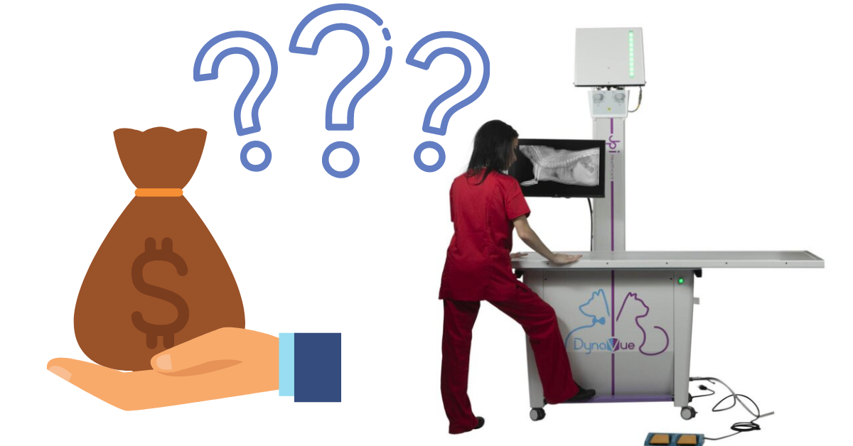
How Much Does Veterinary Fluoroscopy Equipment Cost
For many years, when it came to diagnostic imaging modalities in general practice, many veterinarians were limited to film or digital radiographs. Some general practitioners may have had ultrasound available, but for more advanced imaging modalities, patients were referred to specialty hospitals.
Nowadays, general practitioners may have noticed that imaging technology is becoming more widely available. Even looking at digital radiography, machine prices have come down significantly in the last 10 years.
And it’s not unusual for a practice to offer an additional imaging modality such as fluoroscopy or cone-beam CT, along with interventional radiology procedures, in addition to radiography and ultrasound.
Today, we’ll discuss the availability of fluoroscopy, cost considerations, and other factors to consider when investing in a veterinary fluoroscopy unit.
How to Choose a Fluoroscopy System
When shopping for a fluoroscopy unit, veterinarians will find that there are some veterinary-specific machines available, while others were designed for human medicine. Either option could potentially be useful depending on the specific needs of the practice.
It’s important to consider the specific features of the equipment you are purchasing, as there are big differences between some of the machines. It’s not always an “apples to apples” comparison when price shopping.
Some of the factors to consider include…
Which type of fluoroscopy unit is best? A C-arm is what comes to mind for most vets, as that was the primary system available for many years. And C-arms still have their place, especially when used in the surgery suite. But the downsides are that they can be expensive and very bulky. A table unit is another option. Small, mobile units are also available, especially for large animal practitioners, although they may not carry all the capabilities of larger units.
X-ray output/generator. Fluoroscopy generators can be continuous (constant x-ray output during a study) or pulsed (short bursts of x-rays, similar to the frames of a movie reel). As you can probably imagine, pulsed generators tend to have a lower overall output, which is important for radiation safety. However, exposures can still add up over time, so it’s important to practice radiation safety no matter which unit is being used.
What will your practice use fluoroscopy for? If there is a specific need such as fluoroscopy guidance during major surgeries, that may determine the type of unit your practice purchases.
Where in the practice will the unit be used? Consider whether the power supply in the room needs to be redone or if the room itself needs modifications to meet radiation safety guidelines. Also, does the unit physically fit into the room in which it’s intended to be used? A combination unit, such as one that combines fluoroscopy with digital x-ray or CT functions, could help with space savings in the hospital.
Ease of use and technical support. Efficiency in the hospital is important, even more so during busy times. Machines that are difficult or confusing to use could create challenges with effectively schedule fluoroscopy studies. This could lead to less frequent use of the machine, and therefore less of a return on investment.
Cost considerations. Of course, look at the actual purchase cost. But also consider factors such as financing/payment plan options for cash flow, warranty and maintenance (a warranty or service plan might not be an option when purchasing used equipment), and calculating the return on your financial investment. A practice consultant or financial expert can help with these financial projections and decisions.
How Much Do Veterinary Fluoroscopy Machines Cost?
Once a veterinary practice has evaluated their needs, it’s time to start comparing machines that would be a great fit for the practice. So, how much can a veterinarian expect to pay for a fluoroscopy unit?
The price is incredibly variable depending on the type of unit a vet practice is looking for.
Used or refurbished mini C-arm units are available through resellers starting at around $15,000. On the other hand, some new fluoroscopy units can cost well over $100,000.
There are used and new options sold between these price ranges, too.
How Can a Veterinary Practice Maximize the Return on Their Equipment Investment?
No matter which type of equipment a veterinary practice purchases, it’s important to make sure the equipment actually gets used!
Some of the factors above will help ensure the best ROI. For example, ease of use/efficiency and keeping the machine in good working order are both important.
Staff training, appropriate pricing strategies for fluoroscopy studies, and client education on the value of the service can also contribute to ROI.

Keeping all these factors in mind will help promote regular use of the machine—which carries benefits to patient care and practice profitability alike.
Written by: Dr. Tammy Powell, DVM

Optimize Care with Veterinary Fluoroscopy Technology

In addition to capturing a single moment in time on radiographs, it’s possible to record live, real-time X-ray studies. This is known as fluoroscopy.
So, what is fluoroscopy used for in vet med? And how can vets choose which fluoroscopy machine is right for their practice? Read on to learn more about veterinary fluoroscopy…
How Does Fluoroscopy Work?
Fluoroscopy uses x-rays to create video studies rather than still images. These videos can be viewed in real-time, and depending on the system and software, clips can also be saved as part of the medical record and shared for consultations.
Generators for fluoroscopy can be classified as continuous or pulsed. Continuous generators produce x-ray output for the entire length of the study. Pulsed generators, on the other hand, produce X-rays in short bursts—similar to a frame in a video.
Since fluoroscopy generates images and videos using X-rays, radiation safety protocols must be followed, just as they would while taking traditional radiographs.
This means wearing appropriate PPE and badges (including a ring badge for fluoroscopy), increasing distance away from the machine as much as possible during exposures, using the lowest possible exposures/x-ray output, and keeping hands out of the primary beam during exposures.
Common Uses of Fluoroscopy in Vet Med
While not a replacement for standard radiographs, fluoroscopy can provide additional value. The real-time imaging capabilities of fluoroscopy can capture movement in addition to just still images.
For that reason, fluoroscopy can offer a dynamic, functional view into many parts of the body—especially for conditions that are difficult to capture in the split-second exposure of a regular X-ray study.
Common examples include swallowing studies and diagnosing tracheal collapse. Both of these things are notoriously difficult to catch on regular x-ray images since perfect timing is needed to produce a diagnostic image—an X-ray image that captures just the right moment in time when the abnormality is visible.
Fluoroscopy has additional diagnostic and interventional uses in vet med, too. One example is urinary studies. Moving the patient around in real-time can help to break up bladder sludge to differentiate it from uroliths or abnormalities/thickening of the bladder itself. Real-time fluoroscopy also has value when performing contrast studies of the bladder (to evaluate for bladder ruptures, radiolucent uroliths, etc.) or procedures such as a retrohydropropulsion of urethral stones.
Skull and jaw X-ray studies can be time-consuming and technically challenging. The same is true for diagnosing joint laxity or small fractures within a complex joint. These may require multiple x-ray views—including stress views. Vets may find that fluoroscopy studies add value by showing the anatomy in real-time, from various angles, and with dynamic movement. This could make for a much quicker and easier alternative to taping the patient perfectly into position for each separate shot.
A few more examples of diagnostic uses for fluoroscopy include evaluating for nasopharyngeal polyps and dynamic hernias, as well as real-time movement during GI transit studies.
As for interventional procedures, fluoroscopy may assist during certain orthopedic surgeries or aid in guided aspirates or placement of feeding tubes. The degree of invasiveness of the procedure may determine which type of fluoroscopy unit is required (more on this below).
Fluoroscopy Machines Available to Veterinarians
For a long time, the primary option was C-arm technology, which is expensive and bulky.
Sometimes, a C-arm is the preferred option. For example, when used in the surgery suite (such as with major orthopedic surgeries performed with the aid of fluoroscopy guidance), a C-arm is typically the only option for working around the surgery table.
However, newer and less expensive options are coming to the market. For example, there is a 2-in-1 digital x-ray and fluoroscopy combination unit available from JPI. This is convenient because it takes up less space and there’s no need to buy two separate machines.
The limitations of the x-ray/fluoroscopy combo would be the lack of a horizontal beam and the inability to use fluoroscopy intraoperatively in major surgeries since it is attached to the x-ray table. However, less invasive procedures could be performed on the x-ray table with fluoroscopy guidance.
Each practice would need to decide which option is best for them. However, it is good to see new technology becoming available that could increase the accessibility of fluoroscopy to general practitioners.

Right now, demand for veterinary services is high. So, fluoroscopy could be one additional, unique service for vets to offer to their patients and clients.
Written by: Dr. Tammy Powell, DVM
Watch the swallow with Fluoroscopy!

Fluoroscopy / DR Digital X-Ray Veterinary All In One System
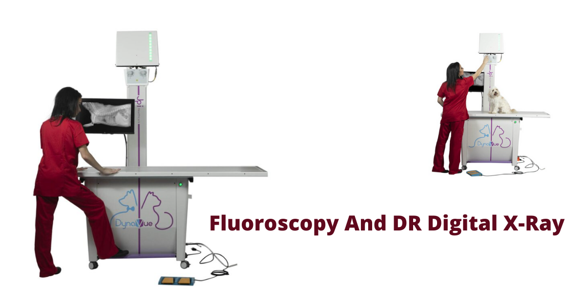
Fluoroscopy And DR Digital XRay, veterinary, All In One System
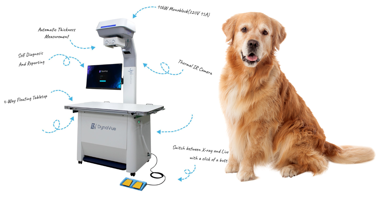
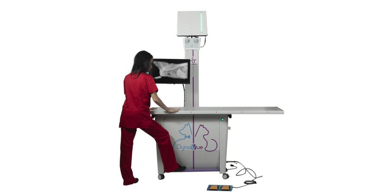
DynaVue is a very popular digital image and radiographic workflow platform used in digital radiography. It offers a complete fluoroscopy and X-Ray system, which comprises an x-ray generator, x-ray imaging table, software platform, and a large capacity Exam Pacs server for storing all the images.
DynaVue Overview
The DynaVue fluoroscopic and digital X-Ray system is based on the principles of noninvasive imaging, which utilizes ionizing radiation for medical diagnosis.
With the help of advanced processing algorithms, the system delivers high-quality images at a very low dose. The system can provide real-time cross-sectional images at a resolution of 400 microns and a speed of 30 frames per second.
The fluoroscopic imaging system is mainly used in minimally invasive surgeries and procedures such as interventional radiology, computed tomography angiography, musculoskeletal procedures, endoscopy, vascular procedures, and intraluminal stenting in veterinary medicine.
DynaVue will prove to be an advantage to veterinary practitioners as it reduces patient exposure to radiation while delivering high-quality images that can be used for various diagnostic purposes. The company offers a wide range of services for customers planning to buy DynaVue, including consultation with specialists and technical support from trained and experienced professionals.
Efficiency Requirements of a Fluoroscopy System
A sound fluoroscopy system will be able to take x-rays from multiple angles. This helps ensure doctors get the best possible images of their patients' internal problems.
The ideal system will take x-rays from three different positions: horizontal, vertical, and angled. In many cases, this is colloquially referred to as 3D imaging because of its appearance on the screen.
A good fluoroscopy system should have several modes built-in that can be used for different types of procedures. For example, an image set that can be used for knee replacement surgery is not appropriate for heart monitoring; therefore, having several different settings is crucial.
DynaVue is the Future Of Portable Fluoroscopy System
DynaVue all in one fluoroscopy and digital X-Ray system meets the exacting demands of modern veterinary and is built to provide precise, high-quality images that can be easily captured and shared with other clinicians.
The system is reliable enough to withstand the high levels of stress during a typical day in a busy veterinary facility. Yet, it is simple enough to be operated by anyone with minimal training.
The system has many features that will help enhance your practice's capability and save money in the long run. These include:
Advanced focusing - This allows for better image control.
Multiple radiography modes - Provides different functionality from one device.
Real-time image processing (8 FPS for fluoroscopy) - Supports remote monitoring.
Minimally invasive fluoroscopy - Helps lower radiation risks for the X-Ray test subjects.
DynaVue combines high-quality X-ray images with superb image quality and a wide range of therapeutic applications to perform real-time minimally invasive procedures under digital control.
It provides superior diagnostic performance to help veterinarians find early-stage diagnostics of diseases and reduce costs associated with long-term treatment.
Efficiency Requirements of a DR Digital XRay System
There are many types of digital radiography systems, and you can find them in various places. Consider the following when choosing your digital radiography system:
Technology
X-ray systems that use computer technology are almost always going to be the most advantageous. These systems will give you the greatest flexibility, such as adjusting the screen brightness or contrast levels or freezing and zooming into your images.
Installation
Your DR digital x-ray system should be easy to install and be able to be set up by 1 or 2 people. It is best if you can connect it to your computers with an Ethernet connection, but at the very least, they should have a direct USB port connection available.
Basic
Advanced x-ray machines offer all kinds of features that can be confusing and sometimes not even used by dentists. It is best to find an x-ray system with all of the basic features such as Auto Exposure, Automatic Battery Charger, Auto Focus, Automatic Film Advance, Automatic Film
Identification and Automatic Film Orientation.
Resolution
You want a minimum screen resolution of 1024x768 pixels to allow you to get sharp images on your screen. You will also need a good amount of contrast on your screen to view your images clearly.
DynaVue provides superior image quality by combining two technologies: True Waveform™ and a photodiode sensor array (PSA).
The PSA provides high-quality images with low noise levels, even at higher frame rates. The True Waveform™ feature enables superior imaging performance during cardiac gating procedures by synchronizing the image acquisition to the patient's heart rate.
For large facilities, the DynaVue system is easily scalable to meet your requirements by adding modules and accessories.
DynaVue excels in small diameter access applications because of its unique True Waveform™ technology, which provides superior imaging performance for soft tissue applications such as small blood vessels, nerve bundles, and lymph nodes.
DynaVue Features Guarantee High Efficiency
DynaVue is a 2-in-1 system that works for soft tissue and fluoro imaging. It is a state-of-the-art imaging solution that enables healthcare professionals to simultaneously see fluoro and soft tissue anatomical structures.
Flexible movement with 4-way floating table: DynaVue DR and Fluoroscopy system is one of the few systems that provides a 4-way floating table. This enables the physician greater freedom of movement, which creates a better visualization of the anatomy.
23" touchscreen display: The large display and high-quality resolution make it easy to conduct your examinations. This device ensures high-quality imaging and an intuitive user experience.
10 kW monoblock type; 15 kW inverter-based system: DynaVue DR and Fluoroscopy systems are developed for advanced diagnostic applications. Up the image quality, up the efficiency, and up the flexibility. Up the power with the Fluoroscopy system and enjoy a full range of possibilities with a single system.
Pulsed fluoroscopy offers reduced dose: The DynaVue DR system uses a backscatter method that reduces patient radiation exposure by up to 50 percent. This system also offers greater visibility throughout the exam. What's most important is that the system can still produce excellent diagnostic images comparable to existing diagnostic imaging procedures.
DynaVue uses ExamVue, a straightforward, easy-to-use, and highly customizable digital radiography software.
ExamVue is the most advanced DICOM 3.0 compliant software for X-ray imaging and reporting. More than 1,000 hospitals worldwide trust it for their daily clinical activities.
The intuitive interface offers seamless frame rate control, imaging and annotation, post imaging processing, and connection to DICOM 3 to allow your team to focus on their work instead of dealing with technology.
Takeaway: The brand new DynaVue is the perfect solution for busy animal hospitals looking to optimize their diagnostic capital while minimizing radiation exposure with a minimal investment in terms of space and equipment.
Written by: Rachel Best
Sources
https://www.ncbi.nlm.nih.gov/pmc/articles/PMC7653184/
https://www.researchgate.net/topic/Minimally-Invasive-Surgery~Diagnostic-Imaging/publications
https://www.dicomstandard.org/
https://www.jpihealthcare.com/examvue-pacs/
https://www.medicalexpo.com/prod/jpi-healthcare-solutions/product-100612-915091.html
Treatment for Blocked Cats: The Power of Fluoroscopy

Urinary obstructions in cats are a life-threatening issue that vets see and treat commonly.
And while most vets have a system for caring for these unfortunate felines, it is exciting to learn about new technology that may provide value during the treatment of these critical patients.
One such development is fluoroscopy technology, which is now more accessible than ever to general practitioners. Here are three ways fluoroscopy may help while unblocking a cat…
Confirming Placement of the Urinary Catheter
Depending on the type of indwelling catheter used, it may be of value to check the placement of the catheter on radiographs.
This is especially true for red rubber catheters—which can double back on themselves or even tie into a knot if too much of the catheter is placed into the urinary bladder—or for any catheter long enough to cause additional trauma if passed so far that the tip contacts and irritates the bladder wall.
While traditional x-rays may be used for this purpose, fluoroscopy offers the advantage of being able to adjust the catheter placement in real time. That way, the catheter can be quickly adjusted prior to placing sutures.
Real-Time Contrast Studies of the Bladder and Urethra
Blocked cats with severely distended urinary bladders are at risk for bladder rupture—whether from the condition itself or from manipulation (pressure or cystocentesis) while the bladder is excessively full.
A large bladder rupture is often obvious since a full bladder will suddenly become difficult or impossible to palpate. However, a small tear or leak may be more difficult to detect.
A positive contrast cystourethrogram can help to identify small ruptures, by looking for contrast outside the borders of the urinary bladder on radiographs.
While traditional radiographs can certainly be used for this purpose, fluoroscopy may prove useful because of the ability to see things moving in real time, and the ability to move the patient and quickly see the abdomen at multiple angles without taking a lot of x-ray shots.
In addition to the bladder, this can be very useful for tracking the movement of contrast through the urethra (to identify obstructing materials, strictures, etc.), versus only seeing a couple of snapshots in time on traditional x-rays.
Since contrast media can be irritating to tissue that is already compromised, a vet must weigh the pros and cons of doing a contrast study right after unblocking a cat. But this procedure could also be useful after the cat has had time to heal, to ensure the bladder is intact before the urinary catheter is removed and the patient is sent home.
Evaluating for Uroliths and “Sludge”
In cats, sludge, sand, or gritty material—which may be a combination of crystals, blood clots, mucus, etc.—can accumulate in the urinary bladder. And while less common than in dogs, uroliths are sometimes diagnosed in cats.
On radiographs, sludge may be confused with a bladder mass or even sometimes with uroliths. Fluoroscopy can help with characterizing this material inside the urinary bladder (and distinguishing it from a bladder mass) by seeing how it moves in real time, as the patient is moved.
Additionally, a vet could use fluoroscopy to track the progress of flushing this material out of the bladder (or urethra) after relieving a blockage.
Conclusion
For many years, traditional radiographs have been included as part of the workup for blocked cats, and a vet can provide excellent care to feline patients with current protocols.

However, as fluoroscopy technology continues to evolve and become more accessible to general practitioners, it’s exciting to see the ways in which this modality can make a vet’s job easier and provide more information for patient care.
Written by: Dr. Tammy Powell, DVM