


Fluoroscopy / DR Digital X-Ray Veterinary All In One System
Fluoroscopy And DR Digital XRay, veterinary, All In One System
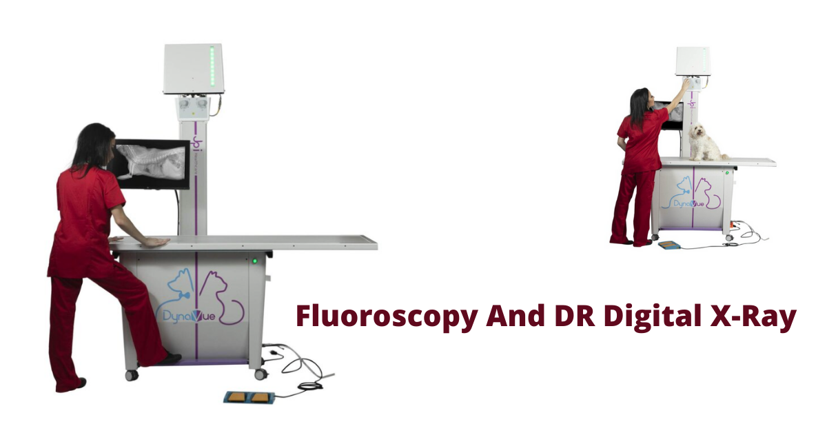
DynaVue is a very popular digital image and radiographic workflow platform used in digital radiography. It offers a complete fluoroscopy and X-Ray system, which comprises an x-ray generator, x-ray imaging table, software platform, and a large capacity Exam Pacs server for storing all the images.
DynaVue Overview
The DynaVue fluoroscopic and digital X-Ray system is based on the principles of noninvasive imaging, which utilizes ionizing radiation for medical diagnosis.
With the help of advanced processing algorithms, the system delivers high-quality images at a very low dose. The system can provide real-time cross-sectional images at a resolution of 400 microns and a speed of 30 frames per second.
The fluoroscopic imaging system is mainly used in minimally invasive surgeries and procedures such as interventional radiology, computed tomography angiography, musculoskeletal procedures, endoscopy, vascular procedures, and intraluminal stenting in veterinary medicine.
DynaVue will prove to be an advantage to veterinary practitioners as it reduces patient exposure to radiation while delivering high-quality images that can be used for various diagnostic purposes. The company offers a wide range of services for customers planning to buy DynaVue, including consultation with specialists and technical support from trained and experienced professionals.
Efficiency Requirements of a Fluoroscopy System
A sound fluoroscopy system will be able to take x-rays from multiple angles. This helps ensure doctors get the best possible images of their patients' internal problems.
The ideal system will take x-rays from three different positions: horizontal, vertical, and angled. In many cases, this is colloquially referred to as 3D imaging because of its appearance on the screen.
A good fluoroscopy system should have several modes built-in that can be used for different types of procedures. For example, an image set that can be used for knee replacement surgery is not appropriate for heart monitoring; therefore, having several different settings is crucial.
DynaVue is the Future Of Portable Fluoroscopy System
DynaVue all in one fluoroscopy and digital X-Ray system meets the exacting demands of modern veterinary and is built to provide precise, high-quality images that can be easily captured and shared with other clinicians.
The system is reliable enough to withstand the high levels of stress during a typical day in a busy veterinary facility. Yet, it is simple enough to be operated by anyone with minimal training.
The system has many features that will help enhance your practice's capability and save money in the long run. These include:
Advanced focusing - This allows for better image control.
Multiple radiography modes - Provides different functionality from one device.
Real-time image processing (8 FPS for fluoroscopy) - Supports remote monitoring.
Minimally invasive fluoroscopy - Helps lower radiation risks for the X-Ray test subjects.
DynaVue combines high-quality X-ray images with superb image quality and a wide range of therapeutic applications to perform real-time minimally invasive procedures under digital control.
It provides superior diagnostic performance to help veterinarians find early-stage diagnostics of diseases and reduce costs associated with long-term treatment.
Efficiency Requirements of a DR Digital XRay System
There are many types of digital radiography systems, and you can find them in various places. Consider the following when choosing your digital radiography system:
Technology
X-ray systems that use computer technology are almost always going to be the most advantageous. These systems will give you the greatest flexibility, such as adjusting the screen brightness or contrast levels or freezing and zooming into your images.
Installation
Your DR digital x-ray system should be easy to install and be able to be set up by 1 or 2 people. It is best if you can connect it to your computers with an Ethernet connection, but at the very least, they should have a direct USB port connection available.
Basic
Advanced x-ray machines offer all kinds of features that can be confusing and sometimes not even used by dentists. It is best to find an x-ray system with all of the basic features such as Auto Exposure, Automatic Battery Charger, Auto Focus, Automatic Film Advance, Automatic Film
Identification and Automatic Film Orientation.
Resolution
You want a minimum screen resolution of 1024x768 pixels to allow you to get sharp images on your screen. You will also need a good amount of contrast on your screen to view your images clearly.
DynaVue provides superior image quality by combining two technologies: True Waveform™ and a photodiode sensor array (PSA).
The PSA provides high-quality images with low noise levels, even at higher frame rates. The True Waveform™ feature enables superior imaging performance during cardiac gating procedures by synchronizing the image acquisition to the patient's heart rate.
For large facilities, the DynaVue system is easily scalable to meet your requirements by adding modules and accessories.
DynaVue excels in small diameter access applications because of its unique True Waveform™ technology, which provides superior imaging performance for soft tissue applications such as small blood vessels, nerve bundles, and lymph nodes.
DynaVue Features Guarantee High Efficiency
DynaVue is a 2-in-1 system that works for soft tissue and fluoro imaging. It is a state-of-the-art imaging solution that enables healthcare professionals to simultaneously see fluoro and soft tissue anatomical structures.
Flexible movement with 4-way floating table: DynaVue DR and Fluoroscopy system is one of the few systems that provides a 4-way floating table. This enables the physician greater freedom of movement, which creates a better visualization of the anatomy.
23" touchscreen display: The large display and high-quality resolution make it easy to conduct your examinations. This device ensures high-quality imaging and an intuitive user experience.
10 kW monoblock type; 15 kW inverter-based system: DynaVue DR and Fluoroscopy systems are developed for advanced diagnostic applications. Up the image quality, up the efficiency, and up the flexibility. Up the power with the Fluoroscopy system and enjoy a full range of possibilities with a single system.
Pulsed fluoroscopy offers reduced dose: The DynaVue DR system uses a backscatter method that reduces patient radiation exposure by up to 50 percent. This system also offers greater visibility throughout the exam. What's most important is that the system can still produce excellent diagnostic images comparable to existing diagnostic imaging procedures.
DynaVue uses ExamVue, a straightforward, easy-to-use, and highly customizable digital radiography software.
ExamVue is the most advanced DICOM 3.0 compliant software for X-ray imaging and reporting. More than 1,000 hospitals worldwide trust it for their daily clinical activities.
The intuitive interface offers seamless frame rate control, imaging and annotation, post imaging processing, and connection to DICOM 3 to allow your team to focus on their work instead of dealing with technology.
Takeaway: The brand new DynaVue is the perfect solution for busy animal hospitals looking to optimize their diagnostic capital while minimizing radiation exposure with a minimal investment in terms of space and equipment.
Written by: Rachel Best
Sources
https://www.ncbi.nlm.nih.gov/pmc/articles/PMC7653184/
https://www.researchgate.net/topic/Minimally-Invasive-Surgery~Diagnostic-Imaging/publications
https://www.dicomstandard.org/
https://www.jpihealthcare.com/examvue-pacs/
https://www.medicalexpo.com/prod/jpi-healthcare-solutions/product-100612-915091.html

Upgrade to Digital X-Ray & Keep Current Table & Generator

DR Digital X-Ray - Easy to Upgrade - Use Your Current Table and Generator
Each veterinary practice relies on various tools and equipment to carry out its services. One of the most important pieces of equipment in treating animals is an X-ray machine.
These machines make it possible to examine the internal structures of an animal to make diagnoses as to the ailments the animals are suffering from.
Broken bones, impacted gastrointestinal tracts, inflammation, and myriad other conditions will all be exposed without the need for invasive surgery with the aid of good x-ray equipment.
In this piece, we'll be taking a look at one of the best x-ray setups designed for use in veterinary practices. This is the DR Digital X-Ray assembly with Plates, Software/Computer, and ExamVue PACS (Server Based) offered by New Vet Equipment.
We'll be taking a step-by-step look at the various components in this assembly, highlighting their positive attributes and how they can positively impact your professional operations. Let's get right into it.
Flat Panel Detector (CareRay Cesium Plate)
Digital Radiography (DR) utilizes x-ray sensitive plates to capture data while the patient examination is going on directly. This data is immediately transferred to a computerized system without needing an intermediate cassette, as happens during Computer Radiography (CR) procedures.
For this reason, DR systems are considered a great choice for those hoping to upgrade from film x-ray techniques.
Flat Panel Detectors (FPDs) are the essential components of these systems and typically use combinations of amorphous silicon detectors fitted with gadolinium or cesium scintillators.
These are responsible for converting x-rays hitting them into light, which is subsequently translated into digital data by thin-film transistors. This data is represented on the acquisition screen for viewing by the vet.
Various advantages come with using Digital Radiography instead of other methods, including:
Superior images that exhibit higher resolutions and overall quality, making diagnoses easier and more accurate for veterinary medicine practitioners.
The ability to enhance images through software tools and algorithms. This capability is not possible when using film x-rays.
Radiation dosages are significantly reduced because the amount of radiation produced by the x-ray generator does not need to be as high as it is when using other radiography methods.
Processing chemicals and film will be a thing of the past, making the entire radiography procedure much simpler.
It's possible to retrofit your equipment to accommodate Digital Radiography equipment without the need to overhaul your entire x-ray setup.
Processing times will be significantly reduced (by up to 5% in some instances).
Thanks to increased efficiency and a more streamlined workflow, higher patient throughput can be achieved.
Digital Radiography systems can be utilized across multiple modalities and systems.
Host Workstation (Desktop/Laptop)
Clients are given the option of choosing between a laptop-based or desktop acquisition computer. These are both sufficiently powerful computers for the tasks they will be called upon to handle, so the choice will largely depend on your particular preferences or space considerations.
The desktop version utilizes an Intel Core i5-6500 with 16GB Ram, 1600Mhz processing speed, and two 1TB hard drives. On the other hand, the laptop version is a ThinkPad E580 running on an Intel Core i7, with 8GB RAM and 500GB hard disk space. It features a 15.6" display. Both these options run on the Windows 10 operating system and come with a 3-year manufacturer's warranty.
Compatible with Portable X-Ray Generator
Portable medical x-ray units are not much different from fixed digital x-ray units in their method of operation except for their size. Even so, they also have certain unique benefits for practitioners of veterinary medicine, including:
Portability: Some veterinary practices can be very busy at times, and a portable x-ray unit makes it possible to conduct an x-ray. This convenience causes less stress to the ailing animal and makes things easier for the x-ray technician as well.
Safer Operation: The use of traditional medical imaging equipment was a source of concern for those exposed to potentially harmful radiation. Portable units come with protective shields mounted on the front of the devices to help prevent patients and operators from exposure to scattered radiation.
Increased Speeds: These units eliminate long waits and processing times, leading to quicker diagnosis, treatment, and also recoveries.
ExamVue PAC Software
The ExamVue PAC software is an imaging management solution that will work seamlessly with your hardware while integrating easily with your office workflow. It is designed to facilitate the viewing, storage, sharing, and management of DR images. It offers a variety of benefits and features, including:
Advanced specialty tools, including Vertebral Heart Score (VHS) and Norberg Angle that make it possible to make quick diagnoses on your x-ray images
Interactive sharing features that allow for the sharing of diagnostic images with clients and colleagues through their Windows tablets or work desks
Integration capacity for up to ten Windows stations within a single office setup
A large variety of annotation tools make it a useful tool for those wishing to share and collaborate with others in the course of their work.
5-Year Warranty
This DR Digital x-ray setup comes with a 5-year warranty to see owners enjoy their use without the worry of system or mechanical failure. These warranties include 5-year drop coverage, five years of remote software support, and five years of hardware support as well.
Shipping and Installation
Depending on your location, you might be eligible for free shipping and installation services. With a quick call or email, you can find out whether or not you qualify for this service.
Final Thoughts
All veterinary professionals know that the success of their efforts relies not only on their personal skill, experience, and training but on the equipment they use. Even the best practitioner of veterinary medicine will perform at less than their best if they are using substandard, inaccurate equipment. To acquire the best digital x-ray assembly for your veterinary practice, visit New Vet Equipment today and place your own order.
Written by: Rachel Best

Veterinary Digital DR X-Ray JPI Directvet System

Veterinary Digital DR X-Ray System Package Deal
Regardless of healthcare professionals' skills and experience, the quality of medical care patients receive will only be as good as the equipment used. This applies not only to human beings and their doctors but to animals and veterinary healthcare practitioners. NewVetEquipment is the proud supplier of high-quality veterinary equipment and is pleased to offer the DirectVet Plus full Veterinary Digital X-Ray System.
Here's a closer look at what this system has to offer
DirectVet Plus X-Ray Table
Veterinarians have to handle pets and animals of all sizes, which means that the x-ray table they use needs to withstand the weight of larger animals. This 32x54-inch system can support more than 300 pounds of weight at a time, and the generator arm is designed to move back and forth over the subject. This makes the system capable of handling the x-ray needs of animals as large as ponies with relative ease.
CubeX 28 X-Ray Generator
CubeX is well-known and respected for its high-frequency, portable generators for veterinary and medical use. These x-ray generators are lightweight and compact. Veterinarians will be pleased with the CubeX 28 due to its user-friendly digital displays, simple design, and soft-touch controls. It comes with two-stage, dynamic auto-line compensation, dual integrated laser pointer, and an easy-to-use inverted control panel that makes it compatible for use with a table.
CareRay Cesium Flat Panel Detector
CareRay Digital Medical Systems is an industry leader in developing, researching, and manufacturing x-ray flat-panel detectors. This system comes with the 17x17-inch cesium flat panel detector that is packed with exceptional performance features, including:
High-definition filming: The high levels of detail these panels can produce make it possible for veterinarians and radiologists to diagnose tuberculosis, inflammation, tumors, and more. Experts will observe and make judgments regarding the biliary tract, abdomen, and urinary tract. These panels also facilitate skeletal viewing, muscular system observations, foreign body identification, and trauma diagnoses.
Image and Video Playback functionality: It comes with a playback function that allows you to save and review videos that highlight the motility and morphology of the organs in the gastrointestinal tract.
Visual contrast functionality: Barium-based imaging is often necessary for the x-raying and observation of the esophagus, angiographies, enemas, sinus fistulas, and other circumstances where observations need to be made compared to surrounding tissues.
Large-format perspective functionality: Veterinarians will be able to get a clear view of the pulsation of the heart and respiratory movement of the subject's lungs. This is made possible by the wide format of the flat panel's 17x17-inch layout. These clear and comprehensive radiographic images make the need for multiple imaging and repositioning unnecessary.
Easy integration: These flat panel x-ray detectors are easy to configure and integrate with whatever system you're using. This broad system compatibility will make the operator's or technician's life much easier.
All CareRay Digital flat panels are fully FDA, CFDA, CE, and ISO13485 certified and are widely used in the veterinary, medical, security, and industrial fields in various capacities. They are highly durable pieces of equipment made out of aluminum alloy and carbon fiber housing.
ExamVue PAC Software
The proper software has to be put in place to support the function of this system in an office setting. ExamVue has developed its simple, fast, and user-friendly software, with a very convenient diagnostic function. ExamVue PAC is server-based, and it comes with a series of advanced specialty tools, including line profiles, freehand, rectangle, polygon, and histogram. Image annotation is also made possible via user text, length, cobb angle, angle, and R/L mark.
Smaller practices and more extensive operations are accommodated by the system, with the capability of supporting up to 10 separate viewers. It has unique features and capabilities, including image stitching, importation, comparisons, processing, storage, and communication. It will work well with any DICOM DIR or Q/R compatible devices.
Host Computer Workstation
Buyers have the option of choosing a laptop or desktop computer as the host workstation. Whichever the case, this piece of equipment will have the computing power and processing muscle to handle high volume and high-complexity operations. The desktop host is an i5-6500 processor with 16 GB RAM and 2 Terabytes of storage with a 3-year warranty. The acquisition laptop option is a ThinkPad E580 running on an Intel Core i7 processor with 8GB RAM and 500GB of storage. These specifications, however, might be subject to change.
5-Year Warranty and Easy Payment Plans
Acquiring the equipment to run a veterinary and radiology service properly is a significant investment. It can be a hurdle for many trying to establish new offices or expand their current operations. To help clients acquire the equipment they need, they may choose between one-time payments or make reasonable monthly payments.
All the equipment that comes with this system is covered under a 5-year warranty, with 5-year drop coverage and software support included. The DirectVet Plus System offers peace of mind to all who choose to make it a part of their operation.
Take note that you may qualify for a deduction on capital equipment purchases under section 179 of the current Internal Revenue Service (IRS) tax codes. Your potential deduction might allow you to treat the full cost of new equipment and software as a deductible expense whether you purchase cash, capital lease, loan, or an Equipment Finance Agreement (EFA). Further information may be found on the IRS section 179 portal.
Final Thoughts
The DirectVet Plus Digital X-Ray System is an effective solution for veterinarians looking for a convenient, powerful, and user-friendly way to handle their x-ray needs. It is a system that doesn't need any additional power supplies, construction, or preparations. It is a self-contained veterinary digital x-ray equipment system that will be ready for use as soon as it's delivered, making it an excellent choice for veterinarians in the process of establishing their practice. Visit NewVetEquipment.com to place your order or have any questions or concerns you might have addressed.
Written by: Rachel Best

5 Tips to Improve Efficiency With Radiographs

Efficiency can increase a veterinary practice’s income by allowing more patients to be seen or more procedures such as radiographs to be performed.
Additionally, it may lead to an increased average charge per patient—which can improve a practice’s bottom line while delivering excellent patient care.
When it comes to radiographs, here are five ways to increase efficiency…
Start With the Best Equipment for Your Practice’s Needs
Having good, well-functioning equipment can really make life easier for a veterinarian and their team. After all, slower machines and image processors can increase the time per shot. And equipment that’s not operating at its best may lead to frustrating retakes—or even to rescheduling a procedure.
To maximize the usefulness of radiography equipment at a veterinary practice, start by taking an inventory of which equipment is there, including: generator, table, plate or cassette, film processor or digital image software, etc.
Next, evaluate each piece of equipment with the following questions:
How is the equipment functioning right now?
If not working well, can repairs or maintenance solve the issue—and what is the cost?
Is there any routine maintenance due to be performed?
Does anything need to be replaced—and is it the whole system, or just one specific component?
Would an upgrade improve efficiency? For example, upgrading from film to digital x-rays can save a lot of time that would otherwise be spent processing films.
Develop Standard Protocols and Techniques
If certain procedures are performed infrequently or don’t have a standard set of protocols to follow, this may lead to confusion, inconsistency, or errors—all of which can waste time and cause frustration.
To make things more efficient, it helps to have standard protocols for team members to follow, which have been properly explained to them. Protocols may include:
Specific instructions for patient positioning for different radiographic views, such as thorax, hip, spine, etc.
Guidelines for effective patient restraint while minimizing the staff’s exposure to radiation. For example, be sure the team knows how to properly use positioning aids such as sandbags and tape.
If patients are sedated, be sure to have a minimum standard for patient monitoring, with prepared monitoring sheets a team member can easily pick up and use for their monitoring notes.
Have a standard technique chart, or make sure the team knows how to properly set up an x-ray study using a digital program that automatically sets technique. This includes explaining how to measure a patient in the position in which they will be radiographed.
Have the Right Resources Available for Radiographic Interpretation
This may include textbooks and other references for what is normal on each radiographic view and what is not.
Access to a second opinion can also be very valuable. Try to create a collaborative environment where veterinary colleagues within the practice can help each other discuss and interpret radiographs. Consider subscribing to an online forum such as the Veterinary Information Network (VIN), where a vet can post their radiographs for a second opinion. Or, consider using a teleradiology consultation service with veterinary radiologists.
Practice Makes Perfect
Efficiency in taking and interpreting radiographs will improve over time, with practice. So even if fitting more radiographs into a busy schedule feels time-consuming at the beginning, it will get to be second nature over time.
The same is true with x-ray image interpretation—many vets become faster and more proficient with practice. Also, be sure to study the radiographs of normal patients, to gain a thorough understanding of all the different ways normal anatomy can look in different sizes and breeds of veterinary patients.
Plan for Conversations With Clients
If pet owners are unsure about proceeding with radiographs—especially when sedation is required—the conversation can take up a lot of time for veterinarians and their team members. By planning what to say and gaining comfort with these conversations, time can be saved while providing patients with the care they need. Also, clients may feel more comfortable if everything is explained in just the right way.
Efficient x-ray conversation techniques may include general principles for good communication (active listening, empathy, non-verbal communication, etc.), as well as proactively discussing the answers to commonly asked questions and concerns. For example, explain how sedation not only makes the radiographs more detailed and accurate—it also makes the procedure less scary and more comfortable for a dog or cat.

Putting all these principles together, a veterinary team may find the whole x-ray process becomes more efficient. This is good for team morale, as it helps to keep the day running as smoothly as possible. It can also increase a practice’s bottom line over time while providing excellent care for patients. For all these reasons, it’s usually well worth it for a veterinary practice to invest in radiographic efficiency!
Written by: Dr. Tammy Powell, DVM