


How Veterinary Dentistry Improves Staff Engagement and Productivity
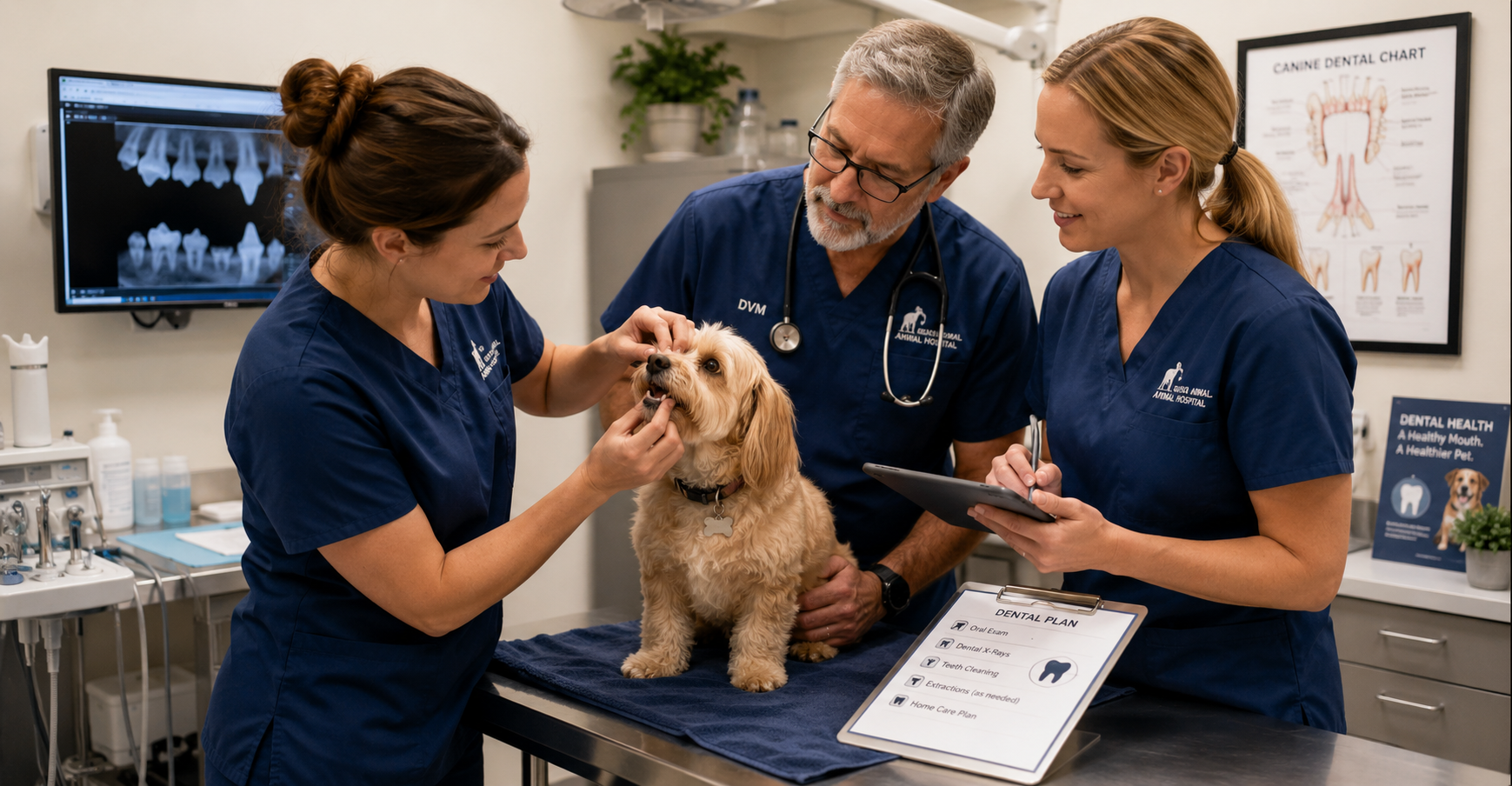
Veterinary Dental Care Supports Team Growth and Retention
Ask most veterinary professionals why dentistry matters, and you'll hear plenty about periodontal disease, oral pain, and improved patient health. What you may hear less often is this:
A strong dental program can also strengthen the veterinary team.
I know how that sounds, but stick with me.
In a profession where burnout, staffing shortages, and compassion fatigue continue to challenge practices of every size, dentistry offers something many teams are searching for:
- Meaningful work
- Skill development
- Collaboration
While dentistry certainly benefits patients and clients, it can also become one of the most rewarding areas of practice for veterinarians, technicians, and support staff alike.
Giving Technicians the Opportunity to Practice at a Higher Level
Veterinary technicians are highly skilled professionals, yet many teams struggle with fully utilizing those skills. Dentistry offers technicians opportunities to become deeply involved in patient care before, during, and after procedures.
Depending on state regulations and practice protocols, technicians often play critical roles in:
- Patient preparation and anesthesia monitoring
- Dental charting and radiography
- Scaling and polishing
- Recovery monitoring
- Client education
Rather than performing the same routine tasks throughout the day, dental technicians can engage in more advanced, hands-on work.
For many team members, that increased responsibility brings greater job satisfaction.
When people are trusted to use their skills, they become more invested in both their work and their patients.
The Moment Things Change: Seeing What’s Hidden
One of the most rewarding moments during a dental procedure often happens when the first radiographs appear on the screen.
A tooth that looked relatively normal externally may reveal severe bone loss. A fractured tooth may show a damaged root. A cat with mild tartar may have advanced resorptive lesions.
These discoveries create valuable learning opportunities for the entire team.
Dental radiography turns procedures into diagnostic investigations rather than routine cleanings. Team members begin asking questions:
- What are we seeing?
- Why is this happening?
- What treatment options are available?
- How can we improve this patient's comfort?
Those moments build clinical confidence and encourage continued learning.
Over time, your team becomes better at recognizing pathology, discussing findings, and contributing to patient care decisions.
Immediate Results Create Meaningful Work
Veterinary medicine can sometimes feel emotionally challenging because progress isn't always obvious.
Chronic disease management takes time. Behavioral improvements may take months. Long-term treatment plans often unfold gradually.
Dentistry is different.
A patient may arrive with severe tartar, inflammation, infection, or oral pain and leave significantly more comfortable the very same day.
The team can see the difference. Clients often notice the difference quickly. And perhaps most importantly, staff members know they helped relieve pain that may have gone unnoticed for months or years.
Those experiences matter.
They remind your team why they entered the field in the first place.
Creating More Predictable Days
Anyone who has worked in veterinary medicine knows that unpredictability is part of the job.
Emergencies happen. Schedules change. Appointments run long.
While no service completely eliminates those challenges, well-organized dental programs can add welcome structure to the workday. Dedicated dental appointments often create:
- Predictable procedure blocks
- Clear team assignments
- Consistent equipment setups
- Efficient patient flow
Your team knows their role. Supplies are prepared. Expectations are clear.
Instead of constantly reacting, your team spends more time working together with purpose.
Investing in Growth Keeps Teams Engaged
Few things improve retention as much as professional growth. Dental programs often encourage continuing education opportunities, such as:
- Dental radiography training
- Anesthesia education
- Dentistry certifications
- Wet labs and hands-on courses
When you invest in these areas, employees often feel valued and supported.
A technician who develops expertise in dentistry may become a mentor, trainer, or internal resource for the rest of the team. That sense of ownership can significantly improve engagement and long-term retention.
In an industry facing staffing challenges, opportunities for growth matter.
Building a Culture Around Better Care
The most successful dental programs become more than a service line. They become part of the practice culture.
Technicians become advocates for dental education. Doctors collaborate on treatment planning. Clients receive more consistent recommendations.
That sense of purpose can strengthen teamwork and create a culture centered around delivering better care.
Dentistry Is About More Than Procedures
It's easy to measure dentistry by production numbers, procedure counts, or equipment investments. But those metrics only tell part of the story. Every dental procedure is also an opportunity:
- To help a pet live more comfortably.
- To strengthen a technician's skills.
- To build confidence within the team.
- To create meaningful work.
- To support professional growth.
Dentistry isn't simply a revenue center. It's a service that allows your team to practice at a higher level while making a real difference in their patients' lives.
And when your team feels challenged, supported, and connected to their work, everyone benefits—including the pets they care for every day.


Building a Sustainable Veterinary Dental Program: The Benefits to Pets, Clients, and Practice Growth
Build a Stronger Veterinary Dental Program
How Better Dental Care Benefits Pets, Clients, Veterinary Teams, and Practice Growth
When veterinarians talk about dentistry, the conversation usually centers on patient health—and for good reason.
Dental disease can cause chronic pain, infection, tooth loss, and a significant decline in a pet’s comfort and quality of life. Helping pets live healthier, more comfortable lives is the primary reason veterinary dentistry matters.
But there is another side of the story that also deserves attention.
A well-developed dental program can strengthen client relationships, help more pet owners follow through with recommended care, create opportunities for team development, and provide a predictable source of revenue that supports the long-term health of the practice.
Veterinary dentistry is one of the rare areas where everyone can benefit: pets, clients, veterinary teams, and the clinic.
Dental Disease Is Often Underdiagnosed
One of the biggest challenges in veterinary dentistry is that many pets do not show obvious signs of discomfort.
Unlike people, dogs and cats often continue eating and following their normal routines even when they are experiencing significant dental pain. They quietly adapt, which can make oral disease difficult for pet owners to recognize.
As a result, serious dental conditions may go unnoticed until they become advanced. Veterinarians commonly discover:
- Advanced periodontal disease
- Tooth-root infections
- Fractured teeth
- Tooth resorption
- Significant bone loss
Many of these conditions can only be fully identified through a comprehensive oral examination and dental imaging.
Because pets are so skilled at hiding oral pain, proactive dental care is especially important.
A strong dental program helps practices identify disease earlier and intervene sooner, before smaller problems become more painful, complicated, and expensive to treat.
Dentistry Creates Opportunities to Improve Patient Health
Unlike procedures that happen only once during a pet’s lifetime, dental care often becomes part of an ongoing healthcare plan.
Oral health requires regular monitoring, preventive care, and treatment throughout a patient’s life. This creates opportunities for:
- Annual dental evaluations
- Professional cleanings
- Dental imaging
- Follow-up procedures
- Preventive care recommendations
- Ongoing client education
Every dental conversation is another opportunity to improve a pet’s quality of life while strengthening the relationship between the veterinary team and the client.
The goal is not simply to perform more procedures. It is to create a proactive approach to oral health that supports patients throughout their lives.
Existing Clients Are Often the Best Opportunity for Growth
Many veterinary practices spend significant time and money attracting new clients.
While new client acquisition is important, existing clients often represent one of the greatest opportunities for sustainable practice growth.
Why?
Because trust has already been established.
When a veterinarian recommends dental care based on examination findings, clients are often more receptive than they would be to an unfamiliar service they have never discussed before.
Dental recommendations are a natural extension of preventive healthcare. The practice is not trying to sell an unnecessary service. It is helping the client understand and address a health problem that already exists.
“Here is what we are seeing.”
“Here is why it matters.”
“Here is how we can help.”
Clear communication helps clients understand the value of treatment and make more confident decisions about their pet’s care.
Dentistry Can Create Predictable, Recurring Revenue
Every veterinary practice needs revenue to operate, invest, and continue providing high-quality care.
One advantage of dentistry is that it can become a relatively predictable service line.
Because dental disease is common and often ongoing, practices can build consistent revenue through:
- Annual dental procedures
- Dental imaging
- Follow-up treatment
- Extractions when medically necessary
- Preventive dental programs
Unlike services that may fluctuate significantly from month to month, dentistry can become a dependable part of practice operations.
That stability can help a practice plan for staffing, equipment, training, and future growth with greater confidence.
Profitable Dentistry Supports Better Veterinary Medicine
Profitability should never take priority over patient care.
However, it gives veterinary practices the resources they need to continue providing excellent medicine.
A profitable dental program can help fund:
- Staff training and continuing education
- New diagnostic equipment
- Facility improvements
- Additional support staff
- Expanded treatment capabilities
- Improved patient-care technology
In many cases, the investments made possible through dental revenue benefit the entire practice, not just the dental department.
The result is a stronger clinic that is better equipped to serve patients, support its team, and help clients make informed healthcare decisions.
Building a Dental Program That Lasts
The most successful dental programs are not built around production goals. They are built around patient care.
Practices that focus on education, early detection, quality diagnostics, and comprehensive treatment recommendations often find that growth follows naturally.
When pets receive better care, clients understand the value of treatment.
When clients understand the value, they are more likely to follow through with recommended care.
That, in turn, gives the practice the resources it needs to invest in its team, equipment, and services.
It becomes a healthy cycle that benefits everyone.
The Real Value of Veterinary Dentistry
At its core, veterinary dentistry is about identifying hidden disease, relieving pain, and improving quality of life.
The business benefits are real, but they are a result of providing valuable care—not the primary objective.
A healthy dental program is not about maximizing revenue. It is about building a sustainable practice that can continue serving pets, supporting veterinary teams, and helping clients make confident decisions about their animals’ healthcare.
Better Dentistry, Better Business: The Value of Digital Dental X-Rays

The Hidden ROI of Veterinary Dentistry: How Digital Dental X-Rays Pay for Themselves
When veterinary practices consider investing in dental equipment, the conversation often starts with patient care…and rightfully so.
Dental disease is one of the most common health issues affecting companion animals, and dental radiographs have become an essential part of providing high-quality care.
But there is another side of the conversation that is equally important: the business case for veterinary dentistry.
Running a successful practice requires more than compassion and clinical expertise. You need to generate enough revenue to pay your team, invest in technology, maintain facilities, and continue delivering exceptional care.
Fortunately, veterinary dentistry is one of the few service areas where improved patient outcomes and financial sustainability often go hand in hand.
The key is making smart equipment investments that support both goals.
Why Dental Radiographs Are the Standard of Care
Many pet owners are surprised to learn that much of a tooth lies below the gumline. In fact, studies have shown that a significant percentage of dental pathology cannot be identified through a visual examination alone.
Without dental radiographs, veterinarians may miss:
- Tooth root abscesses
- Resorptive lesions
- Periodontal bone loss
- Retained roots
- Fractured teeth
- Endodontic disease
Digital dental X-rays allow you to identify problems that would otherwise remain hidden, helping you provide more accurate diagnoses and treatment recommendations.
Today, many veterinarians consider dental radiography a fundamental component of a complete dental procedure rather than an optional add-on.
Looking Beyond the Price Tag
One of the biggest misconceptions about dental equipment is that practices need to spend top dollar to build a successful dental program.
Some premium dental imaging setups can cost $35,000 or more once sensors, generators, software, and accessories are included.
While those systems may offer additional features, you can achieve excellent clinical results with quality digital dental radiography systems in the $12,000-$15,000 range.
The difference matters.
A lower initial investment can significantly shorten the time required to achieve a positive return while still providing the diagnostic capabilities veterinarians need to deliver quality care.
A Simple ROI Example
Let’s look at a realistic scenario:
Assume a practice purchases a digital dental radiography system for $15,000. Now consider a conservative estimate:
Average dental procedure revenue: $600
Dental procedures performed per month: 10
Monthly dental revenue: $6,000
Of course, equipment cost is only one piece of the equation. There are labor, anesthesia, supplies, and overhead expenses to consider. However, even when accounting for those costs, dental procedures often represent a valuable service line for many practices.
If the practice attributes only a portion of that revenue toward recovering the equipment investment, the payback period can still be surprisingly short.
For example:
Equipment investment: $15,000
Contribution toward equipment recovery per procedure: $150
Procedures needed to recover investment: 100
At just 10 dental procedures per month, the equipment could effectively pay for itself in less than a year.
Now compare that to a $35,000 investment.
Using the same assumptions, the practice would need approximately 233 procedures to recoup the equipment cost, significantly extending the payback timeline.
The takeaway is not that higher-priced systems are inherently bad investments. The purchase price directly affects how quickly a clinic can realize a return.
Improving Workflow Efficiency
The ROI of digital dental radiography is not limited to revenue generation. Digital systems can also improve workflow efficiency throughout the practice.
Digital dental X-rays eliminate many time-consuming steps and can help reduce the need for unnecessary retakes.
Benefits often include:
- Immediate image review
- Improved image storage and retrieval
- Easier integration with practice management software
When procedures move more efficiently, you can often complete appointments more predictably and utilize staff time more effectively.
Helping Clients Say “Yes” to Treatment
Client education is one of the most overlooked benefits of digital imaging. When pet owners can actually see pathology on a screen, treatment recommendations become much easier to understand.
A fractured tooth root, severe bone loss, or a resorptive lesion is often far more compelling when accompanied by an image rather than a verbal explanation alone.
The result is better patient care and greater confidence in the veterinary team’s recommendations.
What to Look for When Purchasing Dental Equipment
Not all dental radiography systems are created equal, but the most expensive option is not always the best choice. When evaluating equipment, consider:
- Image Quality: The system should consistently provide diagnostic-quality images with excellent detail.
- Ease of Use: A user-friendly interface can reduce training time and improve adoption among team members.
- Sensor Durability: Dental sensors represent a significant investment. Durability and warranty coverage matter.
- Software Integration: Look for systems that work seamlessly with your existing workflow and practice management software.
- Service and Support: Reliable technical support can make a major difference when issues arise.
- Total Cost of Ownership: Consider not only the purchase price but also future maintenance, replacement costs, and warranty protection.
Better Dentistry, Better Business
Veterinary dentistry should never be viewed solely as a revenue generator. At its core, dental care is about identifying pain, treating disease, and improving quality of life for pets.
But it is also fair, and necessary, to acknowledge the business benefits.
Profitable service lines help you retain talented staff, invest in better technology, expand patient care capabilities, and continue serving your community for years to come.

How to Identify and Correct Underexposed Veterinary X-Rays
In veterinary diagnostics, every image tells a story—but only if that image is clear, properly exposed, and reveals the details you need to see.
Underexposed radiographs are a common and frustrating issue, especially in busy clinics or those with less experienced staff. They don’t just create workflow delays—they can obscure critical findings and compromise patient care.
The good news? Identifying and correcting underexposed veterinary X-rays is entirely within your team’s reach!
Whether you're using a full-body digital x-ray system or a dental x-ray system, here’s what you need to know to spot underexposure and fix it fast.
What Does an Underexposed X-Ray Look Like?
Underexposed radiographs typically appear too light or “washed out.”
Anatomical structures, especially soft tissue or overlapping organs, may lack contrast and definition. This can make assessing for subtle fractures, pulmonary patterns, or abdominal masses difficult.
Let’s look at an example…
Jasper, a middle-aged Labrador, comes in for coughing and mild lethargy. The initial thoracic X-ray appears extremely pale—lung fields look nearly indistinguishable from the soft tissue.
On retake, using corrected exposure settings, the radiograph reveals a suspicious nodule.
That second image could distinguish between catching early pathology and sending the patient home undiagnosed.
What Causes Underexposure in Veterinary Radiographs?
Several factors can contribute to underexposure:
Incorrect technique settings (kVp and mAs) are often the most common culprits. Too low = too little penetration.
Improper patient positioning or size estimation: Thicker body parts may require adjustments to the technique.
Equipment calibration issues: Older or poorly maintained units may not output consistent exposure levels.
Sensor or plate problems: In digital systems, underexposure may also stem from a malfunctioning DR sensor or image plate.
Best Practices to Correct and Prevent Underexposed X-Rays
You can adjust technique settings thoughtfully.
If you’re routinely seeing underexposed images, review your technique chart. Increasing mAs improves image density, while adjusting kVp affects penetration and contrast.
Use a controlled approach—changing one variable at a time—to determine what works best for your unit and patient size.
Use a standardized positioning protocol
Precise radiographic technique starts with consistent positioning. Ensure your team utilizes positioning aids, is familiar with your machine’s baseline settings, and selects the appropriate view for the clinical question. Consider creating quick-reference guides near your X-ray suite.
Train your team to recognize exposure errors.
Empower your technicians to identify underexposure on the spot and retake images if necessary. Time spent training now saves retakes, delays, and diagnostic errors later.
Upgrade to high-quality digital systems.
Modern digital radiography systems often include automatic exposure detection and produce images in seconds, providing more consistency and reducing the likelihood of human error.
If you're using an older system or film-based radiography, consider upgrading to improve efficiency and accuracy.
Why It Matters
Correct exposure is more than a technical detail—it impacts patient care. A radiograph that misses a mass, hides a fracture, or blurs a lung pattern can delay treatment and put your patient at risk.
By optimizing your imaging protocols and investing in equipment that supports high-quality diagnostics, you help ensure that each patient receives the care they deserve. Each Veterinarian has the information they need to make confident decisions.
Would you be interested in upgrading your Imaging Suite?
Explore our catalog to find the right fit for your hospital. Whether you're opening a new practice or modernizing your existing setup, explore our selection of imaging equipment and don’t hesitate to reach out with any questions.

EZ Dent: Advanced Veterinary Dental X-Rays with MyVet USA

Dental radiographs are an integral part of dental procedures in small animal medicine
Without x-rays, it’s not possible to see the full extent of periodontal disease or other problems (FORLs, dentigerous cysts, tooth root abscesses, etc.) that happen below the gumline.
As dental radiography has become prevalent in modern veterinary medicine, there are many companies and manufacturers that offer dental x-ray equipment and software for veterinarians. Today, we’ll be examining some of the features and considerations of the EZ Dent dental x-ray system by MyVet USA—including the x-ray generator, intraoral sensor, and software.
MyVet Imaging
In 2018, the US-based MyVet Imaging became a subsidiary of South Korea-based Rayence, a provider of digital x-ray equipment for human medicine.
Rayence and MyVet Inc. are headquartered in New Jersey for US operations, with veterinary offerings for small and large animal radiography, including digital dental radiography for small animal and equine patients. They advertise innovative dental imaging solutions, including the world’s first veterinary panoramic dental x-ray system and Carbon Nanotube (CNT) technology.
The EzRay VetTM Cart
Despite the intrigue of a panoramic system, many veterinarians prefer the details and consistency of traditional style DR or CR radiographs for small animal dental rads. The EzRay Vet Cart, a mobile intraoral x-ray cart system, is offered by MyVet for this purpose.
Some of the benefits of the EzRay Vet Cart advertised by MyVet include…
Carbon nanotube (CNT) technology, which they claim reduces maximum exposure time by 75% while still maintaining image quality. The beam has a 0.4mm focal spot, and exposure setting controls are said to automatically adjust the exposure setting to maintain image density.
Ergonomic design, featuring a non-drift arm and head that is compact, lightweight, and easy to position with one hand.
Preset technique options.
No warm-up time or waiting time between shots.
A wall-mounted unit is also available.
The EzRay Air VetTM
The EzRay Air Vet is a handheld generator option. This may suit the needs of veterinarians who require the unit to have ultimate mobility, or who have tight spaces in their clinic. MyVet advertises that the portable generator also features CNT technology, is up to 30% lighter than other handheld units, has no warm-up time (for faster workflow), and features a double scatter shield (with internal and external shielding) to protect from scatter radiation.
EzSensor Vet
The EzSensor Vet is an intraoral sensor for veterinary digital radiography, featuring CMOS technology. Available in sizes 1.0, 1.5, and 2.0, the sensor features rounded corners and a slim design to help with positioning. MyVet advertises superior image quality with low noise, as well as water and dust protection and a reinforced fiber optic cable, which helps with durability.
MyVet DentTM Acquisition Software
The DICOM-compliant MyVet Dent acquisition software is designed to be intuitive and work seamlessly with the EzSensor to quickly acquire, process, and manage images. Since it’s a digital system, there’s no need to process films or plates, so the results are fast. MyVet also advertises that the software features customizable options and tools, and easy integration into an existing clinical network.
Additional Considerations
The EzDent dental x-ray system from MyVet may be an affordable option for small animal veterinarians who perform dental procedures. As with any dental x-ray or other equipment purchase, it’s important to ensure you’re obtaining the best equipment for your needs and to calculate to see if the total costs are as expected and within a budget that makes sense for your practice.
To evaluate the equipment quality and user-friendliness, ask for a demonstration or for referrals to other veterinarians who are using the equipment and can give you honest feedback.
To calculate total costs, ask about any additional upfront fees (shipping and installation, for example) and ongoing costs (service and maintenance, warranty renewal, software upgrades, etc.).

By considering all these factors, you’ll know you’re receiving a good value, and you can confidently choose the best option for your dental needs.
Written by: Dr. Tammy Powell, DVM

Midmark vs Dentalaire: Best Dental X-Ray Options for Vets

Midmark Dental X-Ray and Dentalaire Dental X-Ray for Veterinarians
Dental radiography is generally considered part of the standard of care for veterinarians who perform dental cleanings, extractions, and other procedures in their practice.
There are many reasons why dental x-rays are important. Just a few examples include: identifying different types of FORLs to determine the best course of treatment, confirming full removal of all root tips during a difficult extraction, discovering and documenting pathology such as the full extent of damage from periodontal disease and whether the location of a missing tooth is hiding a dentigerous cyst under the gumline.
If you’re new to dental x-rays or looking to replace or upgrade your equipment, here are some important considerations, as well as information on two major equipment providers: Midmark dental x-rays and Dentalaire dental x-rays.
Considerations for Purchasing a Dental X-Ray Unit
Here are a few questions to ask yourself (and discuss with sales reps and colleagues) prior to your purchase…
What equipment do you need? Do you need to purchase EVERYTHING? Or, do you just need a new sensor… a new generator or processer… or new software? If you don’t need an entirely new unit as a whole, purchasing just the pieces you need can save significant money. Just check for compatibility issues if you are purchasing components from a different manufacturer or newer components to pair with older models.
What’s your dental x-ray space like? Do you need a wall-mounted unit or something on a stand? Or even something handheld?
What type of service contract or warranty is provided? What are the ongoing and renewal costs? Is bite damage for sensors covered? And is loaner equipment provided while your equipment is being repaired?
How is the software? Is it user-friendly? Does it provide all the functions you need?
What type of sensor(s) do you need? Are you interested in film, CR (phosphor plates), or DR?
Some practitioners like DR for its speed, but dislike the limited size options and rigidity of the sensors. DR is also more expensive to replace. Film and CR, on the other hand, are generally available in sizes ranging from 0-4, to cover small, medium, and large patients. And CR can still be pretty fast. Opinions and preferences vary, so you’ll need to see what works best for you.
Does your staff know how to take dental x-rays? There’s a learning curve, so ask about training provided by the seller, if available.
Comparing Midmark Dental X-Ray and Dentalaire Dental X-Ray
Midmark and Dentalaire and both excellent providers that offer a wide array of dentistry equipment, including anything you’d need to start doing dental x-rays or update/replace your current equipment.
Here are some of the main selling points advertised by each company:
Generator settings and usability: Both Midmark and Dentalaire note their dental x-ray generators are user-friendly and simple to operate, with intuitive, easy-to-read displays and veterinary-specific options. Both have options to input your own settings or to use pre-programmed techniques. Additionally, Dentalaire notes that exotics and extremity settings are included.
Positioning arm: Both providers claim a well-constructed positioning arm that’s easy to move, with precision braking for preventing drift.
Image quality: Both Midmark and Dentalaire dental x-ray systems are generally well regarded by veterinarians for image quality. Additionally, they each advertise a focal point of 0.4mm for high-resolution images with sharp, clear details.
CR equipment: Midmark and Dentalaire both offer CR sensor plates in all sizes, as well as readers.
DR equipment: DR technology is continuing to evolve!
In fact, Midmark notes that they have the world’s first bite-resistant sensor, and they stand behind it with a 5-year warranty (including one free sensor replacement in case of catastrophic damage).
Dentalaire advertises a sensor that is also resistant to bite damage, especially when paired with their protective boot covering, and they offer a 3-year warranty for upgrades.
For both companies, DR sensors come in both size 1 and size 2.
Software: Both Dentalaire and Midmark have comprehensive and user-friendly software with DICOM capabilities and compatibility with PACS. Midmark advertises that their software integrates with leading practice management systems and offers the ability to enhance images. Dentalaire notes they have customizable patient reports and advanced image comparison available on-screen.
Training: Dentalaire offers four hours of on-site training in dental x-ray positioning. Midmark also offers in-clinic training, and their training is RACE approved for CE credits for both veterinarians and technicians.
Support: Each company offers technical support.
Conclusion
There are several excellent providers of veterinary dental x-ray equipment. Midmark and Dentalaire and two major contenders, and both have great offerings. The one you choose may come down to personal preference.
Consider asking your sales reps or providers for demos, and for references of other veterinarians who have the equipment and can give you the pros and cons they’ve experienced.

Also, ask about any ongoing or additional costs. By having as much information as possible, you can find the equipment that’s the best fit for your individual practice.
Written by: Dr. Tammy Powell, DVM

Positioning for Veterinary Dental Radiography

Many disease processes may go undiagnosed without radiography, which provides a useful diagnostic and monitoring tool. Radiography also allows us to plan our extractions or other treatments more carefully.
We have produced a comprehensive guide to dental positioning but here is a handy overview of what you should be aiming to achieve, especially if you’re using a handheld generator.
Required views for Veterinary X-rays
A full mouth series consists of rostral maxillary and mandibular views, right and left maxillary views, and right and left mandibular views.
The rostral maxillary and mandibular views should include the canine teeth.
Maxillary canine teeth are best imaged in separate oblique views to prevent superimposition of the first and second premolars upon the canine tooth roots.
In addition, of course, you may wish to take additional views for specific suspected or confirmed dental abnormalities.
A portable handheld X-ray generator is a great asset to aid you in getting the full set of views, as it gives full flexibility in positioning the tube head.
However, care must be taken to avoid inadvertent radiation exposure, and all local rules for radiography must of course be followed.
Positioning the dog or cat
When placing the sensor in the patient’s mouth, care needs to be taken so that it is not damaged, particularly with more flimsy films or sensors.
This may mean using positioning aids like small rubber-coated dental wedges, modeling clay placed in a plastic bag, or disposable gauze sponges/paper towel sheets.
A towel under the patient’s neck will also help to keep them straight during radiography. The tongue can lie between the sensor/film and the teeth in cats and small dogs, the soft tissue opacity will not interfere with image production.
When taking radiographs, you will need to bear in mind the position of the skull, the placement of the sensor/film, and the position of the tube head.
Broadly speaking, the two most common positioning techniques are the parallel technique and bisecting angles.
The parallel technique is used for the caudal mandibular premolars and molars. The bisecting angle technique is used for all the maxillary teeth and the rostral mandibular teeth.
Parallel technique
Place the patient in lateral recumbency with the relevant side facing upwards. The film/sensor will be placed intraorally on the lingual surface of the teeth. Use a film/sensor positioning aid as necessary to keep the film/sensor in place and as parallel as possible to the tooth of interest.
The film/sensor must cover the entire area of interest, from crown to root. The tube head or x-ray machine is set at a 90-degree angle (perpendicular to the film) to take the image.
Bisecting angle technique
The parallel technique has limited use, and so bisecting angles must be used to get the full set of views. The theory behind this is that the correct angle stops image distortion.
If the x-ray beam is too parallel to the sensor/film, it will make the image elongated (like a low setting sun casting long shadows).
If the image produced is abnormally short, then the beam has been made too perpendicular to the sensor/film (like a high sun at noon making short shadows).
The right angle between the two of these will create an image that is a true representation of the patient’s dental anatomy.
When obtaining views of the maxillary teeth the patient is placed in sternal recumbency and when the mandibular teeth are imaged the patient is usually in dorsal recumbency.
The sensor/film needs to be intraoral, placed in the area of interest with the patient biting on it.
You will then need to imagine a line running parallel to the sensor film and another that is parallel to the tooth (crown to root). Where these lines intersect, they will form an angle, which you will then need to divide in half. Aim the primary beam perpendicular to this imaginary line, keeping it centered over the tooth of interest.
This should produce a true image of the tooth at the correct height and width.
Tips for imaging specific teeth
Rostral mandibular incisors and canine teeth.
Place the patient in dorsal recumbency and make sure that the palate is parallel to the table. Put the sensor/film between the teeth and tongue using a positioning aid.
Position the tube head 90 degrees, perpendicular to the sensor. In small and medium dogs, it is possible to get the canines in the same image as the incisors. For large dogs, it might be necessary to move the sensor caudally to capture all the roots.
Rostral mandibular premolars
The patient is placed in dorsal recumbency with the skull parallel to the table. The sensor/film will be parallel to the table in the bite between the maxillary and mandibular premolars.
The tube head is aimed perpendicular to the bisecting angle line and centered over the premolars of interest.
Caudal right or left mandibular teeth
The cat or dog is in lateral recumbency with the side of interest facing upwards, ensuring the skull is parallel to the table.
The sensor/film should be intraoral on the lingual side of the tooth of interest. The sensor/film can be orientated in portrait or landscape. Aim the tube head perpendicular to the tooth of interest and sensor or film.
Rostral maxillary incisors and canine teeth
The patient needs to be in sternal recumbency with the skull parallel to the table. The film/sensor is placed between the maxillary and mandibular incisors with the help of a positioning aid to hold it in place.
The bisecting angle line is determined with the tube head perpendicular to it centering over the incisors. A 20–30-degree angle is often used which helps prevent superimposition of the maxillary first and second premolars.
Oblique views of the maxillary canine teeth
With the patient in sternal recumbency, position the skull with padding to ensure it is parallel to the floor. The film/sensor is placed intraorally and caudally towards the opposite arcade. This helps to get the root apex in the shot. You may need a positioning aid to keep it in place.
The tube head is aimed perpendicular to the bisecting angle from a rostrolateral approach (centered over the canine tooth). The tube head should be at a 45-degree angle to the sensor/film, ensuring the sensor/film is large enough to capture the crown and the root.
Right or left maxillary premolars
The patient should be in sternal recumbency with the sensor film placed beneath the maxillary teeth (so that the patient is biting the sensor/film). Positioning devices might be needed to make sure the sensor/film is kept parallel to the table.
The bisecting angle should be determined with the tube head aimed perpendicular to it. In dogs, the tube head is at a 30-45-degree angle, but cats require a steeper angle of 20-30 degrees due to their zygomatic arch.
Teeth with three roots may require a second view to assess their mesial roots, which can be achieved by moving the tube head rostrally while keeping the bisecting angle the same.
Summary
Hopefully, this guide gives you a few pointers when getting started with dental radiography.
One final parting tip that may be of help is if the image is distorted, check the beam angle. If the image is normal but not all areas of interest are visible, check the plate position.

Veterinary Dental Care: Digital Radiology at the Forefront

Transforming Veterinary Dentistry with Digital Radiology
Dental disease is the most prevalent condition in dogs and cats presenting to small animal primary care veterinarians.
Periodontal disease is listed as the most common oral issue observed.
In recent years, digital radiology has revolutionized dental procedures in veterinary practice.
Accessing detailed images of what is lingering below the gingival surface at the click of a button can prove crucial in time-dependent clinical decision-making.
Digital dental radiology can vastly increase procedure efficiency, reduce serious complications, and improve the welfare of many veterinary patients.
Dental radiographic equipment is considered essential in the U.S. veterinary hospital according to the WSAVA global dental guidelines.
However, to appreciate the importance of dental radiology, it is essential to understand its clinical application in treating different dental conditions.
This article explores the use of digital dental X-rays in practice through specific clinical examples and how this technology might benefit your team and your patients.
Periodontal disease
Periodontal disease occurs when subgingival plaque and bacteria cause inflammation of the soft tissue and alveolar bone supporting the tooth.
Common sequelae include oronasal fistulae, abscessation, osteomyelitis, pathological mandibular fractures, and ocular disease.
Evidence is also growing to support the notion that periodontal disease contributes to systemic conditions in some patients.
Periodontal disease is most easily identified in anesthetized patients and has been observed in 44-100% of dogs as young as the age of two years in various studies.
The condition is identified by probing for periodontal pockets around each tooth. However, for teeth with a tight interproximal space (e.g., molars), pathological pockets cannot be reached with a probe and will only be detected via dental radiographs.
Without radiology, several teeth affected by periodontal disease could be missed entirely, with serious consequences for that patient.
Additionally, dental X-rays must be performed in small-breed and toy-breed dogs.
Their mandibular molars occupy a larger area of the jaw than other breeds and severe periodontal disease can cause areas of bone lysis that dramatically weaken the mandible, causing pathological fractures in some cases.
Dental extractions without radiographs risk iatrogenic mandibular fractures in these dogs and cats.
Feline resorptive lesions
Dental radiology is integral to determining the appropriate treatment in cases of TR or tooth resorption.
Where roots are retained without evidence of replacement resorption (dentoalveolar ankylosis), the roots remain painful, and there is a risk of an endodontic infection developing.
However, where a dental X-ray identifies roots that are completely resorbed and replaced by bone, infection is unlikely to occur, and extraction is not recommended.
In these cases, crown amputation is an acceptable treatment. For those feline patients with resorptive lesions where some evidence of the root and periodontal ligament remains, or signs of infection or stomatitis, coronectomy is unsuitable, and surgical extraction may be necessary.
Without dental radiographs, it is impossible to determine the condition of the tooth root and the appropriate course of action.
Performing coronectomy inappropriately can lead to extensive oral pain, root infections, and the requirement for revision dentistry.
In addition, identifying the best approach for extracting affected teeth can save considerable time during a dental procedure and avoid the need for salvage procedures.
PDT or persistent deciduous tooth
Deciduous teeth that fail to exfoliate can lead to overcrowding, malpositioning of permanent adult teeth, and increased plaque build-up, with all the consequences that this entails.
In addition, fractures and pulp exposure of deciduous teeth can cause endodontic disease and osteomyelitis, damaging the adjacent adult teeth in the process. Retained deciduous teeth, similar to FT, should be assessed for root resorption before extraction.
In many cases, where the root is partly resorbed, a surgical extraction approach is required. Non-surgical extraction without radiography risks tooth fracture and retained roots, leading to painful endodontic infections.
Where the root is entirely ankylosed, the clinician can be confident in their decision to amputate the crown, saving them time and stress and eliminating any unnecessary trauma to the patient.
Tooth trauma
Crown fractures with Dentin exposure can be painful and lead to root infections in the same way that pulp exposure can.
But, due to the subtle presentation, clients often chose to ignore these innocuous injuries.
In these cases, dental radiographs can assess the deeper structures for signs of inflammation and infection and demonstrate to the client when further action is warranted. Interventions include root canal therapy or tooth extraction.
However, when the tooth is viable, the exposed dentine can be sealed, and regular dental radiology can be used to monitor for future issues.
Similarly, where teeth are worn down due to excessive chewing, the viability of each tooth can only be assessed through dental radiology.
Other dental anomalies
The application of dental radiology is extensive.
Other uses include the assessment of ‘missing teeth’ for retained roots or unerupted teeth, dentigerous cysts, oral tumors (where CT is not available as a first-line assessment), and in cases of persistent oral pain.
In addition, digital dental radiology can be useful in cases of dental trauma where advanced imaging is not available.
Conclusion
Visual dental assessment is no longer acceptable to assess the extent of dental disease a patient is suffering.
And in many veterinary clinics, it is hard to imagine the future of veterinary dental care without digital radiology.
The WSAVA advises that dental radiographs are essential before and after all dental extraction procedures, allowing rapid assessment of whether tooth extraction is required, the most suitable technique, and the potential risks the clinician might face.
Through dental radiology, complications are reduced, vets work more efficiently, reasons for clinical decisions are evidenced, and the outcome of a procedure is visually documented.
But, most importantly, patient welfare is prioritized in therapeutic planning, and dental radiology is a huge step forward in reducing life-long dental disease and oral pain in veterinary patients.
References:
Fulton, A.J., Fiani, N., Verstraete, F.J. (2014). Canine pediatric dentistry. Veterinary Clinics of North America Small Animal Practice 44(2), 303-24
Gorrel, C. (2015). Tooth resorption in cats: pathophysiology and treatment options. Journal of Feline Medicine and Surgery 17(1), 37-43
Lund, E.M., Armstrong, P.J., Kirk, C.A. (1999). Health status and population characteristics of dogs and cats examined at private veterinary practices in the United States. Journal of the American Veterinary Medical Association. 214(9), 1336-41
Niemiec, B., Gawor, J., Nemec, A., et al. (2020). World Small Animal Veterinary Association Global Dental Guidelines. Journal of Small Animal Practice. 61(7), E36-E161
O'Neill, D.G., James, H., Brodbelt, D.C., et al. (2021). Prevalence of commonly diagnosed disorders in UK dogs under primary veterinary care: results and applications. BMC Veterinary Research 17(1), 69
Wallis, C., Holcombe, L.J. (2020). A review of the frequency and impact of periodontal disease in dogs. Journal of Small Animal Practice 61(9), 529-540
www.rvc.ac.uk/vetcompass/news/the-cat-s-out-the-bag-the-most-common-diseases-in-pet-cats-revealed

Revolutionizing Veterinary Care: Digital Dental Radiology

Exploring the Impact of Digital Dental Radiology in Veterinary Care
Since the discovery of X-rays over a hundred years ago, radiography has proved an invaluable tool in both the human and veterinary medical fields.
But it wasn’t until the mid-1980s that the technology made such a huge leap forward, that it turned the whole discipline upside down.
This is when digital radiography came along.
Up until this point, the process of obtaining radiographs was time-consuming, laborious, and at times, messy and potentially dangerous.
By using X-ray film and manually developing each image, practitioners were able to obtain an image, but if the alignment was wrong, or the settings were incorrect, they had no option but to repeat the whole process again.
With digital radiography, the image obtained can be instantly adjusted and manipulated to aid diagnosis and then stored digitally, allowing rapid recall of previous studies and saving vast amounts of physical space.
Where digital radiography has really come into its own though, is in the field of dentistry.
Veterinary dentistry has undergone a similar transformation in recent decades
Our understanding of the intricacies of how to provide a high-quality service has improved drastically, all aided by being able to look ‘under the surface’. When teaching dentistry, emphasis is placed on the areas below the gum line as often being the source of dental problems.
Therefore, it should be the required focus of treatment, much more so than the visible crown. We are now able to complement this knowledge with the ease and detail that digital dental radiology allows in order to provide a first-class, thorough treatment process for all our patients.
Teeth are regularly likened to icebergs
What you can see is only a small portion of the whole - and with teeth, it’s what is below the gum line that we need to focus on.
What we see on the crowns of the teeth, such as tartar or discoloration, and what we see on the gums, such as recession or gingivitis, are just markers for the damage we cannot see.
As with most things in veterinary medicine, it is always best to treat the source of a problem, rather than just manage the symptoms.
Scaling the tartar off the visible surface will give a good cosmetic appearance, but the bacteria and plaque – the origin of the tartar – will be in the gingival sulcus. If this isn’t cleaned as well, the problem can spread down into the periodontal space and tooth root.
The only way to thoroughly evaluate this area is to use radiography.
More complex pathologies really benefit from radiographic examination
Digital dental radiography can also bring huge advantages to the treatment of one of the most common, yet frustrating, aspects of feline dentistry – the feline resorptive lesion.
By utilizing dental radiographs, we can determine if the lesion is a type one or type two and therefore which of the two diametrically opposite treatment options – full extraction or crown amputation – is appropriate.
If radiography shows that only a crown amputation is needed, we have saved not only time and stress for the surgeon but also prevented unnecessary pain and trauma for the animal which would have occurred had the option chosen been to attempt full extraction.
Without the radiography, we only have half a diagnosis, and the treatment option chosen would be a ‘best guess’.
Imagine working like this in any other aspect of veterinary medicine!
Other things that can only be detected by radiography include:
bone loss
supernumerary teeth (especially if non- or incompletely erupted)
supernumerary roots
abnormally shaped roots
periapical disease
pulp diameter
It’s clear to see how the use of radiography in veterinary dentistry can yield huge benefits. And using digital dental radiography maximizes these benefits and brings its own.
With practice, a full-mouth series of digital radiographs can take a matter of minutes
This speed means that anesthetic time can be drastically reduced – a huge advantage, especially in fragile patients.
From an operator's point of view, digital radiography can provide vastly superior image quality and the ability to enhance certain features and correct radiographic faults.
This will all lead to improved diagnostic ability and treatment of patients.
The images can also be easily shared between practitioners and even sent to imaging specialists without the need to transfer the patient.
A key benefit that is also often overlooked is that digital imaging requires up to 80% less radiation to produce an image than traditional film radiography.
The big advantage in today’s market is a digital system is very reasonable in price.
The integration of digital dental radiography can revolutionize a veterinary practice.
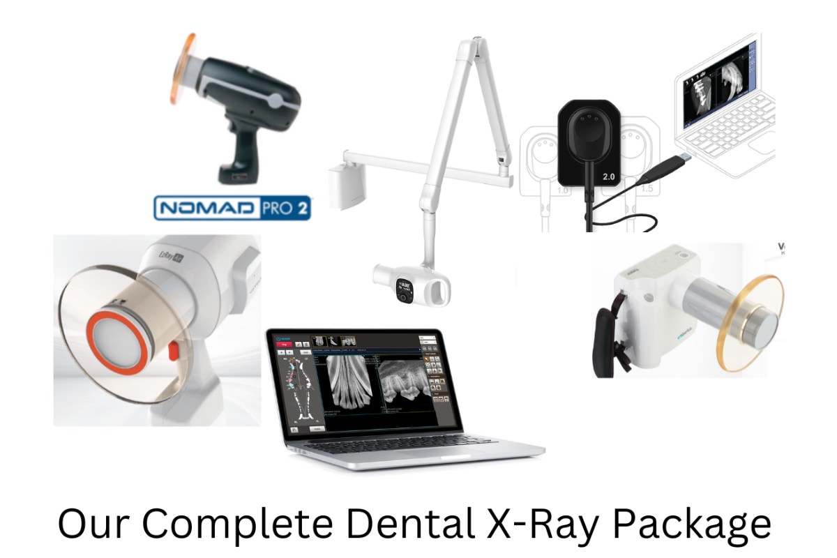
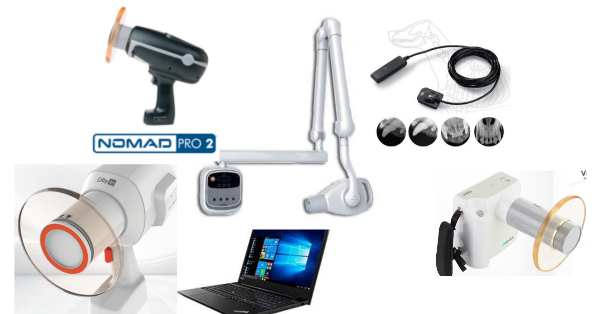
See our complete package system - Click Here
References:
1. Lommer MJ, Vertraete FJ. Prevalence of odontoclastic resorption lesions and periapical radiographic lucencies in cats: 265 cases (1995-1998). J Am Vet Med Assoc 2000;217(12):1866-1869.
2. Heney CM, Arzi B, Kass PH, Hatcher DC, Verstraete FJM. The Diagnostic Yield of Dental Radiography and Cone-Beam Computed Tomography for the Identification of Dentoalveolar Lesions in Cats. Front Vet Sci. 2019 Feb 21;6:42. doi: 10.3389/fvets.2019.00042. PMID: 30847347; PMCID: PMC6393352.
3. Mupparapu M. Digital dental radiography - a review of the solid-state and semi-direct digital detector. Orofac J Sci2011;3(1):40

Veterinary Dental Care with Digital Radiographic Imaging

Elevating Veterinary Dentistry: The Power of Digital Radiographic Imaging
Dental radiography is considered an essential part of human dentistry to aid diagnosis and treatment of dental disorders. The veterinary world is catching up rapidly and dental radiography is fast becoming the standard of care for our veterinary patients.
The production of high-quality dental radiographs requires a number of specific pieces of equipment. First, specific dental X-ray plates should generally be used.
These are small and specifically designed to fit within the oral cavity, minimizing the superimposition of structures within the skull and simplifying radiographic positioning.
They come in a range of sizes from 0 to 4, with size 4 being the largest. Sizes 2 and 4 are the most commonly used. Secondly, a specific dental X-ray generator, either handheld or wall mounted, allows accurate and easy positioning for the various views required.
Traditional analog radiography uses X-ray films with an intensifying screen, set within a light-proof cassette. After exposure to an X-ray beam, this film is then processed using either manual or automatic techniques to provide a high-quality diagnostic image.
The use of digital imaging systems first introduced in the early 2000s has revolutionized dental radiography and has many advantages over older analog systems.
There are two types of digital X-ray set-up - Digital Radiography (DR) and Computed Radiography (CR). DR, or direct, systems use a solid-state sensor plate in place of an X-ray film.
This is linked directly to a computer via either a wire or wirelessly via Bluetooth. CR, or semidirect, systems use a photo-stimulative phosphor (PSP) plate which stores the X-ray exposure.
These are then scanned and translated into a digital image on a computer. Both have advantages and disadvantages, but DR systems are most commonly used in dental radiography systems and are generally accepted as superior.
The advantages of digital dental radiography
While there are many advantages, the most notable include:
Speed - DR systems will produce an almost instant image and the sensor can be left in place making any repositioning for repeat exposures quicker and easier
Reduced number of exposures - Digital radiography systems can adjust for suboptimal exposure settings, meaning repeat exposures due to faults are less likely
Ability to manipulate and magnify images - This allows easier viewing and interpretation of radiographs, picking up more subtle pathologies as the images are more easily interpreted
No degradation over time if stored correctly
No requirement for toxic developing and fixing chemicals
Less space required
Access to telemedicine services
Lower exposure settings - reducing radiation doses to patients and personnel by an estimated 50-80%
Initial problems were reported with reduced image detail compared with analog films, however, these have now long since been resolved.
Another commonly reported disadvantage of digital radiography set-up is that initial costs are somewhat higher than analog systems.
This is certainly true, however, it has been estimated that in a busy veterinary clinic, it would take less than a year to make up for these costs thanks to significantly lower running costs. Recent cost-benefit analyses have shown the investment is worthwhile.
Full-mouth dental radiographs
There are demonstrated benefits of full mouth X-rays as standard for all new patients, or where a clinical condition has significantly changed.
It has been suggested that around 40% more pathology will be detected compared with clinical examination alone.
Radiographs are much more sensitive to detecting periodontal pockets that may be missed by probing alone. They also allow assessment of the thickness and quality of the surrounding bone, reducing the risk of iatrogenic fracture if extractions are attempted, especially in small breed dogs.
Dental radiographs can detect any unusual anatomy such as a curved root that may make extractions more difficult, and post-extraction radiographs can be used to check that no root fragments are remaining.
Especially in cats
Dental radiography is essential when assessing feline mouths where resorptive lesions are present. Without it, it is impossible to differentiate Type 1 lesions that require complete extraction from Type 2 lesions that are better treated with crown amputations.
Diagnosing the lesion type before treatment improves patient outcomes and reduces procedure times. Deciduous teeth, in both cats and dogs, which may have undergone partial resorption can also be properly assessed.
Dental radiographs are useful when assessing fractured or worn teeth for subtle evidence of infection. They are vital in helping to determine whether “missing teeth” are truly missing, fractured crowns with roots remaining or impacted teeth that may lead to serious complications such as dentigerous cysts.
They can also be used to help assess oral masses.
How to make the best use of your dental X-ray system
To make the best use of a dental X-ray system there are a few important considerations;
Correct exposures should be used for different-sized patients and teeth. Some machines will have settings for different teeth programmed in others, others will require the use of a manual exposure chart.
Dental X-ray plates or sensors and correct plate sizes should be used to minimize exposures and allow easier positioning
Good radiation safety should be adhered to at all times following ALARA (as low as reasonably achievable) guidelines
The use of a specific dental X-ray generator is recommended to allow easier and more accurate positioning
Correct radiographic techniques should be utilized - generally, images should be obtained using either a parallel or bisecting angle technique, depending on the teeth and species being imaged. For cats, a near parallel (intra- or extraoral) will be required for maxillary cheek teeth.
Standard views should be obtained for full-mouth radiographs
Dental radiographs should be performed under general anesthesia
All radiographs should be assessed to ensure they are of diagnostic quality
Good training of personnel is vital for both positioning and radiographic interpretation
Digital dental radiography is rapidly emerging as an essential tooth in modern veterinary practice
The whole team should be educated on its benefits to both pets and their owners. Digital radiographic imaging allows early detection of dental disease, simplifies treatment, and improves patient outcomes enhancing veterinary dental care, as well as providing an additional income stream for veterinary businesses.
https://newvetequipment.com/dental-xray-equipment
References:
[1] Niemiec, B. A., Gawor, J., & Viadimír, J. (2017). Practical Veterinary Dental radiography. In CRC Press eBooks. https://doi.org/10.1201/b20288
[2] Niemiec, B. A., & Wright, M. (2011). Digital Dental Radiology. Clinician’s Brief., https://www.cliniciansbrief.com/article/digital-dental-radiology . Accessed 02/08/2023
[3] Bailey, M. (2021). Veterinary dental radiology – an overview. Royal Canin - VetFocus. https://vetfocus.royalcanin.com/en/scientific/veterinary-dental-radiology-an-overview . Accessed 02/08/2023
[4] Haws IJ. The evolution of oral radiography in veterinary medicine. Can Vet J. 2010 Aug;51(8):899-901.
[5] Van Der Stelt, P. F. (2005). Filmless imaging: The uses of digital radiography in dental practice. The Journal of the American Dental Association, 136(10), 1379–1387
[6] DuPont GA. Radiographic evaluation and treatment of feline dental resorptive lesions. Vet Clin North Am Small Anim Pract 2005;943-962.
[7] Niemiec, B. A. (2015). The importance of dental radiography. Today’s Veterinary Practice. https://todaysveterinarypractice.com/dentistry/dental-radiography-series-the-importance-of-dental-radiography/ Accessed 02/08/2023
[8] Niemiec, B. A. (2015). Dental Radiology Series: Techniques for Intraoral Radiology. Today’s Veterinary Practice. https://todaysveterinarypractice.com/dentistry/practical-dentistry-dental-radiology-series-techniques-for-intraoral-radiology/ Accessed 02/08/2023

Veterinary Dental X-ray Features to Look For (That Have Nothing to Do with the X-rays…)

Diagnostic-quality images are the most important feature in a dental x-ray system. While buying something top-of-the-line isn’t always necessary, the images should be of sufficient quality for interpretation, which helps a vet create an appropriate treatment plan.
Assuming the machine produces great x-ray images, other factors can help a veterinary practice decide which system is the best fit for them. Here are a few factors to consider…
Film, CR, or DR
In addition to the generator that produces the x-ray beam, it’s important to think about where the image is picked up and how it’s processed. This article primarily focuses on veterinary digital dental x-ray, which can mean CR (computed radiography) or DR (direct radiography). Film is an option, too, of course. Here are some considerations for each modality…
Film is typically the most time-consuming since it takes time to develop each shot. It also consumes more materials than digital, including the films themselves and processing chemicals. This older technology can cost less initially, but supply costs over time must be factored in.
CR and DR both produce a digital image rather than a physical one. The biggest difference is the way the images are processed. DR sends the image directly to the viewing software just seconds after the digital sensor is exposed, so it is incredibly fast and convenient. The tradeoff is that DR is typically the most expensive modality. But this depends on the specific equipment being compared.
CR often costs a bit less, but it involves an extra step. Phosphor plates are used for the exposure, and they must be run through a processor to obtain the image and then wipe the plate clean for the next exposure. Although this is slower than DR, some veterinary practices improve efficiency by having multiple plate readers to allow more than one to run at a time.
Plate or Sensor Sizes
Many small animal veterinarians see patients ranging from 2-pound chihuahuas and small cats to large breed dogs of 150 pounds or more. Obviously, the teeth in these patients also vary widely in size.
For this reason, it’s desirable to have plates or digital sensors in more than one size—even several different sizes if possible.
There might even be additional applications, such as exotics radiographs, for certain sizes of sensors.
Local Regulations
Some jurisdictions or countries may have regulations on handheld units. It’s important to research first, prior to purchasing any new radiation-producing equipment. Also, the room or suite where the machine would be used should meet all safety regulations.
Setup and Installation
It’s important to consider where dental radiographs will be performed. Depending on the size and arrangement of the designated room, some installation options will fit well, while others will be limited by space constraints.
Common options include wall or ceiling-mounted, stand-mounted, or handheld veterinary dental x-ray units. For busy clinics with more than one prep or x-ray area, a portable handheld unit might be a good fit. When mounting a unit or considering an electrical supply, it’s best to have professional help to make sure everything is secured and safe.
User Friendliness
A user-friendly machine can improve efficiency. This means smoother practice flow, less frustrations and headaches, and potentially a higher number of x-ray studies performed per day or per week (and thus a better return on investment).
Anything that makes dental x-ray studies run more efficiently (minimum number of steps) and intuitively can help. Consider how patient and client information will be input. Look to see if the interface is intuitive and easy to understand. Presets can be very helpful, too.
Think about staff training. It’s no secret that dental radiographs (including that bisecting angle shot) can be a bit tricky at first for anyone who’s new to them. Some companies might offer a veterinary dental radiography training session or CE for team members after the purchase of a new machine.
Software Compatibility and Reliability
Digital veterinary software facilitates the viewing, storing, and sharing of radiographs—including dental ones. Good software makes these functions faster and easier, while software problems can waste time and create headaches.
In addition to basic functions, check how images are formatted, i.e., DICOM, jpeg, etc. Make sure the software is compatible with your practice management software. Ask about technical support, security/privacy/protection against hackers, and how the company handles any issues that arise.
Support
Durability is an important consideration. But even with durable equipment, sometimes accidents or glitches happen.
Protect your investment with warranties, a service plan, and/or 24/7 technical support. But remember that not all plans are created equal. See exactly what the plan covers. One common concern is bite damage (the sensors do go in the patient’s mouth, after all!).
Find out the expected timeline for repairs and whether loaner equipment is available in the meantime. And consider whether replacement parts are likely to be available for the foreseeable future.
Always start by making sure veterinary dental x-ray equipment meets its primary purpose: taking a good quality, diagnostic images.

After that, the purchase isn’t always an “apples-to-apples” comparison. Shop around and see if there’s a good system available with features that benefit your practice and make the process smoother and more efficient.
Written by: Dr. Tammy Powell, DVM

Top 10 Tips for Choosing a Veterinary Dental X-ray Machine
Dental radiographs have become an essential part of veterinary dentistry, often being upheld as the standard of care. That’s because dental x-rays are the only way to diagnose the full extent of dental or periodontal disease.
They help uncover unexpected lesions, assist with planning surgical extractions, and serve as part of the medical record.
Here are 10 factors that can help a busy veterinary practice decide how much to invest and which veterinary dental x-ray equipment is right for them…
Cashflow/return on investment plan. It’s not unusual for veterinary practices to report a significant increase in dental income after investing in dental radiography equipment. That’s because of an increased ability to detect pathology that needs to be treated.
However, each practice has a unique clientele, practice style, patient load and degree of busyness, and financial situation.
Developing a business and financial plan for dentals can help. Think about expected usage of the new equipment, how much to charge in your local area, and other factors. This will help a practice determine how much to invest in their dental radiography equipment.
Image quality and repeatability. Dental x-ray images need to clearly show fine details. A veterinarian must be able to distinguish small changes around the periodontal ligament and root apex, or other fine details, to determine the degree of pathology that is present.
Ask colleagues for advice. See which machines they have, whether they are happy with their purchase, and what they do or do not like about their x-ray system. When planning for a purchase, ask vendors or manufacturers if a test period with a trial machine is available and try it on different size patients to ensure it produces diagnostic images.
Technology: Film, CR, or DR. Although film is becoming less common in all x-ray modalities, there are some practitioners who prefer manually developing dental films and can do so efficiently. But for many veterinarians, digital is the way to go—it’s just a matter of choosing between CR (phosphor plates that use a plate reader to produce a digital image) or DR (a sensor that directly produces a digital image).
Cost is one consideration, as CR technology tends to be less expensive—although it does require the purchase of one or more plate readers. However, DR can be more efficient since there’s no need to run the sensor through any type of developer or plate reader.
Components and sizes. In addition to a generator, the practice will need plates (for CR) or sensors (for DR). Depending on the manufacturer, another important difference between these two technologies comes down to sensor/plate size. CR typically has more of a range of sizes available, which can be helpful for practices that see large dogs, small cats and dogs, and everything in between.
Before purchasing, check which size sensors or plates are available and ensure this will meet your needs.
Mounted versus handheld/portable. Veterinary dental x-ray generators can be mounted to carts or a wall. There are also handheld veterinary dental x-ray generators available. A mobile practice or one with limited space might appreciate a handheld unit, which has a smaller footprint and less wiring/installation concerns. It also allows for flexible angles, potentially without having to move the patient around as much to get all necessary shots.
On the other hand, a handheld unit is at a higher risk of drop damage. And safety must be considered, as veterinary radiology in general is moving toward hands-free restraint and having operators out of the room (if possible) when the image is generated. Some countries or jurisdictions might have restrictions on portable units, too, so be sure to double check before purchasing.
User-friendliness. There’s a learning curve for anyone new to dental x-rays—both for the team members taking the shots, and for the veterinarian interpreting the images. So, no matter which system you purchase, it’s important to invest in some type of training.
That being said, some systems are certainly more intuitive to use than others. Some teams would prefer to have a veterinary digital dental x-ray system with species-specific presets that make it easy to capture all shots at the correct settings.
It also helps to have a generator that is easy to move and set in the right position and angle, whether that’s an ergonomic portable machine or a mounted generator on an arm that stays where it is put.
The durability of ALL components. Common “accidents” in the veterinary setting include drop damage, water damage, and bite damage to the plate or sensor. Look for systems designed to stand up to daily veterinary use.
Warranty, service, and maintenance. Speaking of damage, sometimes accidents happen even when all reasonable precautions are taken. And you don’t want to lose out on your investment if that occurs.
A warranty is very nice to have, although it should cover common veterinary practice-related damage, including bite damage. Ask about warranty costs, coverage, and renewal options.
Also, look for ongoing costs related to service and maintenance, whether loaner equipment is available during extended repairs to equipment, and whether tech support is available.
Veterinary dental x-ray software compatibility. Software is needed for saving images to practice management software or electronic medical records, as well as for viewing and sharing the images. Make sure the dental x-ray equipment you plan to use is compatible with your practice’s software.
Financing and money considerations. Is purchasing the equipment outright an option? Or does financing make more sense in terms of cash flow? Remember to check on tax benefits as well.

Research and discuss everything with decision-makers at the practice. Ask for suggestions from team members who will be using the dental x-ray equipment. Consult business, tax, or financial professionals as needed.
Although any veterinary equipment purchase is a significant investment, it can also be a way to bring new income to the practice—not to mention boost patient care and improve workflow and efficiency.
Written by: Dr. Tammy Powell, DVM

How Much for a Veterinary Digital Dental X-Ray Systems Cost?
An average price range for a new digital dental radiography system would be $8,000-$15,000, depending on which components and features the system includes.
Dental radiographs are now considered a standard part of a comprehensive dental procedure.
Good quality dental x-rays can help a vet discover and treat more pathology, thereby boosting both patient care and revenue.
Although a dental x-ray system is an important equipment investment, a veterinary practice should plan for its purchase appropriately.
This includes knowing how much to budget long-term, exploring financing options, and maximizing the ROI on the new equipment.
It’s important to see exactly what you’re getting for the purchase price. For example, is the generator handheld, or is there a stand, arm, or wall mount included?
For CR plates or digital sensors, how many are included, and in which size(s)?
Also, what type of software are you receiving?
Don’t be afraid to shop around or negotiate with sellers, to receive the best possible price.
However, also keep in mind that the cheapest price isn’t always the best. Quality is key when investing in an important piece of equipment.
Check with colleagues to see which systems they prefer, or ask the seller for references of other vets in your area who have purchased the equipment so you can get some honest feedback.
See if neighboring clinics would mind you stopping by to see the equipment in person and look at the images they’re getting.
What Are the Maintenance Costs of Veterinary Digital Dental X-Ray Equipment?
In addition to the actual purchase price, it’s important to factor in the long-term costs of maintaining your equipment in working order.
Ongoing costs, for both new and used equipment alike, may include…
Warranties. Ask when any initial warranties expire, whether they can be renewed, and how much it will cost to renew. Also, find out exactly what the warranty covers. Ask about drop damage, bite damage (since the sensor will be placed directly in a patient’s mouth!), and any other common incidents that may occur in a veterinary hospital. Not all warranties cover these things.
Ongoing maintenance and repairs. Ask about routine recommended maintenance, as well as costs of the most common types of repairs. If any of this isn’t fully covered by a warranty or service plan, plan ahead for these costs so they don’t catch you by surprise. Also, add these expenses into your total purchase cost. Sometimes, more expensive long-term maintenance can cancel out the cost savings of a lower initial purchase price.
Software upgrades. Timely upgrades are crucial to keeping your equipment working smoothly—for image processing, storage, and sharing. Also, ask about security against hacking or cyber-attacks, since radiographs are part of confidential medical records.
Ask about technical support, and if there is a free 24/7 support line you can call for smaller issues.
Ask about loaner equipment that can be used if any component of your system needs to be repaired. With this option available, you’re less likely to lose revenue if your equipment becomes damaged or needs a repair.
Look Into Equipment Financing Options
Once you’ve calculated all the costs of purchasing and maintaining your new dental x-ray system, it’s also important to consider how you will finance the purchase. This can make or break your monthly cash flow.
The first decision is whether to purchase or rent the equipment. Many vets recommend purchasing if that is an option for your practice. However, each veterinary practice must choose what works best for their finances and practice flow.
Business savings may be a good option to purchase the equipment outright. But sometimes, monthly payment plans are easier on cash flow, since revenue from using the x-ray system could be enough to cover the monthly payment and earn a profit right away.
Of course, this is assuming that the interest rate on the payment plan is reasonable.
For monthly payment plans, check with the equipment seller, or ask about a capital lease (a type of loan for equipment purchases) from banks.
Remember to factor in tax savings (Section 179 of the IRS Tax Code) and seek guidance from a financial professional.
How Can a Veterinary Practice Maximize the Return on Their Equipment Investment?
Of course, you’re probably excited to start using your new dental x-ray equipment. However, maximizing the use of your equipment starts BEFORE the purchase.
If the equipment is confusing or cumbersome to use, it will take more time per study, which means fewer dental procedures can be scheduled. Also, there will be a subconscious reluctance to schedule if the equipment is a pain to use.
So, make sure to invest in a system that is user-friendly and efficient and produces high-quality, diagnostic images.
Also, invest in training for your team, since dental radiographs (especially those bisecting angle shots) can be confusing for anyone new to taking these x-ray studies. That also means training for vets on how to interpret dental images.
While a learning curve should always be expected, good training means less frustration and a shorter time until everyone feels comfortable getting great, diagnostic images—and for vets to feel confident diagnosing pathology on the images.
Finally, plan ahead for how much you will charge for dental x-ray studies, and how this can be worked into the total cost of a dental procedure.

All of these efforts can maximize the ROI on your new digital dental x-ray machine, while ensuring the highest level of patient care—a plan that’s beneficial to everyone.
Written by: Dr. Tammy Powell, DVM

Diagnosing Dental Disease in Small Mammals: A Vet's Guide

Since dental health can be a serious issue that affects a pet’s ability to eat, it’s important to address dental disease when it arises—even in the smallest of patients, like small mammals or pocket pets. While these tiny mouths can be challenging to work with, it is possible to evaluate and treat dental issues in these species.
In recent articles, we’ve discussed dental care in rabbits and guinea pigs. Today, we’ll focus on small rodents such as hamsters, gerbils, mice, and rats.
Which Dental Issues Do Small Mammals Develop?
These species (gerbils, hamsters, mice, and rats) can develop incisor malocclusions, since their incisors are open-rooted and grow continuously.
Unlike rabbits and guinea pigs, these small rodents have closed-rooted molars that do not grow continuously. Fortunately, that means that dental issues of the cheek teeth, while possible, are much less common.
On the other hand, since their molars have fully developed roots and don’t continuously erupt, small rodents may develop periodontal disease secondary to plaque. They may also develop dental caries if fed a diet heavy in sugar, including excessive use of certain commercial rodent treats.
Other oral issues include trauma to the teeth or jaws, food impaction, abscesses and infections, or diseases such as neoplasia. In hamsters, cheek pouch eversions or impactions are especially common.
Normal Dentition in Small Mammals
Mice, rats, gerbils, and hamsters have the dental formula 2(I 1/1, C 0/0, P 0/0, M 3/3), for a total of 16 teeth, with a space called the diastema between the incisors and molars.
The mandibular incisors are about three times as long as the maxillary incisors. The enamel may be white to yellow or orange, depending on the species.
As mentioned above, their incisors grow continuously, while their molars do not.
This dentition is consistent with the lifestyle of these small exotic pets. Their diet primarily includes seeds, roots, tubers, and grains—so unlike rabbits and guinea pigs, they don’t ingest a lot of fiber to the point where they require continuously erupting cheek teeth.
Their incisors wear down due to gnawing and burrowing behaviors. Chewing aids—such as wooden blocks or cardboard that is free of print or dyes—can help promote this natural behavior and keep the incisors in good shape.
Diagnosing Dental Disease in Small Mammals
Symptoms may be subtle at first, and it is common for pet owners to miss disease until it becomes more advanced. Symptoms may include anorexia, any change in behavior (such as hiding or hunching if they are in pain), drooling, and swellings or changes to face symmetry.
Hamsters with cheek pouch impactions may present with swollen cheeks. Or the cheek pouches may be seen protruding from the mouth in the case of eversions.
It’s possible for a veterinarian to do an initial, cursory oral exam on an awake pet using an otoscopic cone, and to evaluate the incisors on an awake pet. This method may uncover obvious abnormalities. But it is likely to miss subtle changes and does not allow for a thorough oral exam.
Anesthesia or sedation are best, both for a full oral examination and for dental radiographs.
Additional tools for small rodents, such as mouth gags and cheek retractors, are needed to get the best view of the teeth and oral cavity. Magnification can also be very valuable.
A dental x-ray study is best performed using a dental x-ray unit, with the sensor placed extra-orally due to the small size of these patients. Mammography film can also be used, as it shows fine details.
Treating Dental Disease in Small Mammals
For incisor malocclusions, the goal is to restore the teeth to their normal length and function. An appropriate type of dental drill is recommended. Avoid nail trimmers, rongeurs, and other cutting tools as these carry a risk of fracturing the teeth during trimming.
Molars should NEVER be trimmed since they have true roots and don’t erupt continuously. However, these cheek teeth may require removal of tartar and treatment of periodontal disease or even extraction of severely diseased or abnormal teeth.
For hamsters with cheek pouch eversions, viable tissue should be put back into normal position and sutured to the cheek. With impactions, the pouch must be emptied out and rinsed with saline.
Antibiotics and pain medications should be prescribed as needed, depending on the pathology.
Any vet treating dental disease in small rodents must have the appropriate sizes and types of dental tools. It’s also possible to use a needle (18 to 25-gauge, depending on the size of the patient) as an elevator during extractions.
Appropriate training is also very important, to ensure these small patients receive the specialized care their need.
Due to their fast metabolism, nutritional support is usually needed until the animal is recovered and eating on its own.
Owner Education is Important
Many clients buy these small pets for their children and don’t necessarily interact with the pets on a daily basis or have a deep understanding of their husbandry needs.

Promoting regular checkups of these patients can help to catch issues early, as well as educate owners on husbandry and on how to tell if their pet is ill.
All of this can serve to better the health of these small rodents and help to catch dental disease or other health issues early, when treatment may be simpler and carry a better prognosis.
Written by: Dr. Tammy Powell, DVM

The Ultimate Guide to Veterinary Dental Cleaning Stations

A comprehensive oral health assessment and treatment (COHAT), also known as a veterinary dental procedure, is an important consideration for any pet’s long-term health.
Keeping the mouth healthy helps protect a dog or cat’s teeth and contributes to a pet’s overall wellbeing. And, it can be a source of revenue for the veterinary practice.
Here are some of the common components of a dental cleaning station, and the roles they play during a COHAT.
The Equipment Needed for Scaling and Polishing
Even for pets who need significant extractions, the dental procedure typically also involves scaling and polishing the remaining teeth, to clean them, keep them in good health for as long as possible, and avoid further tooth loss.
So, it’s safe to say that scaling and polishing are an important part of any dental procedure.
Dental scaling and polishing are often delegated to skilled veterinary technicians and nurses. So, when it comes time to invest in a new dental cleaning station, it’s a good idea for veterinarians and practice managers to review the equipment available and see what is required to meet their practice’s needs.
These common tools on a dental cleaning unit play a role in scaling and polishing:
Ultrasonic scaler. Hand scalers are still used, too, especially for tight spots that need extra attention. And curettes are needed for subgingival cleaning. However, it’s hard to beat an ultrasonic scaler for speed and efficiency when removing large amounts of dental calculus above the gumline. Many include a built-in LED light for easy visualization of the area being cleaned.
Polisher/Low-speed handpiece. This tool is crucial for smoothing over any small defects or microabrasions in the tooth enamel caused during scaling. In other words, polishing should ALWAYS follow scaling. Recent AAHA dental guidelines recommend using disposable prophy angles and individually packaged, fine-grit prophy paste with a polisher.
Air/Water syringe. This tool is useful for flushing away bits of tartar and other debris as you’re working, for drying a tooth prior to applying a sealant, or for irrigation and inspection of any visible subgingival areas after cleaning.
Suction tool. Weak suction can be a convenient way to remove excess water and saliva during a procedure.
Equipment for Drilling and Extractions
Many extractions require drilling into alveolar bone to expose the tooth root or divide the tooth into segments. This allows for safe and efficient extractions, with less risk of leaving root tips or fragments behind.
For this reason, a high-speed drill is an essential component of any veterinary dental cleaning station. The drill should have several burs to choose from, too, since veterinarians see patients of all different sizes and since different bur shapes (rounded or tapered, for example) serve different purposes.
In addition to the drill, hand tools—such as elevators, luxators, extraction forceps, and suture kits—will be used.
Other Factors to Consider In a Dental Cleaning Station
In addition to the tools available on the dental station, here are some considerations that may affect a veterinarian or practice owner’s decision to make a purchase…
The size of the unit. Practices with small spaces may prefer a compact cleaning station, while others might not have a size restriction.
Easy storage and access to tools. No matter the size of the station, it should be easy to store and access the tools right when you need to. No veterinary team member wants to fumble while reaching for a tool or accidentally drop something because it’s difficult to put back.
Water reservoirs. No one wants to run out of water (which is used for many of the tools, including drilling and ultrasonic scaling) during the middle of a procedure, then put things on hold as the reservoir is refilled. For that reason, it may be beneficial to look for a station with a larger distilled water reservoir capacity.
Service, maintenance, and warranty. It’s important to protect any equipment investment and to make sure it’s easy to keep the machine running so a veterinary practice can avoid canceling procedures due to equipment issues.
Also, remember to invest in high-quality complementary equipment. In addition to the dental cleaning station and other dental tools, a complete dental procedure also involves dental x-rays, appropriate anesthetic protocols and equipment, personal protection equipment, and anything else needed to perform a COHAT safely and effectively.
Veterinary dentistry can be a rewarding way to provide excellent patient care and generate income for a veterinary practice—a win-win scenario. To make sure these procedures run as smoothly as possible, it’s important to choose the equipment that’s the best fit for your practice’s needs.
Additional resources:
2019 AAHA Dental Care Guidelines for Dogs and Cats: https://www.aaha.org/globalassets/02-guidelines/dental/aaha_dental_guidelines.pdf
Editor’s Note:
Currently, we offer a complete veterinary dental cleaning station at an affordable price including shipping, delivery, and a 3-year warranty.
The space-saving machine is designed for high-volume use.
It features key tools that are crucial to any veterinary dental procedure—plus, a few exciting special features like advanced cooling for the high-speed drill, a scaler with an endodontics feature, an LED light on the polisher, and more!

Learn more, and contact us with any questions, here: https://newvetequipment.com/cleaning-station
Written by: Dr. Tammy Powell, DVM

Dental Disease and Heart Disease: What’s the Correlation?

Dental disease is one of the most common health conditions affecting dogs and cats, even though much of it is preventable.
Estimates of dental disease prevalence vary, but many experts say that most (more than half of) dogs and cats over three years of age have some form of periodontal disease.
How Dental Disease Progresses to Periodontal Disease
The process begins with plaque, a thin, bacteria-containing film that forms on the teeth. Plaque can be removed by brushing. But if plaque is not removed, minerals in the saliva harden the plaque, which creates tartar (calculus). Tartar is much more difficult to remove, and it can build up to look like “concrete” deposits on the side of a dog or cat’s tooth.
Pet owners may or may not notice tartar on the crown of their pet’s teeth. But the biggest problem comes from the part they can’t see—the tartar that grows below the gumline.
This leads to periodontal disease, a condition that affects the structures that hold teeth in place.
Consequences of Periodontal Disease
The early stage of periodontal disease is gingivitis, inflammation or infection of the gums that is often noticeable as red, swollen, or bleeding gingiva. Gingivitis is reversible if treated in a timely fashion.
After that, further damages from periodontal disease are NOT reversible. This may include damage to the periodontal ligament, cementum, and alveolar bone. These three structures, together with the gingiva, are collectively called the ‘periodontium’.
As periodontal disease progresses, a pet may suffer from gingival recession, tooth loss, alveolar bone loss, and even jaw fractures in severe cases. And one ‘bad’ tooth may affect neighboring teeth, thanks to infection and damage to bone and other surrounding structures.
Bacteremia From Periodontal Disease
Tartar harbors a lot of bacteria, and a mouth affected by periodontal disease carries infections.
These bacteria may enter the bloodstream via areas of the mouth affected by periodontal disease, which may lead to negative health consequences for an affected pet.
Studies demonstrate conflicting conclusions, so the association between bacteremia from periodontal disease and pathology of certain organs (especially the heart, liver, and kidneys) is not as clear-cut as it was once thought to be.
For example, the common condition of MMVD (myxomatous mitral valve degeneration) in small dogs is no longer thought to be caused by periodontal disease. Instead, both are conditions that occur commonly in older, small breed dogs.
The link between periodontal disease and heart disease is more firmly established in human beings. And in pets, there have been associations noted between periodontal disease and pathologic changes to the heart, liver, and kidneys on necropsy.
Because of these associations with organ disease in other parts of the body, and since bacteremia can and does happen in dogs with periodontal disease, it makes sense to take extra precautions for pets with a condition that would make them more at risk from bacteremia.
For example, immunocompromised patients, or patients with certain types of heart conditions may benefit from an injectable dose of prophylactic antibiotics during a dental procedure, whereas it might not be necessary for an otherwise healthy pet.
There’s some speculation that chronic inflammation is responsible for pathology to other organs just as much as (or more than) bacteremia. Maintaining good oral health is the best way to decrease the risks of both infections and inflammation from periodontal disease.
Preventing Dental Issues Is Good for a Pet’s Overall Health
So, what’s the conclusion to draw from all of this information, some of which is conflicting?
Of course, that’s up to each practitioner. But in general, maintaining a healthy mouth will always be a good thing.
Having a chronic disease (such as periodontal disease) anywhere in the body can be a strain on a pet’s health. So appropriate dental care can only serve to improve the overall health of the pet and reduce any risks associated with bacteremia and chronic inflammation.

Additionally, good dental care can also improve quality of life by decreasing oral pain, preventing tooth loss, and decreasing halitosis that could interfere with the human-animal bond. So, preventive dental care is an important part of any pet’s health plan.
Written by: Dr. Tammy Powell, DVM

Veterinary X-Ray Options: Naomi and CareRay Explained

Comparing X-Ray Panels: Naomi and CareRay
Digital radiography equipment continues to evolve, with several types of x-ray detectors or sensors available to veterinarians.
So, which is right for your practice? Do you need the latest technology to provide the best care, or is it best to get the most mileage you can out of previous generations of technology?
The Evolution of Digital X-Ray Detectors
Just like our cell phones and tablets, maybe it feels like there’s always a new type of x-ray technology available, whether you’re looking for small or large animal x-ray equipment, or for general, mobile, or dental radiography.
When trying to research the technology and all the lingo, maybe the choice feels a bit overwhelming.
To start with the basics, here’s a quick overview of the most common digital radiography plates on the market right now…
CR (computed radiography). CR cassettes are also known as phosphor plates. In terms of practical application, the biggest difference between CR and DR is probably the fact that CR requires an extra step—rather than the image being transferred directly from the plates to a computer, the sensors must be read by the CR plate reader.
DR (direct [capture] radiography). DR technology allows images to be read directly from the plate and then show up on a computer screen, which is usually a rapid process. Within DR, there are two main types of plate technologies:
CCD panels. Charge-coupled device (CCD) detectors are generally built into or attached directly to the x-ray table. This technology has been compared to a digital camera in the way it detects and records light.
Flat-panel detectors. Here, the x-rays are converted into an electrical signal (either directly, or indirectly by first converting x-rays into light and then into an electrical signal). The panel is separate from the x-ray table and can be removed in case a horizontal beam is needed.
The advantages of flat panels include lower x-ray doses and better detail and clarity in the images (although opinions on image quality vary amongst practitioners). On the other hand, flat panels tend to cost more than CCD panels.
Note: The abbreviation DR is also used for “digital radiography” in general, in addition to direct capture panels.
Additional X-Ray Equipment Considerations
In addition to the detector that picks up the x-ray beam, successful radiographs also require…
An x-ray generator, which produces the x-ray beam. This may be purchased as part of a new digital x-ray system. Or, older generators and tables may be retrofitted to be compatible with a new digital sensor.
Software. This is what shows the x-ray image that was picked up by the sensor so that a veterinarian can see and read the image. It’s also where images are stored as part of the medical record, or shared electronically with clients, other veterinary clinics, or specialists as needed.
Wired or wireless technology. So far as the sensor or plate goes, some have wire connections while others are wireless.
In addition to considering which sensor technology is a good fit for your practice, it’s important to think about whether or not any other equipment needs to be upgraded, and how compatible the whole system will be together.
When Is It Time to Upgrade?
This depends a bit on a veterinary practice’s needs. Typically, it’s not necessary to replace equipment for the latest model every time new technology is available, and it may be possible to keep older equipment in good working order for many years.
Using the Naomi CCD panels and CareRay Cesium (DR) panels as an example, here are some considerations…
If a clinic is using older Naomi CCD panels that are still producing great, diagnostic images, then it’s typically not necessary to upgrade to a flat panel detector.
The veterinarian is not missing out by hanging onto older technology that still gets the job done.
What if the plates work fine but there’s a software problem—say, the software is no longer supported? Or, what if there’s no longer any technical support or a warranty available for the plates, which sometimes happens with older technology?
Here, the decision to upgrade may come down to personal preference, budget, risk tolerance, and how much time and effort it takes to get readable images from the software.
Of course, if the panels are no longer producing diagnostic images, then it’s probably time to upgrade.
You can always check on your warranty if it’s still in place or ask about the cost of repairs, but sometimes veterinarians find that repairs are costly on older technology and their investment may be better spent on an upgrade. But this strategy will vary depending on each individual business and situation.
No matter which equipment you are currently using (or considering purchasing), remember to also think about things such as availability and cost of technical support, whether a loaner plate is available during repairs, and how the software integrates with your practice software.
Also, see if you can sell your used equipment (or make a trade-in, if your vendor allows) to offset the costs of new equipment.

All of these factors will aid in the decision of whether or not it’s time for an upgrade.
Written by: Dr. Tammy Powell, DVM