

Better Dentistry, Better Business: The Value of Digital Dental X-Rays

The Hidden ROI of Veterinary Dentistry: How Digital Dental X-Rays Pay for Themselves
When veterinary practices consider investing in dental equipment, the conversation often starts with patient care…and rightfully so.
Dental disease is one of the most common health issues affecting companion animals, and dental radiographs have become an essential part of providing high-quality care.
But there is another side of the conversation that is equally important: the business case for veterinary dentistry.
Running a successful practice requires more than compassion and clinical expertise. You need to generate enough revenue to pay your team, invest in technology, maintain facilities, and continue delivering exceptional care.
Fortunately, veterinary dentistry is one of the few service areas where improved patient outcomes and financial sustainability often go hand in hand.
The key is making smart equipment investments that support both goals.
Why Dental Radiographs Are the Standard of Care
Many pet owners are surprised to learn that much of a tooth lies below the gumline. In fact, studies have shown that a significant percentage of dental pathology cannot be identified through a visual examination alone.
Without dental radiographs, veterinarians may miss:
- Tooth root abscesses
- Resorptive lesions
- Periodontal bone loss
- Retained roots
- Fractured teeth
- Endodontic disease
Digital dental X-rays allow you to identify problems that would otherwise remain hidden, helping you provide more accurate diagnoses and treatment recommendations.
Today, many veterinarians consider dental radiography a fundamental component of a complete dental procedure rather than an optional add-on.
Looking Beyond the Price Tag
One of the biggest misconceptions about dental equipment is that practices need to spend top dollar to build a successful dental program.
Some premium dental imaging setups can cost $35,000 or more once sensors, generators, software, and accessories are included.
While those systems may offer additional features, you can achieve excellent clinical results with quality digital dental radiography systems in the $12,000-$15,000 range.
The difference matters.
A lower initial investment can significantly shorten the time required to achieve a positive return while still providing the diagnostic capabilities veterinarians need to deliver quality care.
A Simple ROI Example
Let’s look at a realistic scenario:
Assume a practice purchases a digital dental radiography system for $15,000. Now consider a conservative estimate:
Average dental procedure revenue: $600
Dental procedures performed per month: 10
Monthly dental revenue: $6,000
Of course, equipment cost is only one piece of the equation. There are labor, anesthesia, supplies, and overhead expenses to consider. However, even when accounting for those costs, dental procedures often represent a valuable service line for many practices.
If the practice attributes only a portion of that revenue toward recovering the equipment investment, the payback period can still be surprisingly short.
For example:
Equipment investment: $15,000
Contribution toward equipment recovery per procedure: $150
Procedures needed to recover investment: 100
At just 10 dental procedures per month, the equipment could effectively pay for itself in less than a year.
Now compare that to a $35,000 investment.
Using the same assumptions, the practice would need approximately 233 procedures to recoup the equipment cost, significantly extending the payback timeline.
The takeaway is not that higher-priced systems are inherently bad investments. The purchase price directly affects how quickly a clinic can realize a return.
Improving Workflow Efficiency
The ROI of digital dental radiography is not limited to revenue generation. Digital systems can also improve workflow efficiency throughout the practice.
Digital dental X-rays eliminate many time-consuming steps and can help reduce the need for unnecessary retakes.
Benefits often include:
- Immediate image review
- Improved image storage and retrieval
- Easier integration with practice management software
When procedures move more efficiently, you can often complete appointments more predictably and utilize staff time more effectively.
Helping Clients Say “Yes” to Treatment
Client education is one of the most overlooked benefits of digital imaging. When pet owners can actually see pathology on a screen, treatment recommendations become much easier to understand.
A fractured tooth root, severe bone loss, or a resorptive lesion is often far more compelling when accompanied by an image rather than a verbal explanation alone.
The result is better patient care and greater confidence in the veterinary team’s recommendations.
What to Look for When Purchasing Dental Equipment
Not all dental radiography systems are created equal, but the most expensive option is not always the best choice. When evaluating equipment, consider:
- Image Quality: The system should consistently provide diagnostic-quality images with excellent detail.
- Ease of Use: A user-friendly interface can reduce training time and improve adoption among team members.
- Sensor Durability: Dental sensors represent a significant investment. Durability and warranty coverage matter.
- Software Integration: Look for systems that work seamlessly with your existing workflow and practice management software.
- Service and Support: Reliable technical support can make a major difference when issues arise.
- Total Cost of Ownership: Consider not only the purchase price but also future maintenance, replacement costs, and warranty protection.
Better Dentistry, Better Business
Veterinary dentistry should never be viewed solely as a revenue generator. At its core, dental care is about identifying pain, treating disease, and improving quality of life for pets.
But it is also fair, and necessary, to acknowledge the business benefits.
Profitable service lines help you retain talented staff, invest in better technology, expand patient care capabilities, and continue serving your community for years to come.


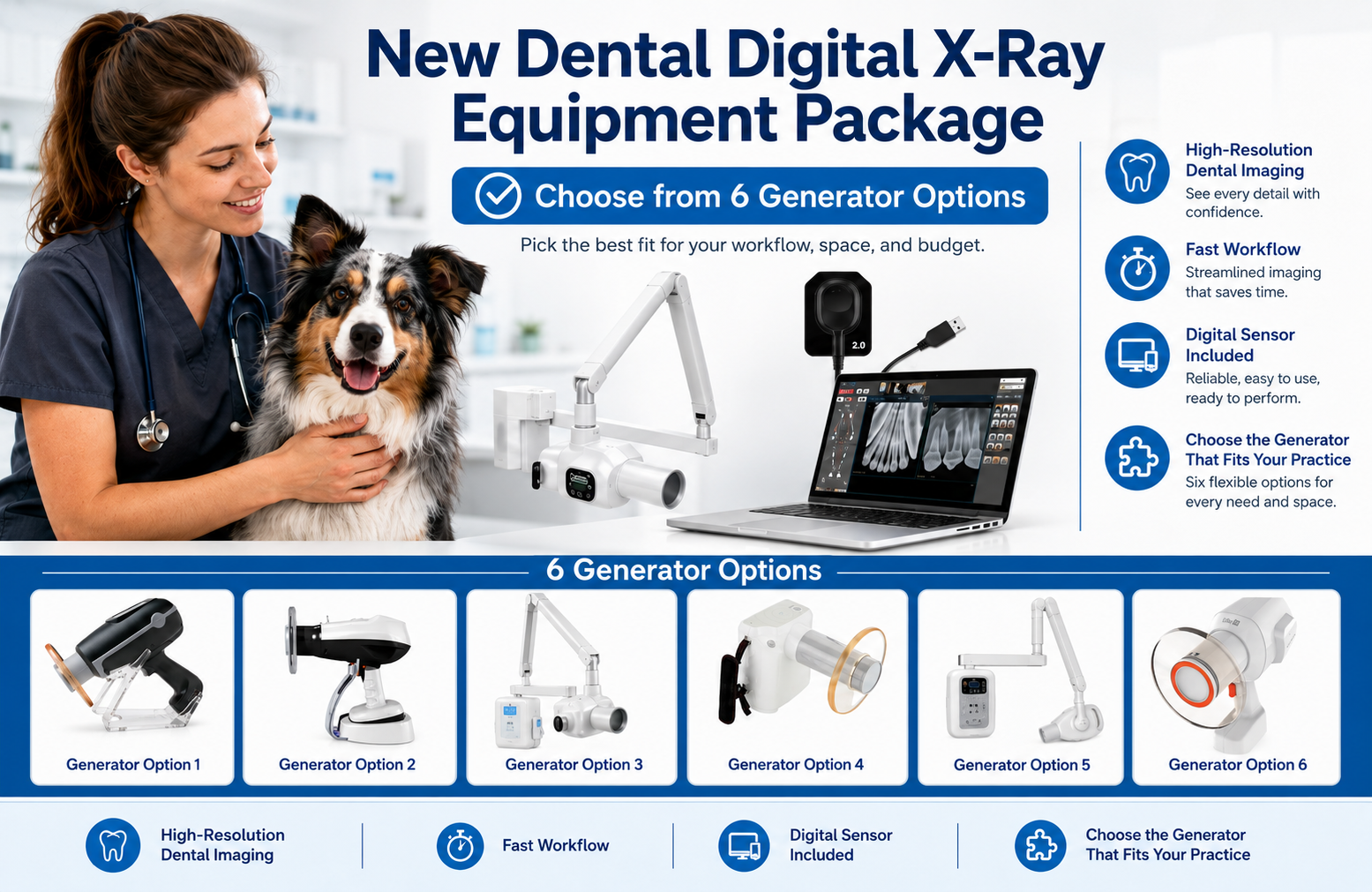
How Much for a Veterinary Digital Dental X-Ray Systems Cost?
An average price range for a new digital dental radiography system would be $8,000-$15,000, depending on which components and features the system includes.
Dental radiographs are now considered a standard part of a comprehensive dental procedure.
Good quality dental x-rays can help a vet discover and treat more pathology, thereby boosting both patient care and revenue.
Although a dental x-ray system is an important equipment investment, a veterinary practice should plan for its purchase appropriately.
This includes knowing how much to budget long-term, exploring financing options, and maximizing the ROI on the new equipment.
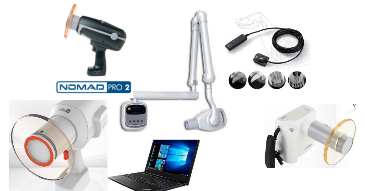
It’s important to see exactly what you’re getting for the purchase price. For example, is the generator handheld, or is there a stand, arm, or wall mount included?
For CR plates or digital sensors, how many are included, and in which size(s)?
Also, what type of software are you receiving?
Don’t be afraid to shop around or negotiate with sellers, to receive the best possible price.
However, also keep in mind that the cheapest price isn’t always the best. Quality is key when investing in an important piece of equipment.
Check with colleagues to see which systems they prefer, or ask the seller for references of other vets in your area who have purchased the equipment so you can get some honest feedback.
See if neighboring clinics would mind you stopping by to see the equipment in person and look at the images they’re getting.
What Are the Maintenance Costs of Veterinary Digital Dental X-Ray Equipment?
In addition to the actual purchase price, it’s important to factor in the long-term costs of maintaining your equipment in working order.
Ongoing costs, for both new and used equipment alike, may include…
Warranties. Ask when any initial warranties expire, whether they can be renewed, and how much it will cost to renew. Also, find out exactly what the warranty covers. Ask about drop damage, bite damage (since the sensor will be placed directly in a patient’s mouth!), and any other common incidents that may occur in a veterinary hospital. Not all warranties cover these things.
Ongoing maintenance and repairs. Ask about routine recommended maintenance, as well as costs of the most common types of repairs. If any of this isn’t fully covered by a warranty or service plan, plan ahead for these costs so they don’t catch you by surprise. Also, add these expenses into your total purchase cost. Sometimes, more expensive long-term maintenance can cancel out the cost savings of a lower initial purchase price.
Software upgrades. Timely upgrades are crucial to keeping your equipment working smoothly—for image processing, storage, and sharing. Also, ask about security against hacking or cyber-attacks, since radiographs are part of confidential medical records.
Ask about technical support, and if there is a free 24/7 support line you can call for smaller issues.
Ask about loaner equipment that can be used if any component of your system needs to be repaired. With this option available, you’re less likely to lose revenue if your equipment becomes damaged or needs a repair.
Look Into Equipment Financing Options
Once you’ve calculated all the costs of purchasing and maintaining your new dental x-ray system, it’s also important to consider how you will finance the purchase. This can make or break your monthly cash flow.
The first decision is whether to purchase or rent the equipment. Many vets recommend purchasing if that is an option for your practice. However, each veterinary practice must choose what works best for their finances and practice flow.
Business savings may be a good option to purchase the equipment outright. But sometimes, monthly payment plans are easier on cash flow, since revenue from using the x-ray system could be enough to cover the monthly payment and earn a profit right away.
Of course, this is assuming that the interest rate on the payment plan is reasonable.
For monthly payment plans, check with the equipment seller, or ask about a capital lease (a type of loan for equipment purchases) from banks.
Remember to factor in tax savings (Section 179 of the IRS Tax Code) and seek guidance from a financial professional.
How Can a Veterinary Practice Maximize the Return on Their Equipment Investment?
Of course, you’re probably excited to start using your new dental x-ray equipment. However, maximizing the use of your equipment starts BEFORE the purchase.
If the equipment is confusing or cumbersome to use, it will take more time per study, which means fewer dental procedures can be scheduled. Also, there will be a subconscious reluctance to schedule if the equipment is a pain to use.
So, make sure to invest in a system that is user-friendly and efficient and produces high-quality, diagnostic images.
Also, invest in training for your team, since dental radiographs (especially those bisecting angle shots) can be confusing for anyone new to taking these x-ray studies. That also means training for vets on how to interpret dental images.
While a learning curve should always be expected, good training means less frustration and a shorter time until everyone feels comfortable getting great, diagnostic images—and for vets to feel confident diagnosing pathology on the images.
Finally, plan ahead for how much you will charge for dental x-ray studies, and how this can be worked into the total cost of a dental procedure.

All of these efforts can maximize the ROI on your new digital dental x-ray machine, while ensuring the highest level of patient care—a plan that’s beneficial to everyone.
Written by: Dr. Tammy Powell, DVM

Diagnosing Dental Disease in Small Mammals: A Vet's Guide

Since dental health can be a serious issue that affects a pet’s ability to eat, it’s important to address dental disease when it arises—even in the smallest of patients, like small mammals or pocket pets. While these tiny mouths can be challenging to work with, it is possible to evaluate and treat dental issues in these species.
In recent articles, we’ve discussed dental care in rabbits and guinea pigs. Today, we’ll focus on small rodents such as hamsters, gerbils, mice, and rats.
Which Dental Issues Do Small Mammals Develop?
These species (gerbils, hamsters, mice, and rats) can develop incisor malocclusions, since their incisors are open-rooted and grow continuously.
Unlike rabbits and guinea pigs, these small rodents have closed-rooted molars that do not grow continuously. Fortunately, that means that dental issues of the cheek teeth, while possible, are much less common.
On the other hand, since their molars have fully developed roots and don’t continuously erupt, small rodents may develop periodontal disease secondary to plaque. They may also develop dental caries if fed a diet heavy in sugar, including excessive use of certain commercial rodent treats.
Other oral issues include trauma to the teeth or jaws, food impaction, abscesses and infections, or diseases such as neoplasia. In hamsters, cheek pouch eversions or impactions are especially common.
Normal Dentition in Small Mammals
Mice, rats, gerbils, and hamsters have the dental formula 2(I 1/1, C 0/0, P 0/0, M 3/3), for a total of 16 teeth, with a space called the diastema between the incisors and molars.
The mandibular incisors are about three times as long as the maxillary incisors. The enamel may be white to yellow or orange, depending on the species.
As mentioned above, their incisors grow continuously, while their molars do not.
This dentition is consistent with the lifestyle of these small exotic pets. Their diet primarily includes seeds, roots, tubers, and grains—so unlike rabbits and guinea pigs, they don’t ingest a lot of fiber to the point where they require continuously erupting cheek teeth.
Their incisors wear down due to gnawing and burrowing behaviors. Chewing aids—such as wooden blocks or cardboard that is free of print or dyes—can help promote this natural behavior and keep the incisors in good shape.
Diagnosing Dental Disease in Small Mammals
Symptoms may be subtle at first, and it is common for pet owners to miss disease until it becomes more advanced. Symptoms may include anorexia, any change in behavior (such as hiding or hunching if they are in pain), drooling, and swellings or changes to face symmetry.
Hamsters with cheek pouch impactions may present with swollen cheeks. Or the cheek pouches may be seen protruding from the mouth in the case of eversions.
It’s possible for a veterinarian to do an initial, cursory oral exam on an awake pet using an otoscopic cone, and to evaluate the incisors on an awake pet. This method may uncover obvious abnormalities. But it is likely to miss subtle changes and does not allow for a thorough oral exam.
Anesthesia or sedation are best, both for a full oral examination and for dental radiographs.
Additional tools for small rodents, such as mouth gags and cheek retractors, are needed to get the best view of the teeth and oral cavity. Magnification can also be very valuable.
A dental x-ray study is best performed using a dental x-ray unit, with the sensor placed extra-orally due to the small size of these patients. Mammography film can also be used, as it shows fine details.
Treating Dental Disease in Small Mammals
For incisor malocclusions, the goal is to restore the teeth to their normal length and function. An appropriate type of dental drill is recommended. Avoid nail trimmers, rongeurs, and other cutting tools as these carry a risk of fracturing the teeth during trimming.
Molars should NEVER be trimmed since they have true roots and don’t erupt continuously. However, these cheek teeth may require removal of tartar and treatment of periodontal disease or even extraction of severely diseased or abnormal teeth.
For hamsters with cheek pouch eversions, viable tissue should be put back into normal position and sutured to the cheek. With impactions, the pouch must be emptied out and rinsed with saline.
Antibiotics and pain medications should be prescribed as needed, depending on the pathology.
Any vet treating dental disease in small rodents must have the appropriate sizes and types of dental tools. It’s also possible to use a needle (18 to 25-gauge, depending on the size of the patient) as an elevator during extractions.
Appropriate training is also very important, to ensure these small patients receive the specialized care their need.
Due to their fast metabolism, nutritional support is usually needed until the animal is recovered and eating on its own.
Owner Education is Important
Many clients buy these small pets for their children and don’t necessarily interact with the pets on a daily basis or have a deep understanding of their husbandry needs.

Promoting regular checkups of these patients can help to catch issues early, as well as educate owners on husbandry and on how to tell if their pet is ill.
All of this can serve to better the health of these small rodents and help to catch dental disease or other health issues early, when treatment may be simpler and carry a better prognosis.
Written by: Dr. Tammy Powell, DVM