


Diagnosing Dental Disease in Small Mammals: A Vet's Guide
Since dental health can be a serious issue that affects a pet’s ability to eat, it’s important to address dental disease when it arises—even in the smallest of patients, like small mammals or pocket pets. While these tiny mouths can be challenging to work with, it is possible to evaluate and treat dental issues in these species.
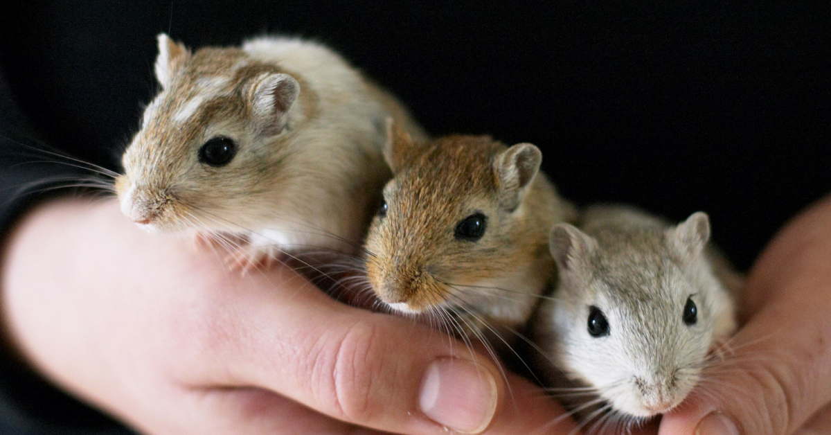
In recent articles, we’ve discussed dental care in rabbits and guinea pigs. Today, we’ll focus on small rodents such as hamsters, gerbils, mice, and rats.
Which Dental Issues Do Small Mammals Develop?
These species (gerbils, hamsters, mice, and rats) can develop incisor malocclusions, since their incisors are open-rooted and grow continuously.
Unlike rabbits and guinea pigs, these small rodents have closed-rooted molars that do not grow continuously. Fortunately, that means that dental issues of the cheek teeth, while possible, are much less common.
On the other hand, since their molars have fully developed roots and don’t continuously erupt, small rodents may develop periodontal disease secondary to plaque. They may also develop dental caries if fed a diet heavy in sugar, including excessive use of certain commercial rodent treats.
Other oral issues include trauma to the teeth or jaws, food impaction, abscesses and infections, or diseases such as neoplasia. In hamsters, cheek pouch eversions or impactions are especially common.
Normal Dentition in Small Mammals
Mice, rats, gerbils, and hamsters have the dental formula 2(I 1/1, C 0/0, P 0/0, M 3/3), for a total of 16 teeth, with a space called the diastema between the incisors and molars.
The mandibular incisors are about three times as long as the maxillary incisors. The enamel may be white to yellow or orange, depending on the species.
As mentioned above, their incisors grow continuously, while their molars do not.
This dentition is consistent with the lifestyle of these small exotic pets. Their diet primarily includes seeds, roots, tubers, and grains—so unlike rabbits and guinea pigs, they don’t ingest a lot of fiber to the point where they require continuously erupting cheek teeth.
Their incisors wear down due to gnawing and burrowing behaviors. Chewing aids—such as wooden blocks or cardboard that is free of print or dyes—can help promote this natural behavior and keep the incisors in good shape.
Diagnosing Dental Disease in Small Mammals
Symptoms may be subtle at first, and it is common for pet owners to miss disease until it becomes more advanced. Symptoms may include anorexia, any change in behavior (such as hiding or hunching if they are in pain), drooling, and swellings or changes to face symmetry.
Hamsters with cheek pouch impactions may present with swollen cheeks. Or the cheek pouches may be seen protruding from the mouth in the case of eversions.
It’s possible for a veterinarian to do an initial, cursory oral exam on an awake pet using an otoscopic cone, and to evaluate the incisors on an awake pet. This method may uncover obvious abnormalities. But it is likely to miss subtle changes and does not allow for a thorough oral exam.
Anesthesia or sedation are best, both for a full oral examination and for dental radiographs.
Additional tools for small rodents, such as mouth gags and cheek retractors, are needed to get the best view of the teeth and oral cavity. Magnification can also be very valuable.
A dental x-ray study is best performed using a dental x-ray unit, with the sensor placed extra-orally due to the small size of these patients. Mammography film can also be used, as it shows fine details.
Treating Dental Disease in Small Mammals
For incisor malocclusions, the goal is to restore the teeth to their normal length and function. An appropriate type of dental drill is recommended. Avoid nail trimmers, rongeurs, and other cutting tools as these carry a risk of fracturing the teeth during trimming.
Molars should NEVER be trimmed since they have true roots and don’t erupt continuously. However, these cheek teeth may require removal of tartar and treatment of periodontal disease or even extraction of severely diseased or abnormal teeth.
For hamsters with cheek pouch eversions, viable tissue should be put back into normal position and sutured to the cheek. With impactions, the pouch must be emptied out and rinsed with saline.
Antibiotics and pain medications should be prescribed as needed, depending on the pathology.
Any vet treating dental disease in small rodents must have the appropriate sizes and types of dental tools. It’s also possible to use a needle (18 to 25-gauge, depending on the size of the patient) as an elevator during extractions.
Appropriate training is also very important, to ensure these small patients receive the specialized care their need.
Due to their fast metabolism, nutritional support is usually needed until the animal is recovered and eating on its own.
Owner Education is Important
Many clients buy these small pets for their children and don’t necessarily interact with the pets on a daily basis or have a deep understanding of their husbandry needs.

Promoting regular checkups of these patients can help to catch issues early, as well as educate owners on husbandry and on how to tell if their pet is ill.
All of this can serve to better the health of these small rodents and help to catch dental disease or other health issues early, when treatment may be simpler and carry a better prognosis.
Written by: Dr. Tammy Powell, DVM

Essential Guide to Guinea Pig Dental Care

Guinea pigs have teeth that grow continuously, and their dental needs are very different from the dental needs of a dog or cat.
However, dental health is still a vital part of a guinea pig’s wellbeing. Here are some important things to know…
Which Dental Problems Do Guinea Pigs Develop?
Congenital malocclusions are possible in guinea pigs, although much less common than in rabbits since guinea pigs have not been bred with dwarf or shortened snout variations.
Acquired dental disease accounts for the majority of dental issues in guinea pigs—with dietary factors being a primary cause, due to uneven or inadequate wearing down of the teeth.
A common problem is an inadequate fiber in the diet, which is normally provided via hay. A vitamin C deficiency can also lead to dental issues since it affects the integrity of the connective tissues that hold the teeth in place—leading to loose and misaligned teeth and subsequent tooth overgrowth.
Any other local or systemic issues (trauma to the jaws or teeth, systemic disease, neoplasia, etc.) that affect a guinea pig’s eating habits could also lead to dental problems.
The uneven or inadequate wearing of the teeth can cause malocclusions, elongated crowns, and sharp edges that can injure or ulcerate the buccal mucosa and cause pain. Overgrowth on the lingual aspect may entrap or irritate the tongue. All of this can make it difficult or impossible for a guinea pig to eat.
Abscesses or infections may also occur, although this is much less common than in rabbits.
Normal Guinea Pig Dentition
In order to recognize what is abnormal in this species, it’s important to know what’s normal for them.
Guinea pigs have open-rooted teeth that continuously grow, and they wear down their teeth by chewing on fibrous foods.
Their dentition is the same in all four quadrants: one incisor, followed by one premolar and three molars. Guinea pigs do not have canine teeth, but instead have a space called the diastema between the incisors and the premolars.
The premolars and molars are anatomically identical and collectively known as the cheek teeth.
Since the mandible is wider than the maxilla, the cheek teeth curve (the mandibular teeth inward and the maxillary teeth outward) to occlude with one another on a sloped plane.
Signs of Dental Disease in a Guinea Pig
Since guinea pigs are prey species, they try to hide signs of a problem for as long as possible. But a pet owner may pick up on changes in eating habits, which develop even in the early stages of malocclusions.
Anorexia is a common symptom of dental problems.
Other symptoms may include weight loss, changes in fecal output (quantity and appearance), dropping food, changes to grooming habits (and subsequent skin issues), salivation, and GI stasis. Facial swellings are possible but less common.
Diagnosing Dental Disease in Guinea Pigs
General anesthesia is best for a thorough dental evaluation.
Cheek teeth abnormalities are very common, more so than incisor malocclusions. So, if an incisor malocclusion is present, it’s a good indicator that a closer look at the cheek teeth is necessary.
A visual examination with a good light source and an otoscopic cone or speculum may reveal a dental issue. However, small or subtle lesions can be missed this way.
Use of an endoscope is very helpful for diagnosing dental problems in guinea pigs, with the added bonus of clear images to show clients so they truly understand the issue.
Radiographs are also important for diagnosing dental issues. And if available, CT is an excellent tool for obtaining diagnostic information.
Treating Dental Disease in Guinea Pigs
Supportive therapy is usually needed, since dental problems impact a guinea pig’s ability to eat, and pets may present in poor condition with weight loss and anorexia. Supportive care commonly includes fluid support, dietary support with a critical care formula, and pain relief.
All of this can help stabilize the patient for anesthesia, prevent complications such as GI stasis, and promote a better recovery and return to normal eating habits.
The main goal of dental treatment is to restore the normal length and shape of the teeth as much as possible and to promote normal occlusion in the process.
A high-speed dental drill is generally used while cutting tools are not recommended because they can cause fractures in the teeth. If needed, extractions of diseased teeth or surgical removal of abscesses can be performed, along with appropriate antibiotic therapy.
Prognosis varies, based on the severity of the disease. Severely overgrown teeth may require multiple treatments are done 1-2 months apart, or even chronic palliative treatments.
To perform safe and effective dental procedures for guinea pigs, it’s important for a veterinarian to gain training and experience, and to have the right tools. Referral to a specialist is a good option, too.
Prevention Is the Best Medicine
Since most dental problems in guinea pigs are related to diet, client education can go a long way toward preventing dental issues in guinea pigs.
Providing instruction on a guinea pig’s dietary requirements is a great step at a new pet visit. Additionally, owners should know to look for anorexia or changes in their pet’s eating habits and to report concerns to their vet right away.
Quick recognition of a problem could help to catch the issue early, resulting in more timely and effective treatment

Written by: Dr. Tammy Powell, DVM