


How Veterinary Dentistry Improves Staff Engagement and Productivity
Veterinary Dental Care Supports Team Growth and Retention
Ask most veterinary professionals why dentistry matters, and you'll hear plenty about periodontal disease, oral pain, and improved patient health. What you may hear less often is this:
A strong dental program can also strengthen the veterinary team.
I know how that sounds, but stick with me.
In a profession where burnout, staffing shortages, and compassion fatigue continue to challenge practices of every size, dentistry offers something many teams are searching for:
- Meaningful work
- Skill development
- Collaboration
While dentistry certainly benefits patients and clients, it can also become one of the most rewarding areas of practice for veterinarians, technicians, and support staff alike.
Giving Technicians the Opportunity to Practice at a Higher Level
Veterinary technicians are highly skilled professionals, yet many teams struggle with fully utilizing those skills. Dentistry offers technicians opportunities to become deeply involved in patient care before, during, and after procedures.
Depending on state regulations and practice protocols, technicians often play critical roles in:
- Patient preparation and anesthesia monitoring
- Dental charting and radiography
- Scaling and polishing
- Recovery monitoring
- Client education
Rather than performing the same routine tasks throughout the day, dental technicians can engage in more advanced, hands-on work.
For many team members, that increased responsibility brings greater job satisfaction.
When people are trusted to use their skills, they become more invested in both their work and their patients.
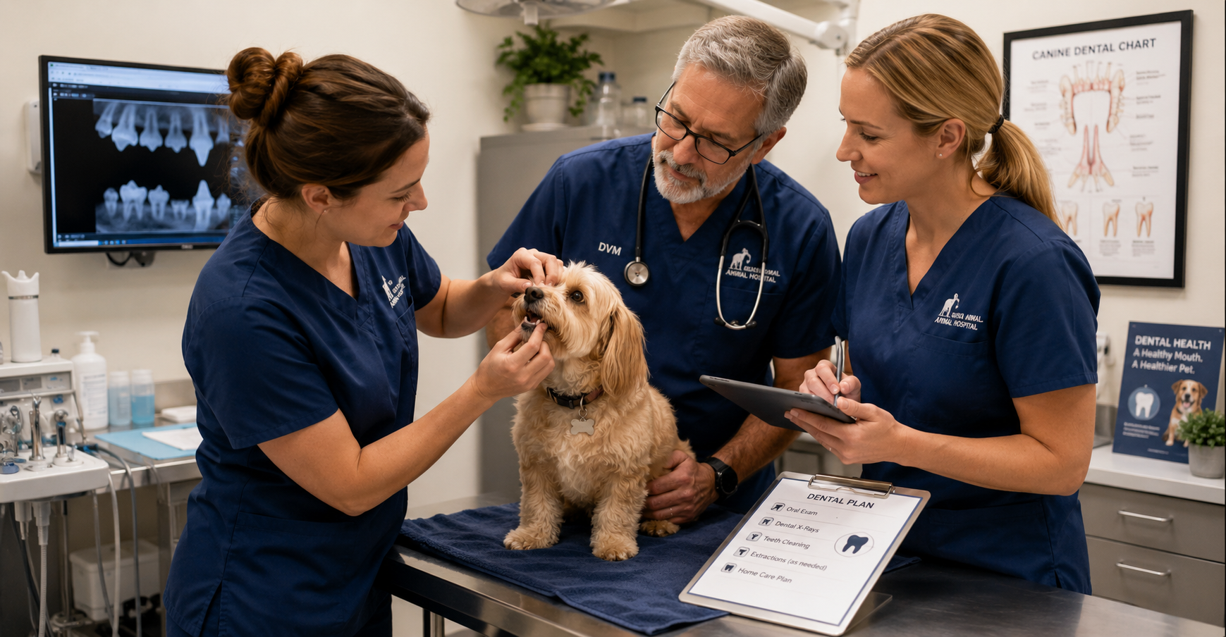
The Moment Things Change: Seeing What’s Hidden
One of the most rewarding moments during a dental procedure often happens when the first radiographs appear on the screen.
A tooth that looked relatively normal externally may reveal severe bone loss. A fractured tooth may show a damaged root. A cat with mild tartar may have advanced resorptive lesions.
These discoveries create valuable learning opportunities for the entire team.
Dental radiography turns procedures into diagnostic investigations rather than routine cleanings. Team members begin asking questions:
- What are we seeing?
- Why is this happening?
- What treatment options are available?
- How can we improve this patient's comfort?
Those moments build clinical confidence and encourage continued learning.
Over time, your team becomes better at recognizing pathology, discussing findings, and contributing to patient care decisions.
Immediate Results Create Meaningful Work
Veterinary medicine can sometimes feel emotionally challenging because progress isn't always obvious.
Chronic disease management takes time. Behavioral improvements may take months. Long-term treatment plans often unfold gradually.
Dentistry is different.
A patient may arrive with severe tartar, inflammation, infection, or oral pain and leave significantly more comfortable the very same day.
The team can see the difference. Clients often notice the difference quickly. And perhaps most importantly, staff members know they helped relieve pain that may have gone unnoticed for months or years.
Those experiences matter.
They remind your team why they entered the field in the first place.
Creating More Predictable Days
Anyone who has worked in veterinary medicine knows that unpredictability is part of the job.
Emergencies happen. Schedules change. Appointments run long.
While no service completely eliminates those challenges, well-organized dental programs can add welcome structure to the workday. Dedicated dental appointments often create:
- Predictable procedure blocks
- Clear team assignments
- Consistent equipment setups
- Efficient patient flow
Your team knows their role. Supplies are prepared. Expectations are clear.
Instead of constantly reacting, your team spends more time working together with purpose.
Investing in Growth Keeps Teams Engaged
Few things improve retention as much as professional growth. Dental programs often encourage continuing education opportunities, such as:
- Dental radiography training
- Anesthesia education
- Dentistry certifications
- Wet labs and hands-on courses
When you invest in these areas, employees often feel valued and supported.
A technician who develops expertise in dentistry may become a mentor, trainer, or internal resource for the rest of the team. That sense of ownership can significantly improve engagement and long-term retention.
In an industry facing staffing challenges, opportunities for growth matter.
Building a Culture Around Better Care
The most successful dental programs become more than a service line. They become part of the practice culture.
Technicians become advocates for dental education. Doctors collaborate on treatment planning. Clients receive more consistent recommendations.
That sense of purpose can strengthen teamwork and create a culture centered around delivering better care.
Dentistry Is About More Than Procedures
It's easy to measure dentistry by production numbers, procedure counts, or equipment investments. But those metrics only tell part of the story. Every dental procedure is also an opportunity:
- To help a pet live more comfortably.
- To strengthen a technician's skills.
- To build confidence within the team.
- To create meaningful work.
- To support professional growth.
Dentistry isn't simply a revenue center. It's a service that allows your team to practice at a higher level while making a real difference in their patients' lives.
And when your team feels challenged, supported, and connected to their work, everyone benefits—including the pets they care for every day.


Building a Sustainable Veterinary Dental Program: The Benefits to Pets, Clients, and Practice Growth
Build a Stronger Veterinary Dental Program
How Better Dental Care Benefits Pets, Clients, Veterinary Teams, and Practice Growth
When veterinarians talk about dentistry, the conversation usually centers on patient health—and for good reason.
Dental disease can cause chronic pain, infection, tooth loss, and a significant decline in a pet’s comfort and quality of life. Helping pets live healthier, more comfortable lives is the primary reason veterinary dentistry matters.
But there is another side of the story that also deserves attention.
A well-developed dental program can strengthen client relationships, help more pet owners follow through with recommended care, create opportunities for team development, and provide a predictable source of revenue that supports the long-term health of the practice.
Veterinary dentistry is one of the rare areas where everyone can benefit: pets, clients, veterinary teams, and the clinic.
Dental Disease Is Often Underdiagnosed
One of the biggest challenges in veterinary dentistry is that many pets do not show obvious signs of discomfort.
Unlike people, dogs and cats often continue eating and following their normal routines even when they are experiencing significant dental pain. They quietly adapt, which can make oral disease difficult for pet owners to recognize.
As a result, serious dental conditions may go unnoticed until they become advanced. Veterinarians commonly discover:
- Advanced periodontal disease
- Tooth-root infections
- Fractured teeth
- Tooth resorption
- Significant bone loss
Many of these conditions can only be fully identified through a comprehensive oral examination and dental imaging.
Because pets are so skilled at hiding oral pain, proactive dental care is especially important.
A strong dental program helps practices identify disease earlier and intervene sooner, before smaller problems become more painful, complicated, and expensive to treat.
Dentistry Creates Opportunities to Improve Patient Health
Unlike procedures that happen only once during a pet’s lifetime, dental care often becomes part of an ongoing healthcare plan.
Oral health requires regular monitoring, preventive care, and treatment throughout a patient’s life. This creates opportunities for:
- Annual dental evaluations
- Professional cleanings
- Dental imaging
- Follow-up procedures
- Preventive care recommendations
- Ongoing client education
Every dental conversation is another opportunity to improve a pet’s quality of life while strengthening the relationship between the veterinary team and the client.
The goal is not simply to perform more procedures. It is to create a proactive approach to oral health that supports patients throughout their lives.
Existing Clients Are Often the Best Opportunity for Growth
Many veterinary practices spend significant time and money attracting new clients.
While new client acquisition is important, existing clients often represent one of the greatest opportunities for sustainable practice growth.
Why?
Because trust has already been established.
When a veterinarian recommends dental care based on examination findings, clients are often more receptive than they would be to an unfamiliar service they have never discussed before.
Dental recommendations are a natural extension of preventive healthcare. The practice is not trying to sell an unnecessary service. It is helping the client understand and address a health problem that already exists.
“Here is what we are seeing.”
“Here is why it matters.”
“Here is how we can help.”
Clear communication helps clients understand the value of treatment and make more confident decisions about their pet’s care.
Dentistry Can Create Predictable, Recurring Revenue
Every veterinary practice needs revenue to operate, invest, and continue providing high-quality care.
One advantage of dentistry is that it can become a relatively predictable service line.
Because dental disease is common and often ongoing, practices can build consistent revenue through:
- Annual dental procedures
- Dental imaging
- Follow-up treatment
- Extractions when medically necessary
- Preventive dental programs
Unlike services that may fluctuate significantly from month to month, dentistry can become a dependable part of practice operations.
That stability can help a practice plan for staffing, equipment, training, and future growth with greater confidence.
Profitable Dentistry Supports Better Veterinary Medicine
Profitability should never take priority over patient care.
However, it gives veterinary practices the resources they need to continue providing excellent medicine.
A profitable dental program can help fund:
- Staff training and continuing education
- New diagnostic equipment
- Facility improvements
- Additional support staff
- Expanded treatment capabilities
- Improved patient-care technology
In many cases, the investments made possible through dental revenue benefit the entire practice, not just the dental department.
The result is a stronger clinic that is better equipped to serve patients, support its team, and help clients make informed healthcare decisions.
Building a Dental Program That Lasts
The most successful dental programs are not built around production goals. They are built around patient care.
Practices that focus on education, early detection, quality diagnostics, and comprehensive treatment recommendations often find that growth follows naturally.
When pets receive better care, clients understand the value of treatment.
When clients understand the value, they are more likely to follow through with recommended care.
That, in turn, gives the practice the resources it needs to invest in its team, equipment, and services.
It becomes a healthy cycle that benefits everyone.
The Real Value of Veterinary Dentistry
At its core, veterinary dentistry is about identifying hidden disease, relieving pain, and improving quality of life.
The business benefits are real, but they are a result of providing valuable care—not the primary objective.
A healthy dental program is not about maximizing revenue. It is about building a sustainable practice that can continue serving pets, supporting veterinary teams, and helping clients make confident decisions about their animals’ healthcare.
Better Dentistry, Better Business: The Value of Digital Dental X-Rays

The Hidden ROI of Veterinary Dentistry: How Digital Dental X-Rays Pay for Themselves
When veterinary practices consider investing in dental equipment, the conversation often starts with patient care…and rightfully so.
Dental disease is one of the most common health issues affecting companion animals, and dental radiographs have become an essential part of providing high-quality care.
But there is another side of the conversation that is equally important: the business case for veterinary dentistry.
Running a successful practice requires more than compassion and clinical expertise. You need to generate enough revenue to pay your team, invest in technology, maintain facilities, and continue delivering exceptional care.
Fortunately, veterinary dentistry is one of the few service areas where improved patient outcomes and financial sustainability often go hand in hand.
The key is making smart equipment investments that support both goals.
Why Dental Radiographs Are the Standard of Care
Many pet owners are surprised to learn that much of a tooth lies below the gumline. In fact, studies have shown that a significant percentage of dental pathology cannot be identified through a visual examination alone.
Without dental radiographs, veterinarians may miss:
- Tooth root abscesses
- Resorptive lesions
- Periodontal bone loss
- Retained roots
- Fractured teeth
- Endodontic disease
Digital dental X-rays allow you to identify problems that would otherwise remain hidden, helping you provide more accurate diagnoses and treatment recommendations.
Today, many veterinarians consider dental radiography a fundamental component of a complete dental procedure rather than an optional add-on.
Looking Beyond the Price Tag
One of the biggest misconceptions about dental equipment is that practices need to spend top dollar to build a successful dental program.
Some premium dental imaging setups can cost $35,000 or more once sensors, generators, software, and accessories are included.
While those systems may offer additional features, you can achieve excellent clinical results with quality digital dental radiography systems in the $12,000-$15,000 range.
The difference matters.
A lower initial investment can significantly shorten the time required to achieve a positive return while still providing the diagnostic capabilities veterinarians need to deliver quality care.
A Simple ROI Example
Let’s look at a realistic scenario:
Assume a practice purchases a digital dental radiography system for $15,000. Now consider a conservative estimate:
Average dental procedure revenue: $600
Dental procedures performed per month: 10
Monthly dental revenue: $6,000
Of course, equipment cost is only one piece of the equation. There are labor, anesthesia, supplies, and overhead expenses to consider. However, even when accounting for those costs, dental procedures often represent a valuable service line for many practices.
If the practice attributes only a portion of that revenue toward recovering the equipment investment, the payback period can still be surprisingly short.
For example:
Equipment investment: $15,000
Contribution toward equipment recovery per procedure: $150
Procedures needed to recover investment: 100
At just 10 dental procedures per month, the equipment could effectively pay for itself in less than a year.
Now compare that to a $35,000 investment.
Using the same assumptions, the practice would need approximately 233 procedures to recoup the equipment cost, significantly extending the payback timeline.
The takeaway is not that higher-priced systems are inherently bad investments. The purchase price directly affects how quickly a clinic can realize a return.
Improving Workflow Efficiency
The ROI of digital dental radiography is not limited to revenue generation. Digital systems can also improve workflow efficiency throughout the practice.
Digital dental X-rays eliminate many time-consuming steps and can help reduce the need for unnecessary retakes.
Benefits often include:
- Immediate image review
- Improved image storage and retrieval
- Easier integration with practice management software
When procedures move more efficiently, you can often complete appointments more predictably and utilize staff time more effectively.
Helping Clients Say “Yes” to Treatment
Client education is one of the most overlooked benefits of digital imaging. When pet owners can actually see pathology on a screen, treatment recommendations become much easier to understand.
A fractured tooth root, severe bone loss, or a resorptive lesion is often far more compelling when accompanied by an image rather than a verbal explanation alone.
The result is better patient care and greater confidence in the veterinary team’s recommendations.
What to Look for When Purchasing Dental Equipment
Not all dental radiography systems are created equal, but the most expensive option is not always the best choice. When evaluating equipment, consider:
- Image Quality: The system should consistently provide diagnostic-quality images with excellent detail.
- Ease of Use: A user-friendly interface can reduce training time and improve adoption among team members.
- Sensor Durability: Dental sensors represent a significant investment. Durability and warranty coverage matter.
- Software Integration: Look for systems that work seamlessly with your existing workflow and practice management software.
- Service and Support: Reliable technical support can make a major difference when issues arise.
- Total Cost of Ownership: Consider not only the purchase price but also future maintenance, replacement costs, and warranty protection.
Better Dentistry, Better Business
Veterinary dentistry should never be viewed solely as a revenue generator. At its core, dental care is about identifying pain, treating disease, and improving quality of life for pets.
But it is also fair, and necessary, to acknowledge the business benefits.
Profitable service lines help you retain talented staff, invest in better technology, expand patient care capabilities, and continue serving your community for years to come.

4 Diagnostic Tools for Your New Veterinary Practice

Launching a new veterinary hospital is an exciting adventure that involves the responsibility of providing top-tier care from day one.
Kick-starting your practice with the proper diagnostic tools is paramount for accurate diagnoses, effective treatments, and building trust with pet owners—all of which are huge contributing factors to a successful veterinary hospital.
Let's explore some key diagnostic equipment that can lay a good foundation for your diagnostic workflow and patient care.
TonoVet Plus Tonometer: Quick and Painless Intraocular Pressure Measurement
Eye health is a core element of veterinary care, and early detection of conditions like glaucoma can prevent irreversible damage. The TonoVet Plus Tonometer provides a non-invasive, anesthesia-free method for measuring intraocular pressure (IOP) in dogs, cats, rabbits, and horses.
Key Features:
Species-specific calibration modes
Red/green positioning lights for accurate alignment
Automated averaging of six measurements
Lightweight and ergonomic design for single-hand operation
To put it into perspective, let’s take a sneak peek at a routine wellness exam, where a 12-year-old indoor cat, Chloe, presents with mild lethargy and a slightly dilated pupil—subtle signs that might easily be overlooked.
Using the TonoVet Plus, your technician quickly detects a noticeable difference in intraocular pressure between the two eyes.
This prompts further investigation and timely intervention, allowing you to address the issue early and provide care that could prevent more serious complications, giving both your patient and the client the benefit of proactive, attentive medicine (what a great first impression!).
Digital X-Ray: Comprehensive Imaging for Accurate Diagnoses
There is no replacement for high-quality imaging in diagnosing such a wide range of conditions. Digital x-ray systems provide clear images with reduced radiation exposure and faster processing times.
Benefits:
Immediate image capture and viewing
Enhanced image quality for better diagnostic accuracy
Reduced need for retakes, minimizing patient stress
Efficient storage and sharing of images
Imagine a cat presenting with nonspecific abdominal pain. A digital x-ray quickly reveals a foreign object in the gastrointestinal tract, allowing for timely surgical intervention and, thankfully, an uneventful recovery!
Dental X-Ray Units: Unveiling Hidden Oral Pathologies
Dental health is easily overlooked but is vital to overall well-being. Dental x-ray units enable veterinarians to detect issues beneath the gum line that are not visible during a standard oral examination.
Advantages:
High-resolution imaging of teeth and jaw structures
Early detection of periodontal disease, tooth resorption, and fractures
Improved treatment planning and client communication
Take Penny, a senior dog here for a routine dental cleaning with no visible issues. Dental X-rays reveal advanced periodontal disease affecting multiple teeth. Early intervention prevents further complications, enhances her quality of life, and restores her love for food.
Laser Therapy Devices: Promoting Healing and Pain Relief
Laser therapy is a non-invasive treatment that accelerates tissue repair and reduces inflammation. It benefits various conditions, including arthritis, wound healing, and post-surgical recovery.
Benefits:
Decreased pain and inflammation
Accelerated tissue regeneration
Improved blood circulation
Non-invasive and well-tolerated by patients
Picture this… Johnny, a rabbit with a non-healing wound, receives a short series of laser therapy sessions. His treatment results in faster healing and reduced discomfort, leading to a more comfortable healing experience and increased owner satisfaction with the care provided.
Investing in Diagnostic Excellence
Equipping your new veterinary hospital with advanced diagnostic tools is a strategic investment in both patient care and practice efficiency.
Advanced diagnostics, such as the TonoVet Plus Tonometer, digital X-ray, dental X-ray, and laser therapy, elevate patient care by enabling earlier and more accurate detection and treatment of health issues, often before clinical signs become apparent.
These tools enable veterinary teams to make data-driven decisions quickly, thereby reducing guesswork and enhancing diagnostic confidence.
Whether they detect subtle changes in intraocular pressure, identify hidden dental disease beneath the gum line, or visualize fractures or internal concerns with radiography, these technologies enable veterinarians to treat proactively rather than reactively.
Ultimately, that means less discomfort, faster recovery, and better quality of life for our patients.

10 Steps To Complete Dental Cleaning Procedures In Pets

Perform a pre-anesthetic checklist, including patient history, exam, lab work, chest X-rays, and prophylactic medications, especially for senior patients.
Ensure all equipment is functioning properly and that all essential instruments are available and sanitized. Ensure the CPR plan and CPR medications are in place, as well as resuscitation status for each patient.
Provide a safe heat source such as a warm air blanket.
Continually check the patient by placing your hand directly on the patient under the heat source. Include this step on your anesthesia monitoring sheet.
Ensure the patient is warm during the pre-anesthetic phase where a patient’s body temperature can lower.
1. During the onset of anesthesia while intubating, use caution when handling the tongue, ensuring it is not pulled forcefully. Gently place the patient in position – note; placing a pet on their back during the entire procedure minimizes anesthesia time and is an effective and expeditious way to perform a dental.
Ensure the tracheal tube is adequately inflated, and an aspiration guard or gauze is in place. Consider the use of intraoral suction to prevent aspiration of water and debris during the procedure.
Continually replace gauze as it becomes saturated with water. Continually check the tracheal tube during the procedure as it may need adjusting depending on the depth of anesthesia and tracheal relaxation
2. While under anesthesia, take before and after photographs of all areas of the mouth to show the pet parent, as well as keep for documentation in the medical record.
3. Begin by irrigating the pet’s mouth with a dilute Veterinary Oral Health Council approved Chlorhexidine based rinse such as CET. Evaluate the oral cavity, and remove obvious calculus at this time to expose gingiva and soft tissues.
4. Perform intraoral regional nerve blocks prior to probing the teeth and gingival tissues, as this procedure may cause pain in the anesthetized patient, requiring a deeper plane of anesthesia.
5. Thoroughly probe and chart each tooth, and perform full-mouth radiographs
6. Clean the teeth, perform extractions, and required orthodontic procedures needed. Ensure hand scalers and a curette are employed to remove subgingival calculus, particularly where periodontal pockets have been identified.
7. Thoroughly irrigate and lavage away oral debris.
8. Polish all surfaces of each tooth and subgingival areas thoroughly and carefully.
9. Rinse the mouth and each tooth with air and water and ensure all debris is removed from the tongue. Take post-procedure photographs.
10. Finally, apply a plaque prevention gel or dental sealant

Recover the patient quietly and employ pain management and anti-inflammatory medication. The post-anesthetic patient should be monitored closely post-surgically.
Written by: Dr. Tammy Powell, DVM

The Best Places to Buy Used Vet Equipment at Great Prices

During the economic fallout of this unprecedented global pandemic, it’s more important than ever for businesses to stretch their dollars as far as they will go.
Buying used equipment is a budget-friendly way to purchase high-quality machines and supplies—so you can operate and upgrade your practice the way you want to.
More importantly, you can buy on a friendly, community-based platform that removes all headaches about negotiating, shipping, and payments—so you can securely get the equipment you need without taking time away from your busy schedule.
In case you’re not already familiar with it, we recommend you visit: usedvetequipment.com.
This website was started because veterinarians needed a marketplace just for their industry—somewhere to easily buy and sell quality used veterinary equipment from trusted colleagues.
Here’s what one happy buyer had to say:
“I purchased a digital x-ray system through usedvetequipment.com. The transaction was smooth. I would not have taken the chance without the third party mediator. Brad did a great job and the sellers were great to work with. I would not hesitate to use this service again.”
—Mark Robinson, DVM Montana
Here are a few advantages that usedvetequipment.com offers to you as a veterinarian buyer…
Quality. On the site, you’ll find products from top manufacturers such as Abaxis, Shoreline, Heska, SurgiVet, Cardell, Welch Allen, Idexx, and much more.
Choices. Whether you’re looking to invest in something as large as a full radiology unit, or something as small as a laryngoscope or warming pad, you can find it on usedvetequipment.com.
There are far too many examples to list here, but other items recently sold include endoscopes, Dopplers, stem cell therapy equipment, surgical tools, cages and floor grates, exam lift tables, and much more.
Easy Payment Options. You’ll be emailed an invoice for convenient online payment.
Good for Animals and the Environment. Reusing and repurposing equipment is not only a great way for the veterinary community to share resources to improve animal care everywhere—it can also help keep materials out of landfills.
Safety and Security. We’ve found the veterinary community to be honest in their descriptions of the equipment they are selling—and the standard buying process lets you inspect the equipment upon arrival before your payment is processed.
Stellar Service. Just check the site, and you’ll find reviews from tons of happy buyers and sellers!
If you’re ready to get started, head over to usedvetequipment.com.
Here are some things to keep in mind as you shop…
Compare your options. If there’s more than one listing for the equipment you need (a common scenario), search around and make obligation-free offers. And if you’re not in a big hurry, search over time to see what else pops up.
Ask Questions. This isn’t a big, faceless online store—instead, it’s all about community! You’re free to reach out to sellers with any questions you may have and to ask us about the details of shipping and payments.
Expect the Best Experience. The goal is for you to feel comfortable (and very happy!) with your purchase.
More interested in SELLING equipment rather than buying? Check out our next article, which will have detailed suggestions for how to successfully sell on usedvetequipment.com.
Until then, take a look around the site to browse what’s available, and be sure to bookmark usedvetequipment.com in case you need to come back to it to make a purchase in the future.
Wishing you good luck in your search—we’re sure you’ll find just the right equipment you need for all the services you’d like to offer at your practice.
A community marketplace like this is the perfect place to buy and sell with colleagues as you build the practice of your dreams.
“… 10,000 times better than any eBay / Amazon kind of purchase! We will definitely look here 1st for future purchases.”
—Sandy Pamplin, Manager Pleasanton Road Animal Hospital San Antonio, Texas

“I recently purchased a film processor through Used Vet Equipment. I was very impressed with the website- pics and descriptions. I received a quick response to my questions. Brad was very professional and thorough in handling the transaction through delivery and tracking info. He made sure I was completely satisfied with the product. I would definitely do business here again! 5-star rating!”
—D Davis, Colorado
Written by: Dr. Tammy Powell, DVM
Advanced Veterinary Dentistry: The Role of Digital X-Rays

Veterinary dentistry has become advanced over recent years, particularly with the advent of oral dental radiographs.
The idea of regular dental care for our pets is new to many pet owners who are beginning to realize the importance of regular exams, teeth cleaning, and oral radiographs.
Practice standards are changing, and pet dentistry has become sophisticated and cutting edge. Similar to when humans have an oral examination and teeth cleaning, the dentist will request radiographs to check for underlying problems or concerns.
The same should be expected for pets when they go to the veterinarian for teeth cleaning. Dental radiology is at the forefront of ever changing veterinary practice standards.
With most of a tooth lying below the gum surface (think of an Iceberg), problems cannot be appreciated until a radiograph is taken and reviewed.
It is virtually impossible to practice veterinary dentistry without dental radiographs, which allow visualization of tooth roots, bone, and surrounding structures not visible otherwise.
Dental radiographs today are not taken with the conventional x-ray unit seen at most veterinary clinics.
Today’s advanced dental x-ray units give exceptional detail and clarity. Dental x-rays are easy to use and typically located adjacent to the patient. They are made using small radiographic films or digital sensors placed inside the patient’s mouth, and can generally be accomplished within 10 or 15 minutes when performed by an experienced and trained technician.
Typically 6-8 images are taken during a routine pet dental cleaning. The radiation risk to the patient from dental radiographs is minimal and outweighs the risk of missing underlying dental problems.
Dental radiographs should be performed early to establish a baseline for future comparison.
Ideally, this can be accomplished at the same time puppies and kittens are undergoing routine spay and neutering and should be performed under anesthesia to effectively diagnose and assist in treating a patient with periodontal disease or underlying pathologic complications.
Anesthesia is important to obtain dental radiographs because the sensor or films must be placed in the pet’s mouth, which would be difficult to accomplish awake.
Studies show that without dental radiographs, up to 75% percent of significant dental problems are missed in pets undergoing routine teeth cleaning.
In addition, the AAHA (American Animal Hospitals Association) Dental Care Guidelines for Dogs and Cats state that preoperative and postoperative dental radiographs are mandated for all tooth extractions.
Dental radiographs taken prior to extractions ensure that a procedure can be properly planned and that no underlying issues such as developmental defects surprise the individual performing the extractions.
Post extraction radiographs ensure that all root fragments are removed and that no inadvertent damage was caused.
A pet can be left in discomfort for years if problems are missed during a dental.
Pets showing dental problems may avoid hard food, have red or bleeding gums, bad breath, have problems picking up and dropping food, make noises when they yawn, chew on one side of the mouth, excessively sneeze, express facial discomfort by pawing at the mouth, excessively salivate, exhibit ropey drool, and more.
Any pet exhibiting these signs should have a dental with oral radiographs to allow the veterinarian to make an accurate diagnosis. Once a diagnosis has been established, the proper treatment plan can then be instituted. Without dental radiographs, an accurate diagnosis is not possible, and pets may needlessly suffer.
Indications for dental radiographs in pets:
Periodontal Disease and Extractions: Radiographs help to determine a treatment plan, identify retained tooth roots, follow each pet’s dental progression as they age, and to assist the operator during extractions to avoid potential injury such as jaw fractures.
Trauma: Identify a fracture or foreign body such as shrapnel or other material.
Defects and Variations in Tooth Density: Identify cavities or resorption lesions in cats.
Endodontic Issues: Determine if a tooth requires a root canal.
Developmental Defects/Anomalies: Detection of supernumerary teeth, missing teeth or teeth that have failed to erupt.
Swellings, Cysts, Tumors: Help determine the source of facial swelling and pain.
Advanced procedures: Pets receiving advanced procedures such as crowns, braces, jaw fracture repair, oral tumor removal.
With modern medical advances in veterinary dentistry, dental radiographs should always be implemented in the complete periodontal exam and treatment of pets undergoing dental cleaning with extractions.
It is otherwise not possible to provide good quality dental care and documentation without utilizing radiographs.

Dental radiography is the number one diagnostic tool in the practice of complete pet dentistry.
Written by: Dr. Tammy Powell, DVM

Veterinary Solutions for Pet Oral Health and Periodontal Care

Periodontal disease is an inflammation and infection in the gums around your pet’s teeth caused by bacteria which adheres to a sticky substance called plaque.
Left untreated, this turns into hard tarter (calculus), which builds up under the gum line and surface of the teeth. Periodontal disease is characterized as gingivitis and periodontitis. Gingivitis is inflammation or reddening of the gums (gingiva), and periodontitis is loss of bone and soft tissue around the teeth leading to non-salvageable loose teeth, malodorous breath, and potential jaw fractures.
This is a very common issue in pets. Periodontal disease is a painful disease and is thought now to be an epidemic. When it starts, periodontal disease shows no outward signs and symptoms, and many pet owners are unaware of the problem in the pet’s mouth until the disease has advanced.
Some symptoms of severe periodontal disease include:
Pawing at the mouth
Red or bleeding gums with bad breath
Loose teeth
Problems picking up food and dropping food
A swelling on the muzzle near the eye caused by an abscessed tooth root (often pet owners think this is a bite).
Making noises when a pet eats or yawns
Head shyness
Bloody or ropey saliva seen in water or toys
Chewing on one side of the mouth
Sneezing or nasal discharge – the nasal cavity can become infected
All breeds and sizes of dogs and cats are at risk of periodontal disease.
This disease not only affects the mouth, but bacteria entering the bloodstream affects the immune system. The risk of associated health complications can be reduced by proper dental hygiene.
Multiple health problems linked to periodontal disease include internal organ problems such as heart and lung disease, liver and kidney disease, bladder infections, pancreatitis, weight loss, and behavioral issues such as biting or suddenly becoming isolated, among others.
Periodontal disease can almost always be prevented or minimized with a combination of home dental care and veterinary dental cleaning.
Once tartar is formed, it cannot be brushed off with a toothbrush. Routine daily teeth brushing can remove the sticky plaque, however, not the tarter.
Trying to brush the teeth when the mouth is inflamed and teeth are loose and Infected will cause your pet to resent brushing and oral care.
Once your pet has undergone a thorough dental and the mouth heals, daily brushing will prevent further dental disease.
A deep thorough subgingival cleaning under the gum line is important and necessary to remove the tarter and infected teeth and must be performed under anesthesia. Non-anesthesia teeth cleaning is solely cosmetic and does not address the source of the problem laying under the gum line. It is important to start brushing as a puppy and kitten and make this a daily routine.
As long as the surfaces of the teeth are cleaned frequently, the gums will stay healthy. The sooner you start to clean your pet’s teeth, the less likely your pet will need extractions.
Periodontal disease prevention includes:
A veterinary examination of your pet’s mouth and teeth at all ages is important. Early detection of problems such as teeth crowding and retained baby teeth can be addressed and resolved.
On-going plaque and tarter control are key to the prevention of oral disease. Teeth brushing daily is the most important way to prevent gum disease. Many pet owners are concerned that they will not be able to perform this task. Many dogs and cats will actually enjoy the daily interaction and like the taste of the dental products.
Look for products with the Veterinary Oral Health Council (VOHC) seal of approval such as toothpaste, oral rinse, and water additives, made to keep your pet’s teeth clean and the gums and bone healthy.
Canned food tends to stick to the teeth more than dry food. Look for dental diets that help pet’s teeth as they chew. A good quality dry kibble works best. Your veterinarian has information about dental based diets.
Offer Dental treats and chews. This is a good way to help prevent periodontal disease in pets. Look for bendable, soft toys and treats and avoid hard treats such as antlers. Use rope toys, rubber balls, and rubber toys where you can hide treats.

Oral health is fundamental to the general health of pets and essential to preventing periodontal disease. Teaching the pet owners that your animal hospital serves to get into the habit of daily dental care with their pet allows bonding time, and can be fun and looked at as “treat time” for their pet.
Written by: Dr. Tammy Powell, DVM

Post-Dental Care for Dogs & Cats: Prevent Periodontal Issues

Much attention is focused on the time leading up to and during dental work in dogs and cats, with much attention being placed on the dental procedure itself.
Without education of the pet parent, preventative care can fall to the wayside, leading to continued periodontal problems in the pet.
Dental calculus can form hours after teeth cleaning, thus making the education of the pet parent a vital part of their pet’s dental health.
Properly educating the pet parent about post-dental preventative care entails a recheck visit a week or sooner after the dental, where the pet’s mouth is rechecked, handouts and dental preventative products are introduced to the pet parent, and a demonstration performed to demonstrate proper oral care techniques.
The best way of preventing gum and tooth disease in dogs & cats is to actively brush the pet’s teeth at least 3 to 4 times weekly.
There are some excellent products, including enzymatic toothpaste-like C.E.T. which can break down much of the tartar and debris on the teeth.
During their recheck examination, pet parents can be shown how to brush their pet's teeth using a standard toothbrush, finger brush, and even gauze.
Emphasis should be placed on emphasizing the importance of making the effort to get the habit of home oral care started.
Other products that the pet parent can offer their dog or cat are chewable products such as C.E.T Hygiene chews, Greenies, and other over the counter dental products such as powders, sprays, and gels, as well as water additives.
During the post dental recheck visit, importance should be placed not only on the aforementioned but mentioning diet as well.
Pets eating canned food may be more susceptible to periodontal disease due to the sugars in the food, as well as the wet food sticking to the teeth.

Regular visits to the veterinarian for an oral health exam can give the pet owner an indication of how well they are maintaining the oral health of their pet.
Written by: Dr. Tammy Powell, DVM

What Dental Radiographs Reveal Beneath the Gumline in Pets

When it comes to dentistry, sometimes teeth are like icebergs—there’s a lot going on “below the surface.” In this case, under the gumline.
Unfortunately, the parts of the tooth you can see don’t always tell the whole story.
That’s where dental radiography comes in.
Here are a few situations when dental x-rays could make your client communications easier, help you provide better patient care, and improve your bottom line…
Finding pesky root tips
If you’re extracting a tooth with a fractured root—whether the root splintered prior to the dental procedure or the tip happened to break off during a difficult extraction—you generally want to be sure you’ve removed each and every piece.
In many cases, root tips left behind can lead to infection, pain, or an abscess, plus another procedure to retrieve the piece and an unhappy client.
Radiographs help because you have confirmation that you’ve removed all pieces of the tooth roots.
For extractions, it can be useful to take “before and after” images that clearly document the successful removal of the tooth and its root(s).
Informing owners of the risk of jaw fractures
Sometimes, periodontal disease has caused so much bone loss that it leads to a pathological fracture in the jaw.
Or, the jaw could have so much bone loss that there’s a high risk of iatrogenic jaw fracture during a dental procedure and extraction.
You can probably imagine what could go wrong without radiographs…
When you extract the bad tooth, a jaw fracture can occur if that area of the mandible is already severely compromised due to disease and bone loss. But without radiographic proof of the underlying condition, the client may be inclined to blame your technique, rather than recognizing the severity of their pet’s periodontal disease.
Taking radiographs beforehand helps to set up realistic expectations of risk.
Discovering retained and impacted teeth
Whenever teeth are missing, dental x-rays are a good idea.
Sometimes, below the gumline, you’ll find retained or impacted teeth. Left in place, they could lead to dentigerous cysts or other problems.
Without radiographs, these “hidden” teeth can be difficult to find and treat.
Detecting damage to neighboring teeth
Maybe there’s one tooth that’s obviously bad… But, bone loss and infection have also affected the integrity of the neighboring teeth.
Dental radiographs will help you determine whether or not neighboring teeth are viable. And, the images provide tangible evidence that a neighboring tooth needs to be extracted.
This visual aid can make it easier to show a client exactly what you’re talking about, and help to reinforce your recommendations.
And more…
Dental radiographs can also help you look for oral neoplasias, feline resorptive lesions, tooth root abscesses, and more.
Sometimes this will help to confirm your clinical suspicions based on the patient’s presentation and symptoms. Other times, the findings may come as a surprise—allowing you to treat before the problem becomes worse.
With all the information dental x-rays provide, they can really help you create a thorough treatment plan that covers everything a patient needs during their dental procedure.

Your treatment recommendations can help to improve your dentistry revenue. And, importantly, your dental images can also help you provide excellent patient care and increase client knowledge and compliance.
Ask us about a free demo for your hospital, to see how dental radiography can fit into your practice space and daily appointment flow.
Written by: Dr. Tammy Powell, DVM

Mastering FORLs: X-Ray Insights for Better Dental Procedures

To extract or not to extract that tooth root?
Is a crown amputation all that’s needed?
When it comes to feline odontoclastic resorptive lesions (FORLs), these may be the questions you’re asking yourself.
FORLs can cause loss of the protective surfaces (enamel, cementum, and dentin) of both the crown and the root(s) of a tooth, resulting in pain.
These lesions come in two varieties:
Type 1: Where the root appears radiographically normal.
Type 2: Where the root is radiographically hard to distinguish from the surrounding bone, with loss of visibility of the periodontal ligament space.
As you know, the treatment for these lesions is different—full extraction of the root(s) for Type 1, versus just a crown amputation for Type 2.
Performing a crown amputation and leaving a root in place for Type 1 could result in problems or complications, not to mention continued pain for the patient.
On the other hand, trying to extract the root of a true Type 2 lesion could be a monumental task and a big headache.
So, it’s very important to determine what type of lesion you’re dealing with. Here’s how…
Diagnosing FORLs
Patient presentation:
A client brings their pet to you because of halitosis, food dropping, and occasional jaw chattering.
During the physical exam, you notice bright red, inflamed gums and gingival hyperplasia near one of the mandibular premolars.
You suspect a FORL—and given that FORLs are one of the most common dental problems in cats, you’re probably right.
You suggest a dental procedure, including dental radiographs, a full cleaning, oral exam, and possible extraction. The client agrees with the plan.
The day of the procedure
After the patient is anesthetized, your technician obtains radiographic images of the entire mouth, which takes just a few minutes to perform.
Then, you look at the radiographs in conjunction with your oral exam.
While probing the tooth in question, you feel a rough patch under the gingival hyperplasia, consistent with a resorptive lesion.
The x-ray confirms your diagnosis.
Determining the best treatment plan
In this case, the patient’s tooth is clearly Type 1, with a periodontal ligament space still visible. So, you perform a full extraction.
While evaluating the dental rads, you also notice the premolar on the opposite side is starting to show radiographic evidence of a lesion.
Even though it’s mild, you call the owner and inform them of the likelihood of progression.
To prevent a painful lesion from developing between dentals, the owner decides to go forward with a pre-emptive extraction of that tooth as well.
Documenting the extraction
In addition to being an integral part of your treatment plan decisions, dental radiographs can also help you document that extraction was performed successfully.
A post-procedure x-ray image is a great way to document that there are no root tips left behind.
And, in cases where a crown amputation was indicated, dental rads provide visual proof that this was an appropriate treatment course.
In this way, dental radiographs not only help you to plan and implement a very high quality of medicine—but they also give you peace of mind and are a valuable part of your medical records.
Talking to clients about dental radiographs
Maybe you already know the benefits of dental radiography—but you’re concerned about discussing the additional cost with clients.
Often, it helps to include the x-rays as part of the dental procedure package. List them on the estimate so the client understands the value they are receiving—just don’t itemize the cost of the radiographs separately.
Be sure to adjust the estimate of the dental package accordingly, to cover your costs. And remember, with a monthly payment plan for your dental radiography system, just a couple of dental procedures per month can cover your investment.

Client education also helps. Explain how the results of the radiographs allow you to provide the best possible treatment and reduce the likelihood of future complications.
With clients who want the best for their pets, it’s a win-win for both of you.
Written by: Dr. Tammy Powell, DVM

How to Explain Halitosis Risks to Concerned Pet Owners

Bad breath, or halitosis, is one of those health problems that’s easy for pet parents to overlook.
Sure, it’s inconvenient. Stinky breath can certainly put a damper on a pet owner’s cuddle time or other interactions with their dog or cat.
However, many pet owners don’t realize that halitosis can mean something much worse… such as periodontal disease, or even an internal problem.
As a veterinarian, you know this very well—but it can be a challenge to communicate the dangers lying “under the surface” when it comes to bad breath.
Explaining halitosis to pet parents
The first step is figuring out where the bad breath is coming from, and explaining the potential risks to clients.
Dental problems such as periodontal disease are the most common cause of bad breath in pets.
But sometimes, halitosis begins somewhere in the body other than the mouth.
Possible sources of halitosis include…
Periodontal disease and other dental issues.
A health condition such as kidney disease (an ammonia-like or unpleasant smell) or diabetic ketoacidosis (more of a sweet smell).
Respiratory diseases.
Digestive issues or dietary indiscretions.
Objects (like branches or chicken bones) stuck somewhere in the mouth.
Oral neoplasias.
Certain toxins.
Based on your patient’s history and physical exam, you’ll determine the most likely cause of the bad breath and decide if diagnostic testing is needed.
If the patient in front of you seems otherwise healthy but has apparent terrible dental disease, you can probably assume the smell (or at least, a large portion of the scent) is coming directly from their mouth.
If a dental procedure is indicated, you’ll likely do pre-op bloodwork, which will help confirm that the patient is otherwise healthy.
A recap of periodontal disease
It all starts with plaque, that thin film that forms on teeth due to food and saliva. We get plaque as humans, too, but fortunately, plaque can be removed by tooth brushing.
If not brushed away, plaque hardens into calculus (tartar) within as little as 24-36 hours. Then, it accumulates more and more over time and harbors bacteria. Calculus can’t be removed by toothbrushing alone.
Soon, this calculus (and its resident bacteria) become very irritating to the gums—and they like to grow below the gum line, where it’s harder to observe the process visually. This results in gingivitis, an early stage of periodontal disease (a disease process affecting the structures that hold teeth in place) where the gums become red and inflamed and may bleed.
From here, as the periodontal disease worsens, it creates pockets in the gumline, destruction of the periodontal ligament, and even bone loss in the maxilla and mandible. In severe cases, this bone loss can cause jaw fractures.
This all leads to pain and tooth loss. And, opportunistic bacteria may enter the bloodstream via inflamed gums, traveling to and causing problems in organs such as the heart or kidneys.
Reminding clients of the dangers of periodontal disease can help them understand their pet’s bad breath is true health and quality of life problem—not just an inconvenience.
Evaluating and treating dental and periodontal disease
Your initial pre-op exam will be done while the patient is awake—ideally with the owner present so you can show them what you’re seeing.
However, awake exams don’t allow for a full oral assessment. And, dogs or cats who are painful might not let you get a good look.
So you’ll make a dental cleaning estimate based on your initial exam but explain to the client that the full assessment will be done during the procedure.
This assessment should include dental radiographs. Since much of the disease process starts below the gumline, x-rays are the only way to see the full extent of damage from periodontal disease.

Some patients will just need routine cleaning, while others may need significant dental extractions. And, you can talk to your clients about continuing dental care at home through tooth brushing and dental treats.
By providing this valuable service, you’ll be not only keeping your canine and feline patients in great health—but also increasing the human-animal bond by preventing halitosis from interfering with the quality time between a pet and their owner.
Disclaimer: This article is for general informational purposes only, and not intended as a guide to the medical treatment of any specific animal.
Written by: Dr. Tammy Powell, DVM

Dental X-Rays: Tips for Accurate Vet Imaging Success

When it comes to evaluating a pet’s teeth, dental x-rays are the best way to see what’s happening “under the surface.” That is, under the gumline.
Dental radiographs help you obtain a full picture of tooth roots, evaluate for periodontal disease and damage, differentiate between different types of FORLs, and look for any other defects that may not be visible with your eyes alone.
Even though the value of dental radiographs (to both your patients and your practice) is hard to deny, there can be a learning curve when your team first begins to use them.
This is important, because in order for your x-ray images to be diagnostic, they need to be good quality.
Fortunately, with practice, dental x-rays become simple and routine—a process your team can quickly replicate for each patient.
Establishing a Dental X-Ray Routine
To make the dental radiograph process easy for you and your team—and as quick as possible for your patients—it’s best to establish a repeatable routine.
This means taking the same shots, in the same order, each time.
You may need a larger number of views depending on the patient’s size and the angle of their mouth, but in general you can start in the same place each time and move through your views systematically until images of all teeth and their roots are captured.
Use your digital software to set up the views in the order you prefer.
Once you establish a routine, it will only take a few minutes to shoot all views for a small patient. And large dogs should take a maximum of 15-20 minutes.
Positioning the Patient
Your patients will be under general anesthesia for their dental procedure, including dental x-rays.
After intubation, leave the patient in sternal recumbency, and simply place a towel under their chin to keep their maxilla parallel to the table. Then obtain your views of the maxillary teeth.
When you finish the views of the maxilla, move the patient to dorsal recumbency, this time using a towel or other prop under their neck to keep the mandible parallel to the table as you obtain your views of the mandibular teeth.
You can use a trough or sandbags to stabilize the patient and keep them from tilting to the side, if needed.
Adjust this plan if you find that another method better suits your needs. For example, you may find that lateral recumbency works best for you for some views.
Whatever system you choose, we recommend repeating the same steps each time and using a method that minimizes the number of times you need to move the patient.
Sensor or Film Placement
Select the right size sensor or film for your patient. For most veterinary patients, this would be either a size 2 (for small dogs and cats, or views focusing on a single tooth) or a size 4 (for large dogs, or for full-mouth/whole quadrant radiographic views).
Whether you’re using a sensor for digital x-rays, or regular dental film, placement is important to ensure you can see all of each tooth root. Remember the roots are much longer than the crowns (twice as long with canine teeth), so position your sensor accordingly.
Finally, determine which side of the sensor or film should be positioned toward the x-ray beam. This may be indicated by a small dot or “dimple” on films. If you have a digital sensor, the cord side should face away from the tube and the cord should exit the front of the patient’s mouth.
Beam Placement
A parallel angle is used for the mandibular premolar and molar teeth. For many pets, this part of the mouth allows the sensor to be placed parallel to the teeth, and the beam to be shot straight forward (perpendicular) into the sensor.
The rest of the mouth, however, prevents this placement (the palate and mandibular symphysis get in the way). So, you’ll need to use a bisecting angle for the rest of your shots (and possibly for the rostral mandibular premolars).
A bisecting angle might seem complicated, but it’s important—choosing the correct angle prevents distortion (lengthening or shortening of the tooth root in your image).
Here is how to determine the bisecting angle:
Position the sensor within the mouth behind the teeth you want to x-ray, as if the patient is biting on it (use gauze or other aids to hold the mouth open so there is no pressure from the teeth that could damage the sensor).
Estimate the angle between the tooth root and the plate.
Divide this angle in half, and draw an imaginary line with your mind.
Position the x-ray beam perpendicular (at a right angle, or 90 degrees) to this line you’ve just imagined.
If the tooth root is too long on your image, raise the angle of the tube relative to the sensor. If the root is too short, lower the angle of the beam (move it more parallel toward the sensor).
Developing a Simplified Technique
Using a simplified technique can allow you to skip the bisecting angle measurement. For example, one described technique uses just three angles for all oral views…
90 degrees for mandibular molars/premolars.
70 degrees for maxillary molars/premolars.
45 degrees for canines and incisors.
Since you can position these angles on your tube head without measuring (many units have predetermined angles marked by the tube head), this makes positioning much easier and faster for you and your staff.
Exceptions, Views of Specific Teeth, and Further Training
These guidelines are a good starting point for many patients. But, you may find you need to adjust your views to accommodate certain dogs and cats, depending on the shape of their mouth and tooth roots.
Cats in particular may need steeper angles than dogs to minimize interference from the zygomatic arch. And some patients will have tooth roots that curve significantly.
For all of these reasons, and for the benefit of hands-on experience, it helps to schedule a demo of your equipment, or to find an in-person training opportunity for your staff. That way, you can maximize your return on investment by using your dental radiography unit to its fullest potential.

Any time spent developing the proper technique for positioning will be well worth it, as you’ll be able to quickly take views that aid your treatment decisions and deliver value to your clients.
Disclaimer: This article is for general informational purposes only, and not intended as a guide to the medical treatment of any specific animal.
Written by: Dr. Tammy Powell, DVM

Veterinary Dental Care: Digital Radiology at the Forefront

Transforming Veterinary Dentistry with Digital Radiology
Dental disease is the most prevalent condition in dogs and cats presenting to small animal primary care veterinarians.
Periodontal disease is listed as the most common oral issue observed.
In recent years, digital radiology has revolutionized dental procedures in veterinary practice.
Accessing detailed images of what is lingering below the gingival surface at the click of a button can prove crucial in time-dependent clinical decision-making.
Digital dental radiology can vastly increase procedure efficiency, reduce serious complications, and improve the welfare of many veterinary patients.
Dental radiographic equipment is considered essential in the U.S. veterinary hospital according to the WSAVA global dental guidelines.
However, to appreciate the importance of dental radiology, it is essential to understand its clinical application in treating different dental conditions.
This article explores the use of digital dental X-rays in practice through specific clinical examples and how this technology might benefit your team and your patients.
Periodontal disease
Periodontal disease occurs when subgingival plaque and bacteria cause inflammation of the soft tissue and alveolar bone supporting the tooth.
Common sequelae include oronasal fistulae, abscessation, osteomyelitis, pathological mandibular fractures, and ocular disease.
Evidence is also growing to support the notion that periodontal disease contributes to systemic conditions in some patients.
Periodontal disease is most easily identified in anesthetized patients and has been observed in 44-100% of dogs as young as the age of two years in various studies.
The condition is identified by probing for periodontal pockets around each tooth. However, for teeth with a tight interproximal space (e.g., molars), pathological pockets cannot be reached with a probe and will only be detected via dental radiographs.
Without radiology, several teeth affected by periodontal disease could be missed entirely, with serious consequences for that patient.
Additionally, dental X-rays must be performed in small-breed and toy-breed dogs.
Their mandibular molars occupy a larger area of the jaw than other breeds and severe periodontal disease can cause areas of bone lysis that dramatically weaken the mandible, causing pathological fractures in some cases.
Dental extractions without radiographs risk iatrogenic mandibular fractures in these dogs and cats.
Feline resorptive lesions
Dental radiology is integral to determining the appropriate treatment in cases of TR or tooth resorption.
Where roots are retained without evidence of replacement resorption (dentoalveolar ankylosis), the roots remain painful, and there is a risk of an endodontic infection developing.
However, where a dental X-ray identifies roots that are completely resorbed and replaced by bone, infection is unlikely to occur, and extraction is not recommended.
In these cases, crown amputation is an acceptable treatment. For those feline patients with resorptive lesions where some evidence of the root and periodontal ligament remains, or signs of infection or stomatitis, coronectomy is unsuitable, and surgical extraction may be necessary.
Without dental radiographs, it is impossible to determine the condition of the tooth root and the appropriate course of action.
Performing coronectomy inappropriately can lead to extensive oral pain, root infections, and the requirement for revision dentistry.
In addition, identifying the best approach for extracting affected teeth can save considerable time during a dental procedure and avoid the need for salvage procedures.
PDT or persistent deciduous tooth
Deciduous teeth that fail to exfoliate can lead to overcrowding, malpositioning of permanent adult teeth, and increased plaque build-up, with all the consequences that this entails.
In addition, fractures and pulp exposure of deciduous teeth can cause endodontic disease and osteomyelitis, damaging the adjacent adult teeth in the process. Retained deciduous teeth, similar to FT, should be assessed for root resorption before extraction.
In many cases, where the root is partly resorbed, a surgical extraction approach is required. Non-surgical extraction without radiography risks tooth fracture and retained roots, leading to painful endodontic infections.
Where the root is entirely ankylosed, the clinician can be confident in their decision to amputate the crown, saving them time and stress and eliminating any unnecessary trauma to the patient.
Tooth trauma
Crown fractures with Dentin exposure can be painful and lead to root infections in the same way that pulp exposure can.
But, due to the subtle presentation, clients often chose to ignore these innocuous injuries.
In these cases, dental radiographs can assess the deeper structures for signs of inflammation and infection and demonstrate to the client when further action is warranted. Interventions include root canal therapy or tooth extraction.
However, when the tooth is viable, the exposed dentine can be sealed, and regular dental radiology can be used to monitor for future issues.
Similarly, where teeth are worn down due to excessive chewing, the viability of each tooth can only be assessed through dental radiology.
Other dental anomalies
The application of dental radiology is extensive.
Other uses include the assessment of ‘missing teeth’ for retained roots or unerupted teeth, dentigerous cysts, oral tumors (where CT is not available as a first-line assessment), and in cases of persistent oral pain.
In addition, digital dental radiology can be useful in cases of dental trauma where advanced imaging is not available.
Conclusion
Visual dental assessment is no longer acceptable to assess the extent of dental disease a patient is suffering.
And in many veterinary clinics, it is hard to imagine the future of veterinary dental care without digital radiology.
The WSAVA advises that dental radiographs are essential before and after all dental extraction procedures, allowing rapid assessment of whether tooth extraction is required, the most suitable technique, and the potential risks the clinician might face.
Through dental radiology, complications are reduced, vets work more efficiently, reasons for clinical decisions are evidenced, and the outcome of a procedure is visually documented.
But, most importantly, patient welfare is prioritized in therapeutic planning, and dental radiology is a huge step forward in reducing life-long dental disease and oral pain in veterinary patients.
References:
Fulton, A.J., Fiani, N., Verstraete, F.J. (2014). Canine pediatric dentistry. Veterinary Clinics of North America Small Animal Practice 44(2), 303-24
Gorrel, C. (2015). Tooth resorption in cats: pathophysiology and treatment options. Journal of Feline Medicine and Surgery 17(1), 37-43
Lund, E.M., Armstrong, P.J., Kirk, C.A. (1999). Health status and population characteristics of dogs and cats examined at private veterinary practices in the United States. Journal of the American Veterinary Medical Association. 214(9), 1336-41
Niemiec, B., Gawor, J., Nemec, A., et al. (2020). World Small Animal Veterinary Association Global Dental Guidelines. Journal of Small Animal Practice. 61(7), E36-E161
O'Neill, D.G., James, H., Brodbelt, D.C., et al. (2021). Prevalence of commonly diagnosed disorders in UK dogs under primary veterinary care: results and applications. BMC Veterinary Research 17(1), 69
Wallis, C., Holcombe, L.J. (2020). A review of the frequency and impact of periodontal disease in dogs. Journal of Small Animal Practice 61(9), 529-540
www.rvc.ac.uk/vetcompass/news/the-cat-s-out-the-bag-the-most-common-diseases-in-pet-cats-revealed

Revolutionizing Veterinary Care: Digital Dental Radiology

Exploring the Impact of Digital Dental Radiology in Veterinary Care
Since the discovery of X-rays over a hundred years ago, radiography has proved an invaluable tool in both the human and veterinary medical fields.
But it wasn’t until the mid-1980s that the technology made such a huge leap forward, that it turned the whole discipline upside down.
This is when digital radiography came along.
Up until this point, the process of obtaining radiographs was time-consuming, laborious, and at times, messy and potentially dangerous.
By using X-ray film and manually developing each image, practitioners were able to obtain an image, but if the alignment was wrong, or the settings were incorrect, they had no option but to repeat the whole process again.
With digital radiography, the image obtained can be instantly adjusted and manipulated to aid diagnosis and then stored digitally, allowing rapid recall of previous studies and saving vast amounts of physical space.
Where digital radiography has really come into its own though, is in the field of dentistry.
Veterinary dentistry has undergone a similar transformation in recent decades
Our understanding of the intricacies of how to provide a high-quality service has improved drastically, all aided by being able to look ‘under the surface’. When teaching dentistry, emphasis is placed on the areas below the gum line as often being the source of dental problems.
Therefore, it should be the required focus of treatment, much more so than the visible crown. We are now able to complement this knowledge with the ease and detail that digital dental radiology allows in order to provide a first-class, thorough treatment process for all our patients.
Teeth are regularly likened to icebergs
What you can see is only a small portion of the whole - and with teeth, it’s what is below the gum line that we need to focus on.
What we see on the crowns of the teeth, such as tartar or discoloration, and what we see on the gums, such as recession or gingivitis, are just markers for the damage we cannot see.
As with most things in veterinary medicine, it is always best to treat the source of a problem, rather than just manage the symptoms.
Scaling the tartar off the visible surface will give a good cosmetic appearance, but the bacteria and plaque – the origin of the tartar – will be in the gingival sulcus. If this isn’t cleaned as well, the problem can spread down into the periodontal space and tooth root.
The only way to thoroughly evaluate this area is to use radiography.
More complex pathologies really benefit from radiographic examination
Digital dental radiography can also bring huge advantages to the treatment of one of the most common, yet frustrating, aspects of feline dentistry – the feline resorptive lesion.
By utilizing dental radiographs, we can determine if the lesion is a type one or type two and therefore which of the two diametrically opposite treatment options – full extraction or crown amputation – is appropriate.
If radiography shows that only a crown amputation is needed, we have saved not only time and stress for the surgeon but also prevented unnecessary pain and trauma for the animal which would have occurred had the option chosen been to attempt full extraction.
Without the radiography, we only have half a diagnosis, and the treatment option chosen would be a ‘best guess’.
Imagine working like this in any other aspect of veterinary medicine!
Other things that can only be detected by radiography include:
bone loss
supernumerary teeth (especially if non- or incompletely erupted)
supernumerary roots
abnormally shaped roots
periapical disease
pulp diameter
It’s clear to see how the use of radiography in veterinary dentistry can yield huge benefits. And using digital dental radiography maximizes these benefits and brings its own.
With practice, a full-mouth series of digital radiographs can take a matter of minutes
This speed means that anesthetic time can be drastically reduced – a huge advantage, especially in fragile patients.
From an operator's point of view, digital radiography can provide vastly superior image quality and the ability to enhance certain features and correct radiographic faults.
This will all lead to improved diagnostic ability and treatment of patients.
The images can also be easily shared between practitioners and even sent to imaging specialists without the need to transfer the patient.
A key benefit that is also often overlooked is that digital imaging requires up to 80% less radiation to produce an image than traditional film radiography.
The big advantage in today’s market is a digital system is very reasonable in price.
The integration of digital dental radiography can revolutionize a veterinary practice.
See our complete package system - Click Here
References:
1. Lommer MJ, Vertraete FJ. Prevalence of odontoclastic resorption lesions and periapical radiographic lucencies in cats: 265 cases (1995-1998). J Am Vet Med Assoc 2000;217(12):1866-1869.
2. Heney CM, Arzi B, Kass PH, Hatcher DC, Verstraete FJM. The Diagnostic Yield of Dental Radiography and Cone-Beam Computed Tomography for the Identification of Dentoalveolar Lesions in Cats. Front Vet Sci. 2019 Feb 21;6:42. doi: 10.3389/fvets.2019.00042. PMID: 30847347; PMCID: PMC6393352.
3. Mupparapu M. Digital dental radiography - a review of the solid-state and semi-direct digital detector. Orofac J Sci2011;3(1):40