


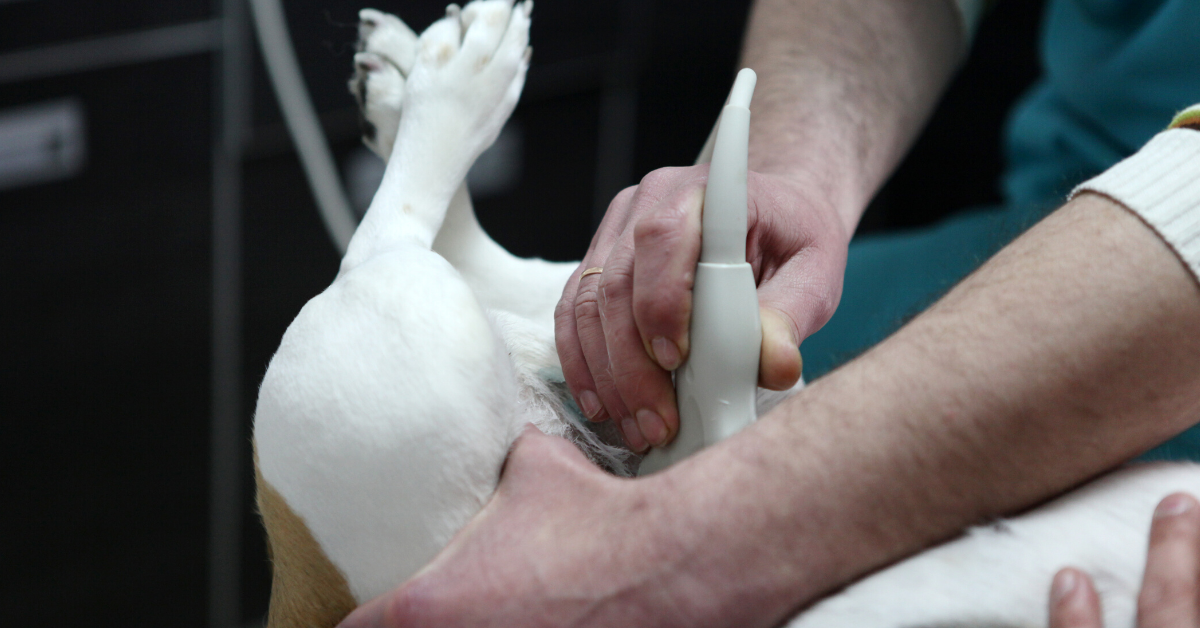
Mastering Ultrasound-Guided Cystocentesis in Dogs and Cats

There are several ways to collect a urine sample from your patient, including free catching, using a urinary catheter, and doing a cystocentesis.
A cystocentesis—which involves obtaining a urine sample via a needle placed directly into the bladder—may be the preferred method in many cases.
This is especially true when you need a sterile urine sample, such as for culture and sensitivity testing for UTIs. And in general, a cystocentesis will provide more accurate results than a free catch urine sample, since it reduces contamination from cells or pathogens of the skin.
It’s possible to perform a cystocentesis by palpating the bladder or using anatomical landmarks alone. However, an ultrasound can make the process easier by removing the guesswork and allowing you to visualize everything.
Here’s how it works…
How to perform an ultrasound-guided cystocentesis for dogs and cats
Prepare for the procedure.
Use an appropriate size syringe (usually 3ml to 12ml) based on the size of your patient.
Choose a needle size and length based on your patient size. A 22g needle works well for many patients.
Use a towel, trough, or other cushioning to make your patient more comfortable during the procedure. This may help minimize their movement.
Use appropriate patient restraint.
For some dogs and cats, this could mean having a trained team member hold them. For very anxious or fractious patients, this might mean chemical restraint.
Dorsal recumbency is generally the best way to position your patient.
Be sure to clean the area of skin where you’ll be performing the cystocentesis. If needed, consider clipping the fur, too.
Estimate the bladder location.
You’ll use your ultrasound to see the bladder, but anatomical landmarks will give you a good starting point for where to place the probe. Try one of these methods…
Locate the caudal four mammary nipples and mentally draw an ‘X’ between them. The bladder will often be near the cross point of the ‘X’.
Place a small amount of alcohol on the caudal abdomen so it pools. Often, the bladder is located directly under where the alcohol naturally pools on the midline.
Visualize the bladder.
Place your probe onto the caudal abdomen. You should be able to see the urinary bladder on the screen—it’s a fluid-filled structure that has the appearance of a ‘black balloon’ with gray or white walls.
Check for any abnormalities.
You could focus on just doing the cystocentesis. However, while you’re visualizing the bladder, it may help to do a cursory exam (or even a more in-depth evaluation, if indicated) to look for any problems contributing to the patient’s urinary symptoms.
For example, note if you see any thickenings, masses, calculi, or other issues in the urinary bladder.
Obtain the urine sample.
Place the needle directly through the abdominal wall, just in front of the ultrasound probe.
Aim the needle slightly caudally.
You should see the needle on your ultrasound screen. Use this visual to guide you, to be sure you get a good, clean sample rather than aspirating part of the bladder wall.
Prepare your sample for analysis.
Right after the cysto, replace the needle on the syringe with a sterile one.
Then, divide and prepare the sample in a timely manner (the fresher the sample, the better for testing) for any tests you need to perform, whether that’s in-house or at an outside lab.
In many veterinary hospitals, cystocentesis is safely performed several times per day, and complications are very rare. However, as with all medical procedures, some risks exist.
These may include…
Hematuria (usually mild and transient).
Much rarer complications such as bladder injury, leakage of urine into the abdomen, puncture of internal organs or blood vessels, or vagal reactions (retching, panting, hypersalivation, collapse).
Seeding of tumors.
If you suspect your patient has a bladder tumor such as transitional cell carcinoma, it may be best to avoid a cystocentesis. Otherwise, the needle could ‘seed’ the tumor as you withdraw your sample from the bladder, allowing cancer to spread into the abdominal cavity.
An ultrasound may help you to notice abnormalities such as bladder tumors during the procedure, in which case your recommendation to the client may change.

Fortunately for most pets, a cystocentesis is a quick outpatient procedure that provides a lot of valuable information. And many dogs and cats don’t show any side effects at all.
For patients with urinary symptoms, a cystocentesis is often the way to go for the most accurate diagnostic testing results and to get to the bottom of the issue faster for healthy pets and happy clients.
Written by: Dr. Tammy Powell, DVM

How to Explain Halitosis Risks to Concerned Pet Owners

Bad breath, or halitosis, is one of those health problems that’s easy for pet parents to overlook.
Sure, it’s inconvenient. Stinky breath can certainly put a damper on a pet owner’s cuddle time or other interactions with their dog or cat.
However, many pet owners don’t realize that halitosis can mean something much worse… such as periodontal disease, or even an internal problem.
As a veterinarian, you know this very well—but it can be a challenge to communicate the dangers lying “under the surface” when it comes to bad breath.
Explaining halitosis to pet parents
The first step is figuring out where the bad breath is coming from, and explaining the potential risks to clients.
Dental problems such as periodontal disease are the most common cause of bad breath in pets.
But sometimes, halitosis begins somewhere in the body other than the mouth.
Possible sources of halitosis include…
Periodontal disease and other dental issues.
A health condition such as kidney disease (an ammonia-like or unpleasant smell) or diabetic ketoacidosis (more of a sweet smell).
Respiratory diseases.
Digestive issues or dietary indiscretions.
Objects (like branches or chicken bones) stuck somewhere in the mouth.
Oral neoplasias.
Certain toxins.
Based on your patient’s history and physical exam, you’ll determine the most likely cause of the bad breath and decide if diagnostic testing is needed.
If the patient in front of you seems otherwise healthy but has apparent terrible dental disease, you can probably assume the smell (or at least, a large portion of the scent) is coming directly from their mouth.
If a dental procedure is indicated, you’ll likely do pre-op bloodwork, which will help confirm that the patient is otherwise healthy.
A recap of periodontal disease
It all starts with plaque, that thin film that forms on teeth due to food and saliva. We get plaque as humans, too, but fortunately, plaque can be removed by tooth brushing.
If not brushed away, plaque hardens into calculus (tartar) within as little as 24-36 hours. Then, it accumulates more and more over time and harbors bacteria. Calculus can’t be removed by toothbrushing alone.
Soon, this calculus (and its resident bacteria) become very irritating to the gums—and they like to grow below the gum line, where it’s harder to observe the process visually. This results in gingivitis, an early stage of periodontal disease (a disease process affecting the structures that hold teeth in place) where the gums become red and inflamed and may bleed.
From here, as the periodontal disease worsens, it creates pockets in the gumline, destruction of the periodontal ligament, and even bone loss in the maxilla and mandible. In severe cases, this bone loss can cause jaw fractures.
This all leads to pain and tooth loss. And, opportunistic bacteria may enter the bloodstream via inflamed gums, traveling to and causing problems in organs such as the heart or kidneys.
Reminding clients of the dangers of periodontal disease can help them understand their pet’s bad breath is true health and quality of life problem—not just an inconvenience.
Evaluating and treating dental and periodontal disease
Your initial pre-op exam will be done while the patient is awake—ideally with the owner present so you can show them what you’re seeing.
However, awake exams don’t allow for a full oral assessment. And, dogs or cats who are painful might not let you get a good look.
So you’ll make a dental cleaning estimate based on your initial exam but explain to the client that the full assessment will be done during the procedure.
This assessment should include dental radiographs. Since much of the disease process starts below the gumline, x-rays are the only way to see the full extent of damage from periodontal disease.

Some patients will just need routine cleaning, while others may need significant dental extractions. And, you can talk to your clients about continuing dental care at home through tooth brushing and dental treats.
By providing this valuable service, you’ll be not only keeping your canine and feline patients in great health—but also increasing the human-animal bond by preventing halitosis from interfering with the quality time between a pet and their owner.
Disclaimer: This article is for general informational purposes only, and not intended as a guide to the medical treatment of any specific animal.
Written by: Dr. Tammy Powell, DVM
The Veterinarian’s Guide to Diagnosing Heart Tumors in Pets

Cancers of the heart are uncommon in dogs and cats. When they do occur, it’s important to differentiate a tumor from other conditions that could cause generalized or focal enlargement of the heart and cardiovascular symptoms.
Which Cancers Occur In Cardiac Tissues?
A cancerous lesion based at the heart could be a primary tumor, or due to metastasis.
Primary tumors that occur at the heart include:
Hemangiosarcomas—the most common heart cancer in dogs, usually occurring at the right atrium. These occur most frequently in larger dogs with longer noses, such as Golder Retrievers, Doberman Pinschers, and German Shepherds.
Chemodectomas, also known as aortic body tumors or heart base tumors. Generally thought to be slow-growing, these tumors occur most commonly in brachycephalic breeds like Boxers, Bulldogs, and Boston Terriers.
Myxomas.
Sarcomas.
Ectopic thyroid tumors.
Lymphoma/lymphosarcoma—the most common heart cancer in cats.
Other cancers are possible, but these are the most commonly diagnosed types. Breeds may vary as noted above, but most pets with tumors of the heart are middle-aged or older.
How Are Heart Tumors Diagnosed?
If slow-growing, tumors near the heart base are often an incidental finding, seen on thoracic radiographs that are taken for another reason.
Other times, diagnostics are pursued because of clinical symptoms—which are often sudden in onset.
Once a mass is large enough to push on the heart and major blood vessels, many cardiovascular symptoms are possible, such as:
Coughing
Ascites
Lethargy
Weakness
Vomiting
Loss of appetite
Difficulty breathing
Collapse
Sudden death
Note: If a dog presents with some of these symptoms, especially sudden weakness and collapse, a quick ultrasound scan may help to identify pericardial effusion and aid in guiding a needle for emergency pericardiocentesis.
Finding Heart Masses Early
As with most cancers and disease processes, discovering a problem earlier rather than later can allow for more treatment options.
General screening radiographs or ultrasound checks—such as with a senior wellness health check—can be a good opportunity to discover heart base tumors before they cause cardiovascular dysfunction and symptoms.
An echocardiogram performed via ultrasound can help to provide more information on the location, size, and invasiveness of the mass.
In some cases, a presumptive diagnosis may be made based on the appearance and location of the mass on an ultrasound study, along with the patient’s signalment. If possible to perform without undue risk, an ultrasound-guided aspirate of the mass can provide more information about which type of tumor is present.
Treatment Options For Cancers Of The Heart
Treatment will be based on the type of neoplasia, how fast the mass is growing, whether metastasis is present, and whether or not the pet is symptomatic.
When a tumor of the heart is diagnosed, a good next step is to screen for metastasis and concurrent conditions via chest x-rays (if not already done), bloodwork, lymph node evaluation, and abdominal ultrasound.
Once a diagnosis is made, treatment options may include:
Periodic monitoring with a cardiologist (especially for slow-growing chemodectomas/heart base tumors) prior to pursuing more invasive treatments.
Surgery to remove the tumor.
Pericardiectomy to remove the pericardium and prevent life-threatening cardiac tamponade or pericardial effusion.
(A pericardiocentesis may be necessary on an emergency basis prior to diagnostics in a pet who presents with acute symptoms. After that, a planned pericardiectomy can help to prevent further emergency episodes of fluid buildup around the heart.)
Chemotherapy, often in conjunction with surgery.
Radiation therapy—either conventional, or via Cyberknife therapy.
Even though cancers of the heart are uncommon in pets, they can be scary to pet owners because of the possibility of sudden onset of serious clinical symptoms (especially with hemangiosarcomas).
By performing diagnostics, referring to specialists as needed, and giving your clients as much information as possible, you can help them make an informed decision for their pet.

Disclaimer: This article is for general informational purposes only, and not intended as a guide to the medical treatment of any specific animal.
Written by: Dr. Tammy Powell, DVM
Ultrasound-Guided Cystocentesis: How and Why to Do One
There are several ways to collect a urine sample from your patient, including free catching, using a urinary catheter, and doing a cystocentesis.
A cystocentesis—which involves obtaining a urine sample via a needle placed directly into the bladder—may be the preferred method in many cases.
This is especially true when you need a sterile urine sample, such as for culture and sensitivity testing for UTIs. And in general, a cystocentesis will provide more accurate results than a free catch urine sample, since it reduces contamination from cells or pathogens of the skin.
It’s possible to perform a cystocentesis by palpating the bladder or using anatomical landmarks alone. However, an ultrasound can make the process easier by removing the guesswork and allowing you to visualize everything.
Here’s how it works…
How to perform an ultrasound-guided cystocentesis for dogs and cats
Prepare for the procedure.
Use an appropriate size syringe (usually 3ml to 12ml) based on the size of your patient.
Choose a needle size and length based on your patient size. A 22g needle works well for many patients.
Use a towel, trough, or other cushioning to make your patient more comfortable during the procedure. This may help minimize their movement.
Use appropriate patient restraint.
For some dogs and cats, this could mean having a trained team member hold them. For very anxious or fractious patients, this might mean chemical restraint.
Dorsal recumbency is generally the best way to position your patient.
Be sure to clean the area of skin where you’ll be performing the cystocentesis. If needed, consider clipping the fur, too.
Estimate the bladder location.
You’ll use your ultrasound to see the bladder, but anatomical landmarks will give you a good starting point for where to place the probe. Try one of these methods…
Locate the caudal four mammary nipples and mentally draw an ‘X’ between them. The bladder will often be near the cross point of the ‘X’.
Place a small amount of alcohol on the caudal abdomen so it pools. Often, the bladder is located directly under where the alcohol naturally pools on the midline.
Visualize the bladder.
Place your probe onto the caudal abdomen. You should be able to see the urinary bladder on the screen—it’s a fluid-filled structure that has the appearance of a ‘black balloon’ with gray or white walls.
Check for any abnormalities.
You could focus on just doing the cystocentesis. However, while you’re visualizing the bladder, it may help to do a cursory exam (or even a more in-depth evaluation, if indicated) to look for any problems contributing to the patient’s urinary symptoms.
For example, note if you see any thickenings, masses, calculi, or other issues in the urinary bladder.
Obtain the urine sample.
Place the needle directly through the abdominal wall, just in front of the ultrasound probe.
Aim the needle slightly caudally.
You should see the needle on your ultrasound screen. Use this visual to guide you, to be sure you get a good, clean sample rather than aspirating part of the bladder wall.
Prepare your sample for analysis.
Right after the cysto, replace the needle on the syringe with a sterile one.
Then, divide and prepare the sample in a timely manner (the fresher the sample, the better for testing) for any tests you need to perform, whether that’s in-house or at an outside lab.
In many veterinary hospitals, cystocentesis is safely performed several times per day, and complications are very rare. However, as with all medical procedures, some risks exist.
These may include…
Hematuria (usually mild and transient).
Much rarer complications such as bladder injury, leakage of urine into the abdomen, puncture of internal organs or blood vessels, or vagal reactions (retching, panting, hypersalivation, collapse).
Seeding of tumors.
If you suspect your patient has a bladder tumor such as transitional cell carcinoma, it may be best to avoid a cystocentesis. Otherwise, the needle could ‘seed’ the tumor as you withdraw your sample from the bladder, allowing cancer to spread into the abdominal cavity.
An ultrasound may help you to notice abnormalities such as bladder tumors during the procedure, in which case your recommendation to the client may change.
Fortunately for most pets, a cystocentesis is a quick outpatient procedure that provides a lot of valuable information. And many dogs and cats don’t show any side effects at all.
For patients with urinary symptoms, a cystocentesis is often the way to go for the most accurate diagnostic testing results and to get to the bottom of the issue faster for healthy pets and happy clients.
Written by: Dr. Tammy Powell, DVM

Diagnosing Tumors of the Heart in Dogs and Cats

Cancers of the heart are uncommon in dogs and cats. When they do occur, it’s important to differentiate a tumor from other conditions that could cause generalized or focal enlargement of the heart and cardiovascular symptoms.
Which cancers occur in cardiac tissues?
A cancerous lesion based at the heart could be a primary tumor, or due to metastasis.
Primary tumors that occur at the heart include:
Hemangiosarcomas—the most common heart cancer in dogs, usually occurring at the right atrium. These occur most frequently in larger dogs with longer noses, such as Golder Retrievers, Doberman Pinschers, and German Shepherds.
Chemodectomas, also known as aortic body tumors or heart base tumors. Generally thought to be slow-growing, these tumors occur most commonly in brachycephalic breeds like Boxers, Bulldogs, and Boston Terriers.
Myxomas.
Sarcomas.
Ectopic thyroid tumors.
Lymphoma/lymphosarcoma—the most common heart cancer in cats.
Other cancers are possible, but these are the most commonly diagnosed types. Breeds may vary as noted above, but most pets with tumors of the heart are middle-aged or older.
How are heart tumors diagnosed?
If slow-growing, tumors near the heart base are often an incidental finding, seen on thoracic radiographs that are taken for another reason.
Other times, diagnostics are pursued because of clinical symptoms—which are often sudden in onset.
Once a mass is large enough to push on the heart and major blood vessels, many cardiovascular symptoms are possible, such as:
Coughing
Ascites
Lethargy
Weakness
Vomiting
Loss of appetite
Difficulty breathing
Collapse
Sudden death
Note: If a dog presents with some of these symptoms, especially sudden weakness and collapse, a quick ultrasound scan may help to identify pericardial effusion and aid in guiding a needle for emergency pericardiocentesis.
Finding heart masses early
As with most cancers and disease processes, discovering a problem earlier rather than later can allow for more treatment options.
General screening radiographs or ultrasound checks—such as with a senior wellness health check—can be a good opportunity to discover heart base tumors before they cause cardiovascular dysfunction and symptoms.
An echocardiogram performed via ultrasound can help to provide more information on the location, size, and invasiveness of the mass.
In some cases, a presumptive diagnosis may be made based on the appearance and location of the mass on an ultrasound study, along with the patient’s signalment. If possible to perform without undue risk, an ultrasound-guided aspirate of the mass can provide more information about which type of tumor is present.
Treatment options for cancers of the heart
Treatment will be based on the type of neoplasia, how fast the mass is growing, whether metastasis is present, and whether or not the pet is symptomatic.
When a tumor of the heart is diagnosed, a good next step is to screen for metastasis and concurrent conditions via chest x-rays (if not already done), bloodwork, lymph node evaluation, and abdominal ultrasound.
Once a diagnosis is made, treatment options may include:
Periodic monitoring with a cardiologist (especially for slow-growing chemodectomas/heart base tumors) prior to pursuing more invasive treatments.
Surgery to remove the tumor.
Pericardiectomy to remove the pericardium and prevent life-threatening cardiac tamponade or pericardial effusion.
(A pericardiocentesis may be necessary on an emergency basis prior to diagnostics in a pet who presents with acute symptoms. After that, a planned pericardiectomy can help to prevent further emergency episodes of fluid buildup around the heart.)
Chemotherapy, often in conjunction with surgery.
Radiation therapy—either conventional, or via Cyberknife therapy.
Even though cancers of the heart are uncommon in pets, they can be scary to pet owners because of the possibility of sudden onset of serious clinical symptoms (especially with hemangiosarcomas).

By performing diagnostics, referring to specialists as needed, and giving your clients as much information as possible, you can help them make an informed decision for their pet.
Disclaimer: This article is for general informational purposes only, and not intended as a guide to the medical treatment of any specific animal.
Written by: Dr. Tammy Powell, DVM

Vertebral Heart Score: How and Why to Calculate One

A vertebral heart score (VHS) is a measurement used to determine if a patient’s heart is enlarged.
The VHS can provide valuable insight to complete your clinical picture, especially at times when you hear a heart murmur, for a general pre-anesthetic check, or to look for cardiac changes on breeds that are predisposed.
It’s a simple, non-invasive way to get more information and to monitor for changes over time.
How to calculate a vertebral heart score
Obtain a high-quality lateral thoracic radiograph. Be sure…
To collimate.
That the patient is straight and not twisted or at an angle.
That you can see enough detail, especially the thoracic vertebrae and the borders of the cardiac silhouette.
Make your first measurement along the longest axis of the cardiac silhouette, from the ventral border of the carina of the mainstem bronchus to the apex of the cardiac silhouette.
You don’t need to measure this in centimeters or inches—instead, just hold up a sheet of paper to your x-ray viewer and make a mark for your measurement, or use calipers. Or, with your digital x-ray system, use the VHS function to mark the measurement right on your screen.
Now, hold the measurement up to the vertebrae, starting with the cranial edge of T4—this is the 4th vertebra to have a rib connected. Count the number of vertebrae (including the vertebral body and the following disc space) that fall into the long axis measurement.
Measure to a decimal place of 0.1 (for example, let’s say your first measurement was 5.2 vertebrae).
Make your second measurement. This time, you’ll measure along the short axis, which is the widest part of the cardiac silhouette. It should be perpendicular (at a 90-degree angle) to your first measurement.
Repeat the process of transferring this measurement to the thoracic vertebrae, starting with the cranial edge of T4. For this example, let’s say your measurement was 4.5 vertebrae.
Add these two numbers together.
In this example, your VHS would be 5.2 + 4.5 = 9.7
How to interpret the results
For dogs, a normal vertebral heart score is less than 10.7 (with an average range of 8.5 to 10.5). For cats, an average VHS is 7.5 (with a range of 6.8 to 8.1).
Anything larger than that is considered abnormal.
Of course, this doesn’t tell you exactly what’s going on—you’ll need further information such as an echocardiogram to determine whether there is DCM, HCM, or some other issue causing the enlargement of the cardiac silhouette.
However, the VHS is a great screening tool to let you know to investigate further. And, if you have a patient who already has cardiac disease, a VHS can help you monitor their disease progression over time, and provide tangible information to a client who wants to know how their pet is doing on treatment.
Keep in mind that, as with any diagnostic tool, there is some individual variation. Some pets—especially certain breeds such as boxers—may fall outside the normal VHS range even if they’re completely healthy.
So, look at the overall clinical picture when assessing a patient. But with that in mind, a VHS is a quick, easy, valuable addition to your toolkit when it comes to evaluating and monitoring cardiac health.
Vetebral heart score tool video

Disclaimer: This article is for general informational purposes only, and not intended as a guide to the medical treatment of any specific animal.
Written by: Dr. Tammy Powell, DVM