


Negotiating a Former Veterinary Hospital Lease: What Every Startup Owner Should Ask For

Lease Negotiation Advice for a Former Veterinary Hospital
Opening your own veterinary practice is exciting! It’s also one of the biggest financial commitments you’ll ever make.
```Looking into a recently closed veterinary hospital instead of an empty retail space or new construction can save you hundreds of thousands of dollars in build-out costs.
But here’s the catch: Just because something is sitting inside the building doesn’t mean it comes with the lease.
Everything from kennel banks to oxygen lines to surgery lighting should be discussed before you sign.
Many landlords are surprisingly willing to negotiate, especially if the alternative is leaving a specialized veterinary property vacant.
Here are the biggest items every startup owner should discuss before signing the lease.
```1. Ask Exactly What Equipment Is Included
```One of the biggest misconceptions is assuming everything inside the building belongs to the landlord.
Sometimes it does. Sometimes it belongs to the previous practice owner. Sometimes it’s owned by a financing company that intends to remove it.
Before negotiating anything else, request a complete inventory of what’s included in the lease. Questions to ask include:
- Which items transfer with the lease?
- Which items will be removed?
- Are any pieces still under financing agreements?
- Is there documentation proving ownership?
- Can any remaining equipment be purchased separately?
Getting these answers in writing helps avoid unpleasant surprises after you’ve already committed to the property.
```2. Negotiate to Keep Kennels and Cage Banks
```Kennels are among the most expensive pieces of permanent veterinary infrastructure.
High-quality stainless steel kennel systems can cost tens of thousands of dollars, depending on size and configuration.
If the cages are already installed and in good condition, keeping them can dramatically reduce your startup budget. Here are some things to evaluate:
- Rust or corrosion
- Door alignment
- Drainage
- Noise levels
- Isolation capabilities
- Recovery cage sizes
Replacing an entire kennel ward after move-in is rarely money well spent if the existing units remain functional.
```3. Don’t Forget the Exam Tables
```Exam tables often remain behind when hospitals close. While they may not be glamorous, quality stainless steel tables can last for decades.
Look for things like:
- Hydraulic or lift functionality
- Stability
- Rust or surface damage
- Cabinet storage underneath
Even if you eventually plan to upgrade, keeping existing tables allows you to prioritize spending elsewhere.
```4. Confirm the Oxygen Lines Are Operational
```Medical gas infrastructure is one of the biggest hidden values inside an existing veterinary hospital. Installing new oxygen piping after move-in can be costly and disruptive.
Ask the landlord:
- Are the oxygen lines still functional?
- When were they last inspected?
- Are maintenance records available?
- Has any portion been disconnected?
- Are shutoff valves clearly labeled?
5. Existing Plumbing Could Save Months of Construction
```Veterinary hospitals require significantly more plumbing than traditional commercial spaces. Think about:
- Wet tables and dental stations
- Grooming tubs
- Laundry
- Laboratory sinks
- Surgical scrub stations
Relocating plumbing becomes expensive quickly. During your walkthrough, identify existing water supply and drain locations.
Even if your floor plan changes slightly, keeping major plumbing where it already exists can dramatically reduce renovation costs.
```6. Inspect Surgery Lighting Carefully
```Good surgical lighting is essential. If existing lights remain, be sure to:
- Test brightness and positioning
- Confirm replacement parts remain available
- Verify electrical safety
If they’re older but functional, they may provide years of additional service while you allocate your startup budget elsewhere.
```7. Ask About Backup Power
```Emergency power is increasingly valuable. If the building already has a generator, determine:
- Who owns it?
- Is it included?
- When was it serviced?
- What systems does it power?
- Is there a maintenance contract?
Losing power during surgery or while storing temperature-sensitive medications can become far more than an inconvenience.
```8. Negotiate Existing Signage
```Exterior monument signs and building signage are expensive to replace. Ask whether you can:
- Reuse existing sign structures
- Replace only the sign face
- Install new branding
- Keep existing lighting
Often, the structure itself has significant value even if the branding changes.
```9. Ask for a Tenant Improvement (Build-Out) Allowance
```A tenant improvement allowance, sometimes called a TI allowance, is money the landlord contributes toward renovating the space. Depending on market conditions, landlords may help pay for:
- Flooring
- Paint
- Electrical upgrades
- HVAC improvements
- Walls
- Plumbing modifications
- Lighting
The amount varies considerably based on location, lease length, and market demand, but it’s always worth asking. A longer lease term can sometimes justify a larger landlord contribution.
```10. Clarify Who Owns Improvements
```This is one of the most overlooked parts of lease negotiations. If you install:
- New cabinetry
- Dental equipment
- Digital radiography
- Surgical lights
- Kennels
- Laboratory casework
An experienced commercial real estate attorney can help ensure the language in the lease protects your investment.
```11. Bring in the Right Experts
```Even if a building appears “move-in ready,” don’t rely solely on appearances. Before signing:
- Hire a commercial building inspector.
- Have a licensed electrician inspect the electrical capacity.
- Ask a plumber to evaluate existing water and drainage systems.
- Bring in an HVAC contractor.
- Have medical gas systems inspected if oxygen lines are present.
- Consult a veterinary equipment specialist to evaluate existing equipment and estimate its remaining service life.
A few hundred dollars spent on inspections can save tens of thousands of dollars in unexpected repairs later.
```A Few Final Notes
```Former veterinary hospitals offer a space already designed for the way veterinarians work.
Remember that every item left behind has value. Don’t assume it’s included, and don’t be afraid to ask for it. A careful lease negotiation can preserve more of your startup budget for the investments that truly set your practice apart: exceptional patient care, a strong team, and the equipment that will support your vision for years to come.
```

How to Find Vacant Veterinary Hospitals Before Everyone Else

A practical guide to finding vacant and off-market veterinary properties before they reach the broader market.
If you're hoping to open your own veterinary practice, buying an existing hospital isn't the only path to consider.
Across the country, former veterinary practices sit vacant after owner retirements, corporate consolidations, relocations, or business closures. Many are already equipped with important necessities, potentially saving hundreds of thousands of dollars compared to building from scratch.
A Former Veterinary Hospital May Already Include:
```- Exam rooms and treatment areas
- Kennels
- Plumbing
- Medical-grade electrical infrastructure
The challenge isn't finding these properties once they're publicly listed. The challenge is finding them before everyone else does.
The good news? With a little detective work, strong networking, and the smart use of AI tools, you can find opportunities that many buyers never see.
Start With Google Maps and Street View
One of the simplest ways to locate former veterinary hospitals is by virtually exploring the communities where you'd like to practice.
Using Google Maps, search for terms such as:
- Veterinary hospital
- Animal hospital
- Veterinary clinic
- Emergency veterinarian
As you browse Google Maps, pay attention to clinics marked as “Permanently Closed.” While not every listing is accurate, it creates an excellent starting point.
Next, switch to Street View and look for clues such as:
- Empty parking lots
- Removed or faded signage
- Vacant-looking buildings
- “For Lease” or “Available” signs
- Buildings that still feature veterinary architecture, such as outdoor runs or multiple exam-room entrances
Even if a building has already changed ownership, it may still be available for lease or redevelopment.
Search Commercial Real Estate Websites
Many former veterinary hospitals eventually appear on commercial real estate marketplaces, although they aren't always advertised as veterinary facilities.
Instead of searching only for “veterinary clinic,” try keywords such as:
- Medical office
- Healthcare building
- Former medical office
- Commercial office
- Freestanding medical building
- Specialty healthcare property
Some listings intentionally avoid mentioning the property's previous veterinary use. This is where some of that detective work comes in.
Floor-plan photos may reveal treatment areas, kennels, surgical suites, or specialized plumbing.
Build Relationships With Commercial Real Estate Brokers
Commercial brokers often hear about available properties before they're officially listed. Introduce yourself to brokers who specialize in:
- Medical offices
- Healthcare properties
- Commercial redevelopment
Let them know you're specifically looking for former veterinary hospitals.
Many brokers will happily add you to their contact list and notify you when a property matching your criteria becomes available.
The more specific you are about your preferred city, square footage, budget, and timeline, the easier it becomes for brokers to think of you when opportunities arise.
Join Veterinary Facebook Communities
Veterinary professionals often hear about closures long before the public does.
Facebook groups can be valuable sources of local information, including:
- Practice owners nearing retirement
- Clinics preparing to relocate
- Hospitals reducing locations
- Corporate closures
- Equipment liquidation announcements
Rather than immediately asking, “Does anyone know of a clinic for sale?” become an active participant in these communities. Over time, conversations naturally reveal opportunities.
Stay Connected With Veterinary Practice Brokers
Although practice brokers are known for selling operating hospitals, they frequently know about clinics that never officially reach the market.
Some owners simply want to retire. Others close after being unable to recruit veterinarians.
Maintaining relationships with veterinary-specific brokers increases the likelihood of hearing about these opportunities early. Practice brokers often maintain extensive buyer networks and may become aware of off-market opportunities before public listings appear.
Talk With Veterinary Equipment Dealers
Equipment representatives visit practices every day. They often know when practices are:
- Closing or downsizing
- Relocating
- Renovating
- Preparing for ownership transitions
While they can't always disclose confidential information, developing relationships can position you to hear about opportunities as they become public.
Let AI Do the Research
Artificial intelligence isn't replacing your search; it's helping you search smarter.
Tools such as ChatGPT, Perplexity, and Claude can dramatically reduce the amount of manual research required to identify potential acquisition targets.
For example, you can ask AI to:
- Find recently closed veterinary clinics in a specific city or state
- Search local news for hospital closures
- Summarize corporate announcements involving practice consolidations
- Monitor news related to veterinary real estate
- Organize findings into spreadsheets
- Build prospect lists for outreach
- Draft personalized introduction emails to property owners or brokers
Example AI Research Prompt
```“Research veterinary clinics, animal hospitals, and veterinary practices that permanently closed, relocated, merged, or became vacant within a 50-mile radius of Columbus, Ohio, between [START DATE] and [END DATE].
For each location, provide:
- Business name
- Full street address
- Type of veterinary facility
- Closure, relocation, or vacancy date
- Current property status, if known
- Building size and acreage, if available
- Asking price or lease rate, if listed
- Property owner, listing broker, and contact information
- Source links
- A brief explanation of the evidence confirming the location is closed or vacant
Search commercial real estate listings, veterinary industry publications, local news, business closure announcements, social media pages, Google Business profiles, county property records, broker websites, auction notices, and archived web pages.
Separate the results into:
- Confirmed permanently closed or vacant veterinary properties
- Clinics that relocated, merged, or were acquired
- Possible closures that require further verification
Do not include clinics that are temporarily closed, operating under a new name at the same location, or lacking credible evidence of closure.
Prioritize properties that still appear suitable for use as a veterinary hospital. Present the findings in a table and clearly label any missing or uncertain information.”
```AI won't uncover confidential deals, but it can help connect publicly available information far faster than manually searching dozens of websites.
Like any research tool, however, AI-generated results should always be verified against original sources before making business decisions.
Remember: The Best Opportunities Rarely Advertise Themselves
The most desirable former veterinary hospitals often fail to appear on major listing websites. Instead, they're discovered through relationships, conversations, and consistent outreach.
The buyers who find the best deals aren't necessarily searching harder; they're searching in more places.
Through traditional networking and modern research tools such as AI, you can dramatically expand the number of opportunities you uncover while staying ahead of competing buyers.
Sometimes the next great veterinary hospital isn't hidden at all; it simply hasn't been discovered yet.

The Hidden Opportunity: Turning a Closed Veterinary Hospital Into Your Dream Practice

Before You Build: Consider Buying or Leasing a Closed Veterinary Clinic
For many veterinarians, owning a practice feels like the ultimate career goal. But then reality sets in.
Commercial construction costs continue to climb. Build-outs can take months. Equipment purchases add up quickly. Before long, the dream of opening your own hospital can start to feel financially out of reach.
But what if the perfect practice is already sitting there, waiting for someone to unlock its potential?
Across the country, veterinary hospitals occasionally close for reasons such as:
- Corporate restructuring
- Lease changes
- Owner retirement
- Relocation
- Shifts in local demand
While seeing a neighborhood clinic close its doors can be disappointing for pet owners, it can also create a valuable opportunity for independent veterinarians who are ready to build something of their own.
Instead of starting with four empty walls, you may be able to breathe new life into a space that was already designed to care for animals.
Why a Former Veterinary Hospital Can Be a Gold Mine
Building a veterinary hospital from scratch is expensive—not just because of the equipment, but because of everything hidden behind the walls.
Think about what may already exist in a former veterinary clinic:
- Exam rooms
- Treatment areas and surgical suites
- Kennel runs
- Pharmacy shelving
- Specialized plumbing
- Medical-grade electrical systems
- Reception and waiting areas
These are not just conveniences. They represent significant investments that have already been made.
Repurposing an existing veterinary facility can dramatically reduce renovation time and construction costs. This may allow you to focus more of your budget where it matters most: your patients, your team, and the technology that will help your practice grow.
Not Everything Needs to Be Brand New
One of the biggest misconceptions about opening a veterinary practice is that everything has to be purchased new.
The reality?
Many existing fixtures inside a former veterinary hospital may still have years of useful life remaining.
Depending on your lease agreement or purchase terms, you might inherit items such as:
- Kennels
- Cabinetry
- Exam room counters
- Surgical tables
- Reception desks
- Storage shelving
- Built-in workstations
- Isolation rooms
Every piece you can safely reuse is money that can be invested elsewhere. That does not necessarily mean you should keep everything, though.
Know Where to Invest
While cabinets and kennels can often serve a new practice well, diagnostic technology is an entirely different story.
Older imaging equipment may no longer provide the speed, image quality, or reliability that today’s veterinary teams expect. Outdated systems can also become more expensive to maintain over time.
When opening a practice, it may be wise to upgrade key technologies such as:
- Digital radiography
- Dental imaging systems
- Ultrasound
- Anesthesia equipment
- Patient monitoring equipment
- Infusion pumps
These investments can improve efficiency, enhance patient care, and help create a modern client experience from day one.
Think of it this way: Keep what still works well, and upgrade the tools that directly affect diagnostics, workflow, reliability, and patient outcomes.
A Faster Path to Opening Your Doors
Starting with an existing veterinary hospital can also save something every entrepreneur values: time.
Instead of waiting months for construction permits, plumbing, electrical work, and room layouts, much of the infrastructure may already be in place.
That means you can spend more time preparing your team, building client relationships, and planning your grand opening—and less time staring at construction schedules.
For veterinarians transitioning from relief work, associate positions, or a mobile practice, this approach can significantly shorten the path from an idea to opening day.
Every Closed Clinic Represents a New Beginning
A building that once cared for pets does not have to become another empty storefront. With the right vision, it can become a thriving independent veterinary hospital again.
Families often appreciate seeing a familiar veterinary location come back to life. Former clients may be excited to have veterinary care available in their neighborhood again. Your team also gets the opportunity to build something meaningful without having to start completely from scratch.
Sometimes the best opportunity is not finding an empty building. It is finding one that already knows how to be a veterinary hospital.

How Bank of America Practice Solutions Can Help You Start Your Veterinary Hospital

How BofA Practice Solutions Helps Vet Startups Succeed
If you’re a veterinarian dreaming of opening your hospital, you’re in good company! Starting a practice is one of the most rewarding (and challenging) things you can do in your career. One of the first questions most veterinarians have when they start exploring this idea is:
How in the world am I going to pay for this?
The good news? You don’t have to do it all on your own.
Bank of America is a proud financial partner of the American Veterinary Medical Association and the American Animal Hospital Association, and their Practice Solutions Program is the perfect first step to answering this question.
Bank of America’s Practice Solutions program is a popular, veterinarian-friendly financing option that has helped countless DVMs and specialists get started on their new journey.
They offer customizable loans for veterinary professionals seeking to start a new practice, expand their existing one, or acquire an existing practice.
Let’s walk through what you need to know and answer some of the most common questions veterinarians ask about loans and the BofA Practice Solutions program.
Why Do So Many Veterinarians Use the Bank of America Practice Solutions Program?
First, a bit of context: Bank of America has an entire division dedicated to veterinary, dental, and medical professionals, and they genuinely understand the unique challenges and expenses associated with opening a veterinary hospital.
Here are some reasons why so many veterinarians choose this program:
100% financing available: Many veterinarians don’t have significant cash reserves after vet school, internships, or years as an associate. BofA offers loans that cover the full cost of your project, including equipment, construction, inventory, and even working capital to get you through the first few months.
Long repayment terms.: They offer terms that help keep your monthly payments manageable, which can be critical while you grow your client base.
Specialized expertise: The loan officers in the Practice Solutions program specialize in working with medical professionals. They understand zoning, hospital layouts, and the reality of veterinary revenue cycles, so you won’t have to explain why an x-ray table is necessary or why you’re budgeting for a wet table.
What Are the Interest and Payoff Options Like?
Another common question that’s asked is: “Can I pay it off early if my practice starts doing well?”
The short answer is: Usually, yes — and often without a penalty.
Bank of America’s Practice Solutions loans typically allow you to pay more than your monthly payment, or even pay off the loan entirely ahead of schedule, without any prepayment penalty. This is a considerable advantage if your hospital grows faster than expected and you want to reduce your interest costs over time.
Speaking of interest…these loans do accrue interest from the moment funds are disbursed (like any commercial loan). But because the terms are longer and the rates are designed for medical professionals, the payments are structured to be as manageable as possible. You’ll make monthly payments that include both principal and interest.
Suppose you’re concerned about total interest paid over the life of the loan. In that case, you can always pay extra toward principal when you’re able, and every extra payment chips away at the balance and reduces future interest.
What Else Should You Consider When Starting Your Hospital?
Here are a few other common questions veterinarians ask when thinking about a startup:
Do I need a business plan?
Yes — and the Practice Solutions team can even help you refine it. A solid business plan shows lenders (and yourself) that you’ve thought through your market, your services, and your numbers.
What do the interest rates look like?
Practice loan rates are very competitive and vary depending on loan purpose and term, so speak with a Practice Specialist who can help with your specific financing needs. There are also options to lock in your rate so you won't have to worry about potential rate increases.
What can the load cover?
It can cover construction, build-out, equipment, inventory, signage, IT, and even some operating expenses for the first few months.
How long does it take?
The financing process can take anywhere from a few weeks to a few months, depending on how ready you are and how complex your project is.
What if I don’t have perfect credit?
They understand that most veterinarians are still paying off student loans. Strong credit helps, but your experience, business plan, and projected income are also taken into account.
Starting your veterinary hospital can feel daunting, but with the proper support and financing, it’s achievable. Programs like Bank of America’s Practice Solutions exist because they recognize how much veterinarians contribute to their communities, and they want to help you succeed.
If you’ve been dreaming of building a practice that reflects your vision and values, don’t let the fear of financing stop you.
A good loan program paired with a solid plan can make your dream hospital a reality — and give you the flexibility to grow (or even pay it off early) as your business thrives.

Connecting with Clients: Online Tools for New Vet Practices

These days, many of your future clients will meet you online long before they walk through your doors. That first digital impression matters—and you can shape it from day one.
From building an engaging social media presence to sending thoughtful newsletters and creating a client-friendly website, here’s how to develop a digital presence that builds trust, grows your client base, and reflects the heart of your practice.
Let’s dig in!
Social Media 101 for Veterinary Practices: Engaging Clients Online
Social media is one of the easiest ways to showcase your team’s personality and connect with pet owners. Platforms like Facebook and Instagram help you stay at the top of your mind, educate your audience, and create an inviting sense of community. The best part? You don’t have to post every day to make an impact.
Tips for Using Social Media Effectively
Be authentic – Share real photos of your team and patients (with permission), highlight fun moments, and show what a visit to your hospital is like.
Keep it educational – Offer simple, practical tips pet owners can use. Topics like “How to Tell if Your Pet Has Dental Disease” or “What to Pack for a Pet Emergency Kit” are great starting points.
Encourage interaction – ask questions, run polls, and invite clients to share photos or stories of their pets. Who doesn’t love every opportunity to share a picture of their pets?
Post consistently – Don’t make it more than it needs to be. Aim for one to three posts per week. It’s more about showing up regularly than flooding your followers with content.
Here are a few social media post ideas to help get you started:
Introduce your team members with short bios.
Share behind-the-scenes glimpses of the clinic.
Highlight a “Pet of the Week.”
Provide seasonal safety reminders or wellness tips.
Celebrate pet-related holidays or awareness months.
With just a few thoughtful posts each week, your social media can become a trusted, feel-good resource that clients enjoy following.
Why Newsletters Matter: Keeping Clients Connected to Your Practice
If social media is more of a casual daily conversation, your newsletter is more like a monthly coffee chat. It’s your chance to share important updates, milestones, reinforce your expertise, and keep your hospital top-of-mind between visits.
Newsletters are effective for a few different reasons:
They go directly to your clients’ inboxes, where they’re more likely to be seen.
They offer more room for storytelling and education than a typical social post.
They strengthen the relationship between your practice and clients by providing consistent, thoughtful communication.
Need help getting things kicked off? Consider starting with some basics like:
Helpful pet care tips
Updates about your hospital (new services, staff additions, changes in hours)
Special promotions or reminders for preventive care
A heartwarming client story or team spotlight
Most new practices find that sending a newsletter once a month strikes a good balance between being frequent enough to stay connected and not so often that it feels overwhelming.
Platforms like Mailchimp or Constant Contact make designing and sending newsletters easy, even if you’re not a designer or marketer.
Building a Strong Online Presence for Your Veterinary Practice
Your website is often the first place clients go to learn about your hospital. A well-designed, informative, and easy-to-navigate site sets the tone for what they can expect—and plays a critical role in gaining their trust.
If a website looks dated and unkept, clients often assume they can expect a similar experience with their hospital visit.
Let’s look at some tips for a User-Friendly Website:
Clear contact information that’s easy to find
Online appointment requests or booking options are a plus
A list of services and what makes your hospital unique
Photos of your clinic and team to create a welcoming feel
Testimonials or client reviews
A layout that works well on both desktop and mobile devices
Improving Your Website’s Visibility
Good visibility means showing up when pet owners search for services online. That’s where search engine optimization (SEO) comes in. It sounds technical, but a few basic strategies go a long way:
Use phrases like “veterinarian in [Your City]” in your page titles and content.
Regularly update your website with new content, such as blog posts or seasonal tips.
Claim and maintain your Google Business Profile.
Make sure your site loads quickly and works well on mobile devices.
Investing time in a strong website and local SEO strategy can help clients find you more easily and feel confident choosing your hospital.
Your website, social media, and newsletter work together to help people get to know you, trust you, and stay connected.
Whether you’re sharing helpful advice, posting a fun photo of the team, or emailing out the latest clinic updates, a regular communication cadence reflects the care and professionalism your hospital proudly represents!

Building a Strong Veterinary Team: Recruitment Guide

A big part of opening a new veterinary hospital is creating a team and environment that fosters excellent patient care and client satisfaction.
Your team is the backbone of your practice and will be the key to building lasting relationships with pet owners.
Let’s examine key details that will help you recruit and retain top veterinary talent, implement strong training programs, and enhance client interactions, setting your new hospital up for long-term success!
Hiring the Right Team: Tips for Recruiting and Retaining Veterinary Staff
We know veterinary hospitals face increased challenges in hiring and retaining skilled team members.
With the industry experiencing staff shortages and high turnover, we must be thoughtful and creative regarding smart recruiting and fostering a positive workplace culture.
Let’s look at three tips to consider in your recruitment strategy:
1. Crafting a Compelling Job Listing
Your job post is potential employees' first impression of your hospital. Make it stand out by including:
Clear Job Title – Be specific about the role (for example: “Experienced Veterinary Technician – Fear-Free Certified Preferred”).
Your Hospital’s Mission and Culture – Highlight what makes your practice unique, whether focusing on work-life balance, mentorship opportunities, or a Fear-Free environment.
Competitive Benefits – Outline salary range, CE allowances, mentorship, flexible scheduling, and wellness perks.
Growth Opportunities – Show candidates how they can advance in their careers within your practice.
2. Finding and Attracting Top Talent
Leverage Veterinary-Specific Job Boards – Post on the AVMA Veterinary Career Center, AAHA’s job board, and VetMed Careers.
Utilize Social Media and Networking – Many professionals find jobs through LinkedIn, Facebook, veterinary groups, and word-of-mouth.
Offer Employee Referral Bonuses – Encourage your team to refer skilled candidates by offering incentives.
3. Retaining Staff and Reducing Turnover
Hiring the right people is only half the battle—keeping them is just as important.
Encourage Work-Life Balance – Consider options like rotating schedules, mental health days, and wellness initiatives that can prevent burnout.
Foster a Supportive Culture – Implement recognition programs, open communication, and leadership training to help staff feel valued.
Invest in Professional Development – Employees seek an employer supporting continuing education, certification programs, and career advancement opportunities.
A happy and engaged veterinary team leads to better patient care, a positive work culture, and higher client satisfaction.
The Power of Staff Training
A well-trained team is necessary for a smooth-running practice. From avoiding unnecessary mistakes to improving patient care and client interactions, ongoing education and structured training programs will go a long way toward your hospital's success.
Here are a few things you might consider in your staff training plans:
1. Certifications and Specialized Training
Encouraging staff to pursue certifications improves patient care and enhances their job satisfaction. Some valuable certifications include:
Veterinary Technician Specialty (VTS) – For techs desiring to specialize in emergency, dentistry, anesthesia, etc.
Fear-Free Certification – Helping all team members improve patient handling and reduce stress for pets (and the pet’s parents).
CPR and Emergency Response Training – Ensures preparedness for critical situations.
2. Continuing Education (CE) for Growth
Providing CE opportunities keeps your team up to date with industry advancements.
Offer CE Stipends – Support staff in attending veterinary conferences, online courses, or workshops.
Host In-House Training Sessions – Bring specialists for hands-on learning experiences and valuable Q&A.
Encourage Cross-Training – Teaching employees additional skills enhances flexibility and teamwork.
3. The Benefits of In-House Training Programs
A structured onboarding and training program improves efficiency and reduces errors. Consider the following:
Shadowing and Mentorship Programs – Pairing new hires with experienced staff eases the transition and helps with change management. Avoid using this method as a “punishment” and implement a program like this early on to help the team get acquainted.
Standardized Protocols and Checklists – Ensure consistency in anesthesia monitoring and client communication.
Monthly Lunch-and-Learns – Keep training ongoing with short, interactive sessions. This is an excellent method for continuing education and team building.
An investment in training elevates patient care and strengthens team morale and retention.
Mastering Client Communication: Enhancing the Customer Experience
Effective client communication is a cornerstone of a successful veterinary hospital. Pet owners must feel informed, respected, and comfortable entrusting their pets to your care.
Here are a few tips to consider establishing a foundation with your new team:
1. Clear and Compassionate Communication
Use Simple, Jargon-Free Language – Avoid overwhelming clients with technical and medical terms they may be unfamiliar with.
Confirm Understanding—Instead of asking, “Do you have any questions?” ask, “What questions do you have?” to avoid a simple “yes or no” answer and encourage open conversation.
Provide Visual Aids and Handouts—Diagrams, videos, and written instructions are very helpful for reinforcing key information.
2. Avoiding Client Shaming and Judgment
Assume the client's good intentions. Clients may not always make the best choices for their pets, but fostering a nonjudgmental environment helps build and maintain trust.
Avoid: “You should have brought your pet in sooner.”
Say Instead: “I’m glad you brought them in today. Here’s what we can do to help.”
Avoid: “You can’t afford this treatment?”
Say Instead: “Let’s explore some options that fit your budget.”
By replacing judgment with options, you build stronger relationships and improve compliance with treatment plans.
3. Enhancing Client Satisfaction to Boost Loyalty
Happy clients become lifelong customers and refer others to your hospital.
Be Proactive with Follow-Ups – A quick call or text after surgery or a new diagnosis shows you care.
Personalize the Experience – Use pet names, remember past visits, and celebrate milestones like birthdays.
Solicit Feedback and Improve – Offer surveys to identify areas for improvement and address concerns.
Mastering client communication leads to better compliance, fewer misunderstandings, and more referrals.
Launching a thriving veterinary hospital requires more than medical expertise—building a strong, well-trained team and creating positive client experiences.
By putting thoughtful effort into innovative hiring practices, ongoing staff training, and compassionate client communication, your practice will undoubtedly thrive in patient care and business success!

Diagnosing Bladder Stones in Pets: What You Need to Know
Sometimes, diagnosing urinary bladder stones in dogs and cats is simple: one set of x-rays and the mineral-dense uroliths glow bright white on your viewing screen.
Other times, it’s not so straightforward… especially for small or radiolucent stones.
Here’s how radiographs and ultrasound can be used to help you find pesky, hard-to-view bladder stones.
Are bladder stones on your differential diagnosis list?
Bladder stones may be at the forefront of your mind if you see a dog or cat who’s…
Having blood in their urine.
Urinating more frequently, and in short streams.
Having urinary accidents in the home.
Straining or experiencing pain during urination.
Excessively grooming around their genitals.
Usually, a client will bring their pet into you for these concerns, and your physical exam will help to determine that there’s no urinary obstruction.
With urinary bladder stones, you may notice some discomfort on palpation of the caudal abdomen. On a cat or small dog, you may even feel stones or crepitus in the area of the bladder.
Some patients, on the other hand, may exhibit minimal symptoms and their physical exam may be normal (sometimes bladder stones are an incidental finding).
Either way, most pets will need some type of imaging to confirm that bladder stones are there. Radiographs are a great place to start…
Finding uroliths via radiographs
In addition to any other needed tests—such as a urine analysis or bloodwork—radiographs are often recommended for pets with urinary symptoms, in order to look for uroliths or other abnormalities.
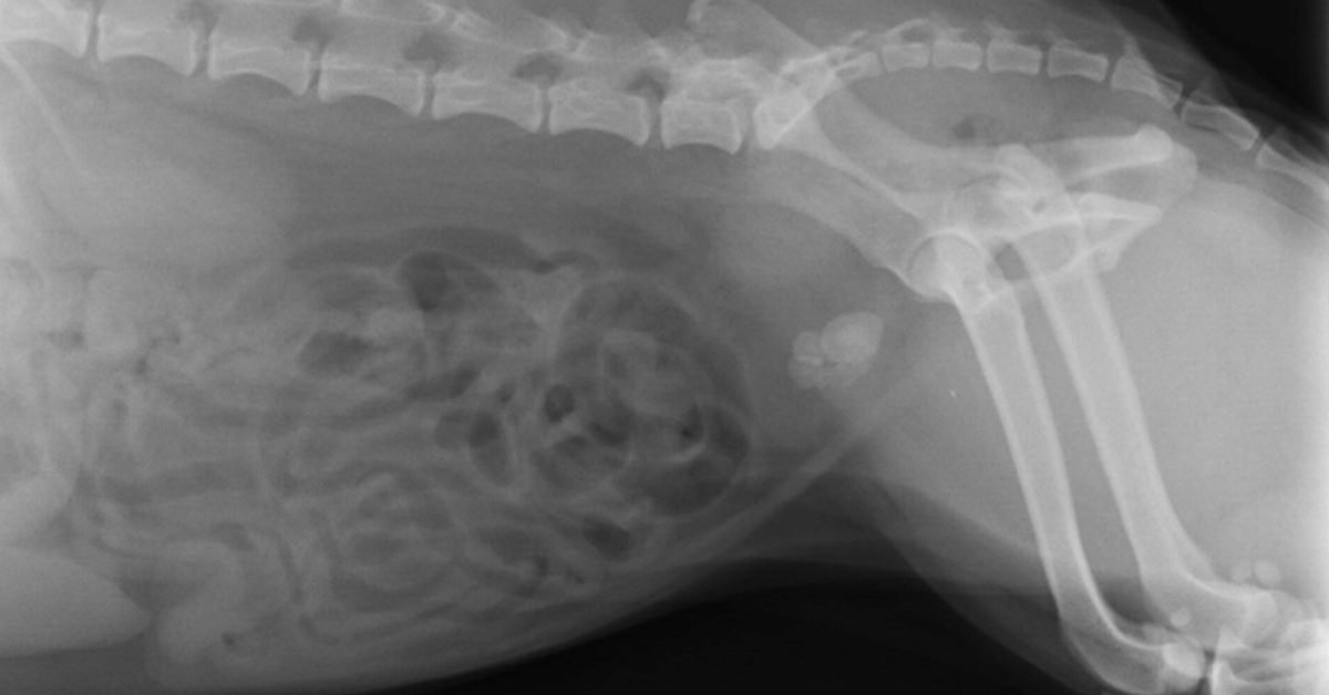
Typically, you’ll start with two simple views of the abdomen—a lateral and a VD.
Take a close look at the urinary bladder to look for radiopaque stones, which should show up as a white opacity relative to soft tissues thanks to their mineral composition.
Stones can range in size from small sand-like grains to more than two inches across. There may be just one or two stones present… or a small group… or even more than 100.
Remember to check the urethra for small stones that could be stuck—especially if the patient is straining or in pain during urination.
For better visualization of the entire urethra in male dogs, consider taking another lateral view with the hindlimbs pulled forward.
Also, check the kidneys and the areas of the ureters. While much less common in pets than in people, occasionally uroliths may be seen higher up in the urinary tract.
If you see stones now—you have your answer.
If you suspect urinary bladder stones but still don’t see them, a contrast study may allow better visualization.
For finding bladder stones, a double-contrast study is ideal.
This includes using both a positive contrast agent (soluble iodinated contrast medium) and a negative contrast agent (room air, or ideally carbon dioxide to reduce the risk of an air embolus) in the bladder together.
Anesthesia or sedation may be needed for the patient’s safety and comfort since the contrast agents are administered via a urinary catheter.
In addition to radiographs, an ultrasound is a useful tool…

Finding uroliths via ultrasound
An ultrasound study is another great option for finding bladder stones, especially radiolucent ones.
The fluid-filled bladder provides contrast for the ultrasound waves so that stones can be found (and often emphasized by acoustic shadowing).
Besides radiolucent stones, you may also see…
Bladder stones (radiopaque or radiolucent) that were too small to visualize radiographically (smaller than 1-3mm).
Other problematic issues in the bladder, such as ‘sludge’ buildup in cats with crystalluria.
Damage to the urinary bladder itself, such as inflammation.
The condition of the upper urinary tract—the kidneys and ureters.
Unexpected findings, such as tumors or anatomical abnormalities of the bladder.
Because of this, ultrasound imaging is a valuable tool for helping you diagnose and treat problems of the bladder, including urinary stones.
Follow-up
Depending on your findings, you may recommend a diet change for dissolvable stones, or a cystotomy to remove the stones.
For dissolution, follow-up imaging can help to track the patient’s progress and see whether or not the stone is dissolving.
When a cystotomy is recommended, remember to use imaging on the day of surgery…
Take pre-op radiographs to confirm the stones are still there, and that your urinary catheter is in place.
Include post-op views to confirm and document that all stones were successfully removed.
Since most stones are radiopaque, standard radiographs are a good option for follow-ups—and typically the imaging choice on the day of surgery.
But ultrasound can also be used in conjunction with other diagnostic tests to monitor the health of the urinary system long-term and to look for early signs of a problem such as a reoccurrence of stones.

Catching stones early, when they’re small, may allow less invasive treatment options such as voiding urohydropropulsion.
With the right combination of imaging modalities, you can help your clients stay on top of treating, monitoring, and preventing urinary bladder stones in their pets.
Written by: Dr. Tammy Powell, DVM
Disclaimer: This article is for general informational purposes only, and not intended as a guide to the medical treatment of any specific animal.

Buy Veterinary Digital X-ray Equipment with Confidence

How to Buy Veterinary Digital X-ray Equipment without Experiencing Buyer's Remorse
Have you ever bought something and regretted the purchase?
I think I have at one time or another. However, it’s one thing to regret ordering a cheeseburger and fries, but quite another when I regret buying a $50,000 car or truck.
When I make a big purchase, I want to feel good about my decision. I want to make sure that I made the right choice.
I want you to feel great about buying from me! I understand it is no small thing for a Veterinarian to spend $20,000 or $30,000 on digital x-ray equipment, and I want you to feel great about buying from us.
So, here is my “purchase without buyer's remorse” plan:
The 30-day satisfaction guarantee, or your money-back offer. When you make a digital x-ray system purchase, I will give you 30 days so that you feel comfortable and confident that you made the right choice.
And if you are not satisfied, return your digital x-ray equipment for a full refund.
Yes, that is correct! A FULL refund!!
I know that after the vet digital x-ray equipment is installed in your clinic, and once you learn how to use the software, you will be very happy with the results you get.
I can offer this guarantee because I am very confident with the quality of the digital x-ray equipment we sell, and I want you to be completely satisfied.
Here is the fine print:
Equipment must be returned in original packaging.
Equipment cannot be returned if damaged by the user during your 30 days.
The buyer pays the cost of shipping/packing/insurance of all returned equipment.
You must give us a reasonable chance to correct any dissatisfaction.
The main reason I am offering this “30-day satisfaction guarantee or your money back” is to give you peace of mind.
I understand I’m not as big as Idexx, Cuattro, or Sound-Eklin. Perhaps that makes you think twice about trying us out.
But I am confident that our equipment is as good, if not better, and now we have a “30-day satisfaction guarantee or your money back” - which the big boys do not offer!

The bottom line is simple:
I am offering quality digital x-ray equipment for the veterinary industry at an amazing price, with a great warranty, and now a 30-day guarantee.
I want to do the right thing, treat you with respect, and help you be successful in your veterinary practice.
Call or text me - Brad Haven, Jr. - 530-355-5886

Diagnosing Tumors of the Heart in Dogs and Cats

Cancers of the heart are uncommon in dogs and cats. When they do occur, it’s important to differentiate a tumor from other conditions that could cause generalized or focal enlargement of the heart and cardiovascular symptoms.
Which cancers occur in cardiac tissues?
A cancerous lesion based at the heart could be a primary tumor, or due to metastasis.
Primary tumors that occur at the heart include:
Hemangiosarcomas—the most common heart cancer in dogs, usually occurring at the right atrium. These occur most frequently in larger dogs with longer noses, such as Golder Retrievers, Doberman Pinschers, and German Shepherds.
Chemodectomas, also known as aortic body tumors or heart base tumors. Generally thought to be slow-growing, these tumors occur most commonly in brachycephalic breeds like Boxers, Bulldogs, and Boston Terriers.
Myxomas.
Sarcomas.
Ectopic thyroid tumors.
Lymphoma/lymphosarcoma—the most common heart cancer in cats.
Other cancers are possible, but these are the most commonly diagnosed types. Breeds may vary as noted above, but most pets with tumors of the heart are middle-aged or older.
How are heart tumors diagnosed?
If slow-growing, tumors near the heart base are often an incidental finding, seen on thoracic radiographs that are taken for another reason.
Other times, diagnostics are pursued because of clinical symptoms—which are often sudden in onset.
Once a mass is large enough to push on the heart and major blood vessels, many cardiovascular symptoms are possible, such as:
Coughing
Ascites
Lethargy
Weakness
Vomiting
Loss of appetite
Difficulty breathing
Collapse
Sudden death
Note: If a dog presents with some of these symptoms, especially sudden weakness and collapse, a quick ultrasound scan may help to identify pericardial effusion and aid in guiding a needle for emergency pericardiocentesis.
Finding heart masses early
As with most cancers and disease processes, discovering a problem earlier rather than later can allow for more treatment options.
General screening radiographs or ultrasound checks—such as with a senior wellness health check—can be a good opportunity to discover heart base tumors before they cause cardiovascular dysfunction and symptoms.
An echocardiogram performed via ultrasound can help to provide more information on the location, size, and invasiveness of the mass.
In some cases, a presumptive diagnosis may be made based on the appearance and location of the mass on an ultrasound study, along with the patient’s signalment. If possible to perform without undue risk, an ultrasound-guided aspirate of the mass can provide more information about which type of tumor is present.
Treatment options for cancers of the heart
Treatment will be based on the type of neoplasia, how fast the mass is growing, whether metastasis is present, and whether or not the pet is symptomatic.
When a tumor of the heart is diagnosed, a good next step is to screen for metastasis and concurrent conditions via chest x-rays (if not already done), bloodwork, lymph node evaluation, and abdominal ultrasound.
Once a diagnosis is made, treatment options may include:
Periodic monitoring with a cardiologist (especially for slow-growing chemodectomas/heart base tumors) prior to pursuing more invasive treatments.
Surgery to remove the tumor.
Pericardiectomy to remove the pericardium and prevent life-threatening cardiac tamponade or pericardial effusion.
(A pericardiocentesis may be necessary on an emergency basis prior to diagnostics in a pet who presents with acute symptoms. After that, a planned pericardiectomy can help to prevent further emergency episodes of fluid buildup around the heart.)
Chemotherapy, often in conjunction with surgery.
Radiation therapy—either conventional, or via Cyberknife therapy.
Even though cancers of the heart are uncommon in pets, they can be scary to pet owners because of the possibility of sudden onset of serious clinical symptoms (especially with hemangiosarcomas).

By performing diagnostics, referring to specialists as needed, and giving your clients as much information as possible, you can help them make an informed decision for their pet.
Disclaimer: This article is for general informational purposes only, and not intended as a guide to the medical treatment of any specific animal.
Written by: Dr. Tammy Powell, DVM

COVID-19 and Your Veterinary Practice- COVID-19 Safety Tips

What we know right now about pets and COVID-19
Currently, pets are not considered to be involved in the transmission of SARS-CoV-2. A handful of pets around the world tested positive (compared to over 4 million human beings), and it is thought that these dogs and cats contracted the virus from a human.
So, pets may have some risk of being infected from us, but at this time there’s no evidence of zoonosis from pets.
Seeing patients during a pandemic
While veterinary medicine is generally considered an essential service, you’ve probably had to change the number of appointments you see—and the manner in which you see your clients and patients—during the coronavirus pandemic.
The CDC recommends using your professional judgment to provide care to pets while minimizing human-to-human exposure and conserving PPE (personal protective equipment).
This could mean…
Seeing urgent care visits only while postponing routine and elective visits.
Using telehealth consultations in lieu of in-person visits whenever possible.
Requesting that clients call ahead of time to schedule their pet’s visit if possible.
Implementing “curbside” service, where one of your team members picks up the pet from the client’s car in the parking lot. The pet owner stays outside and communicates by phone as you examine and treat their pet.
If clients must enter the facility, insisting they wear a face mask and practice social distancing.
If a client is ill with COVID-19 and their pet must be seen, they should have someone else (a healthy person from outside their household) bring in their pet, and also inform you that their pet has been exposed. It’s okay to ask clients while scheduling an appointment, and upon arrival at the clinic, if the pet has had any exposure to a person with known or suspected COVID-19.
Practice frequent hand washing and disinfection of rooms and equipment.
Have clients leave all unnecessary items (such as toys) at home to reduce the risk of spread via fomites.
In some cities or states, local regulations may have more detailed orders in terms of which visits and procedures can be performed. Check your location’s latest updates and mandates.
The same goes for telehealth consults: The FDA temporarily relaxed some of its telehealth guidelines for veterinarians as of late March 2020. However, that may change as businesses start to resume normal practices—and your state may have different rules.
Employee wellness and etiquette
The CDC recommends that staff members who are ill should stay home. Additionally, any team members who arrive at work with symptoms of illness—or become ill while at work—should be separated from others to reduce exposure, and sent home.
Staff members who are ill should not return to work for at least 10 days, until their symptoms have improved, and they are free of a fever (> 100.4 degrees) for 72 hours without using fever-controlling medications.
Social distancing should be maintained within veterinary practices. And face masks should be worn to prevent the spread of respiratory droplets.
As much as reasonably possible, team members should avoid sharing equipment and workspaces. Any shared or frequently used equipment or spaces should be disinfected often.
If a team member is ill, or sent home due to suspicion of COVID-19, other employees should be informed of possible exposure in the workplace, while maintaining confidentiality (in compliance with the Americans with Disabilities Act).
Employees at a higher risk—such as older adults, pregnant individuals, or individuals with certain underlying health conditions—should limit exposure to clients and patients with confirmed or suspected COVID-19 or a history of exposure.
Screening pets for COVID-19
As of this point in time, the CDC, USDA, and AVMA do not recommend routine testing of pets for COVID-19.
However, if you encounter a pet with relevant symptoms and known exposure to the virus, testing may be indicated after more common causes of the symptoms are ruled out.
Contact your local or state public health official, the animal health official, or state veterinarian, who can guide you through the decision and procedures for testing.
When obtaining test samples (usually oral, nasal, or fecal/rectal swabs), strict PPE and disinfection protocols should be used to avoid exposure to the virus.
PPE availability and usage
PPE may include face masks, gloves, eye protection, surgical caps, and gowns or coveralls. These items play an important part in protecting you and your staff from potential SARS-CoV-2 exposures.
Due to shortages of PPE in the human healthcare setting, it’s recommended that veterinarians look at CDC guidelines for PPE usage, use these resources conservatively, and consider reusable PPE where appropriate.
Limiting appointments—such as deciding to only see urgent care visits and postpone elective procedures—also plays a key role in the conservation of PPE for both human healthcare professionals and veterinarians alike.
Checking for updates
Since this is a novel virus and an unprecedented time in terms of global pandemic procedures, knowledge of the situation and recommendations are constantly evolving.

In order to stay up to date—and to stay in compliance with the latest legal requirements and safety recommendations—look for updates from your local and state authorities.
Check the CDC, USDA, and AVMA for more details regarding these recommendations—and be sure to follow them for updates.
Also, look to the AVMA website for additional resources, such as financial and wellness strategies for veterinarians.
Written by: Dr. Tammy Powell, DVM

How to Talk to Pet Owners about Radiographs

“Does he really need an x-ray? Can’t you just give him some medicine and see how he does?
“Why is this so expensive? You just want to make money while my cat is sick!”
As a veterinarian, you’ve probably heard all of this and more.
Of course, most clients want the best for their pets, and many will understand why you want to perform certain tests like radiographs. However, even the most loving of pet parents may show some hesitation or “sticker shock” when you present the estimate…
Often, this isn’t personal. Many pet owners just aren’t prepared for a sudden expense, even if they want to do the best for their furkids. Here are some tips that may help take the conversation in a more positive direction…
Stay calm during the initial reaction
If an owner’s initial response is similar to the examples listed above, try to ignore the knee-jerk reaction to defend yourself. Get comfortable with allowing a moment of uncomfortable silence.
Often, clients will try to fill the silence themselves and offer more of their thoughts and questions. But even if they don’t do this, a quiet moment may give everyone the chance to take a breath and start over, calmly.
Don’t pile onto an owner’s guilt
Of course, you’ll need to explain why the radiographs are important for your treatment decisions, and what dangers exist for the pet if radiographs are delayed.
However, if a pet owner understands this and is just having trouble affording the x-rays, they’re probably already feeling guilty. This causes them to lash out.
In this case, having empathy during the conversation can really help. Think about your tone of voice, body language, and anything else that may help elicit a productive conversation.
If you make an owner feel understood and accepted, they’re likely to come back to you as soon as they CAN afford the tests, rather than seeking care elsewhere.
Bundle your radiographs when possible
Often, estimates are presented as “a la carte,” with different options presented by cost, line by line.
While this may work well in some situations, the client may try to pick and choose, asking which items can be eliminated in order to decrease the cost of treatment.
Instead, try estimates that list a total cost, with radiographs included. Some situations where this may work well include:
Dental procedures with dental rads.
Senior wellness testing, with bloodwork and screening rads.
A blocked cat, with x-ray views included as part of the treatment plan.
For three-view thoracic radiographs, bundle them together rather than listing the individual “per view” charges separately.
Of course, you can still list each item of the treatment plan, so the client knows the value they are receiving. But, list it as a total cost, rather than an itemized estimate.
Explain the benefits and limitations of radiographs
Many pet owners nowadays, especially Millennials, want to be fully involved in their pet’s care—that’s part of the reason why so many owners seek answers from Dr. Google prior to coming for a vet visit.
Those clients will want to receive answers about their pet’s condition right away—so, let the client know that radiographs will help to provide the answers they’re looking for. And since radiographs are available the same day, they can get answers to their concerns very quickly.
On the flip side, also prepare an owner that a radiograph might show normal results. If the pet is feeling well, then the client gets peace of mind. If the pet is ill, further testing may be required—by preparing the owner for this possibility ahead of time, you can avoid upset reactions as much as possible.
Ask about their biggest concern
Even though you probably already know the biggest concerns and questions clients have about x-rays, it never hurts to ask an owner why they’re hesitant. They may appreciate you listening to them, and it saves you time so you can address their biggest concern.
Some examples you may encounter include:
Finances. In this case, you can suggest third-party financing options, such as Care Credit.
Fear for their pet’s comfort, or having to leave their pet at the hospital. Here, discuss the procedure. If sedation is used, explain how this can minimize pain. Or, if digital radiographs are used, explain how this allows the views to be done fast so the pet can go home sooner.
Show clients the value you offer, even after the x-rays are finished
Be sure to go over the results of the radiographs with your client. Try using a quiet exam room rather than the treatment area, so they won’t be distracted.
Give the client a brief “x-ray orientation,” so they know which end is toward the pet’s head and what is abdomen versus chest, etc. Then, explain your findings.
Whenever possible, bring a normal x-ray for comparison, so clients can really see the difference with an abnormal finding.

If you have digital images, share a copy with the client that they can look at after the visit. Offer to let them record you talking about the findings. That way, when the client goes home and shares the findings with their partner or family, everyone is more likely to understand and be on board with your recommendations.
With a little planning of the whole x-ray experience from the client’s perspective, you can make things easier on them—while at the same time, increasing compliance and getting better care for the pets you see.
Written by: Dr. Tammy Powell, DVM

Identifying Trauma in X-Rays of Hit-By-Car Patients

It’s been a slow day, and you just got back from lunch. Your first-afternoon appointment is waiting, and you look through their chart, preparing to go into the room…
All of a sudden, the clinic doors burst open. You hear a big commotion upfront…
You walk up to see what happened and discover a distraught pair of pet owners holding carrying their Border Collie, who’s just been hit by a car.
How to avoid further surprises…
Of course, as a veterinarian, you’re probably used to something like this happening from time to time—panicked pet parents arriving at your clinic with a dog or cat in need. And you’re probably well-prepared to triage and decide what to do next…
However, you’ll want to avoid unpleasant surprises down the line—liked missed diagnoses—by ensuring that you find and plan for unexpected injuries.
For example, maybe that Border Collie has an obviously broken leg and some nasty road rash.
You’ll address those injuries, but first, you’ll explain to the owners that there are other conditions you need to check if their fur kid has just been hit by a car—conditions that could be life-threatening.
What to look for on the x-rays
Your physical exam will help direct you on what to look for. For example, if you feel a painful joint with crepitus, or hear crackles or decreased lung sounds when you auscult the chest, those will be areas you want to explore with radiographs.
But, even if the lungs sound fine and you don’t find any other obvious abnormalities, it’s always good to evaluate the thorax and abdomen radiographically.
Sometimes, x-rays will need to wait…
For example, if the pet is in critical condition, they need stabilization first. And, you’ll use your judgment on whether sedation is needed and safe, for clearer and more diagnostic x-ray images, as well as for relief from pain and stress.
Then, when you read the radiographs, it’s important to evaluate EVERYTHING on your set of x-ray images.
The impact of being hit by a car can cause trauma to many different parts of the body all at once, including serious internal injuries.
Here are a few things to check for on your radiographs…
Pulmonary contusions. A strong trauma to the thorax can cause dangerous bleeding in the lungs, often visible on radiographs as alveolar or interstitial opacities.
A ruptured bladder and uroabdomen. Look for signs of fluid in the abdomen, especially if you notice bloody urine. Try to visualize the bladder.
Internal bleeding or damaged organs. If an organ such as the spleen has been injured, you may notice a hemoabdomen (although radiographically, this would be difficult to distinguish from a uroabdomen). In the case of ruptured intestines, you may notice spots of free air in the abdominal cavity.
Broken bones, including the ribs and vertebrae. Visually trace along the bones one-by-one to be sure no lesions are missed.
Dislocations, such as a dislocated hip. Also, check for tail-pull injuries, especially in cats who may get their tail caught under a car’s wheels.
Diaphragmatic hernia. Look for evidence of intestines or other abdominal organs in the thorax.
Air or fluid in the pleural space. Seen as gas opacity, or fluid opacity.
Skull and jaw radiographs if needed—in case of suspected head trauma.
Of course, other abnormalities are possible, too. As you know, pets don’t always “read the book” and clinical practice can be full of surprises! If you’re unsure of something on the films or digital images, it may be good to recheck it down the line to be sure no problems are brewing internally.
The number of views you take will vary depending on the size of the pet.
For emergency situations such as this, it may be worth having digital radiographs, for faster results without waiting for films to develop.
Also, it’s important to take orthogonal views. That way, you get the full pictures and lesions are less likely to be missed.
Re-evaluate and repeat radiographs as needed
This will depend on what you see on the first set of images, in addition to how the pet is doing in real life. Use your best clinical judgment to determine if and when repeat radiographs are needed, and keep the pet under close observation.
If there’s any doubt, consider using contrast studies to obtain more information, too.
By developing a plan for radiographs—and remembering to check everything on the images—you’ll increase the chances of finding unexpected injuries and addressing them earlier. Or, in case the radiographs are normal, you may be able to give more peace of mind to the worried pet owners.

Disclaimer: This article is for general informational purposes only, and not intended as a guide to the medical treatment of any specific animal.
Written by: Dr. Tammy Powell, DVM