


Diagnosing Bladder Stones in Pets: What You Need to Know
Sometimes, diagnosing urinary bladder stones in dogs and cats is simple: one set of x-rays and the mineral-dense uroliths glow bright white on your viewing screen.
Other times, it’s not so straightforward… especially for small or radiolucent stones.
Here’s how radiographs and ultrasound can be used to help you find pesky, hard-to-view bladder stones.
Are bladder stones on your differential diagnosis list?
Bladder stones may be at the forefront of your mind if you see a dog or cat who’s…
Having blood in their urine.
Urinating more frequently, and in short streams.
Having urinary accidents in the home.
Straining or experiencing pain during urination.
Excessively grooming around their genitals.
Usually, a client will bring their pet into you for these concerns, and your physical exam will help to determine that there’s no urinary obstruction.
With urinary bladder stones, you may notice some discomfort on palpation of the caudal abdomen. On a cat or small dog, you may even feel stones or crepitus in the area of the bladder.
Some patients, on the other hand, may exhibit minimal symptoms and their physical exam may be normal (sometimes bladder stones are an incidental finding).
Either way, most pets will need some type of imaging to confirm that bladder stones are there. Radiographs are a great place to start…
Finding uroliths via radiographs
In addition to any other needed tests—such as a urine analysis or bloodwork—radiographs are often recommended for pets with urinary symptoms, in order to look for uroliths or other abnormalities.
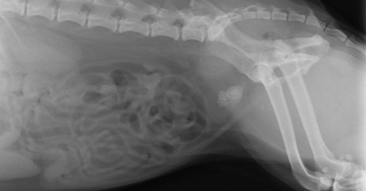
Typically, you’ll start with two simple views of the abdomen—a lateral and a VD.
Take a close look at the urinary bladder to look for radiopaque stones, which should show up as a white opacity relative to soft tissues thanks to their mineral composition.
Stones can range in size from small sand-like grains to more than two inches across. There may be just one or two stones present… or a small group… or even more than 100.
Remember to check the urethra for small stones that could be stuck—especially if the patient is straining or in pain during urination.
For better visualization of the entire urethra in male dogs, consider taking another lateral view with the hindlimbs pulled forward.
Also, check the kidneys and the areas of the ureters. While much less common in pets than in people, occasionally uroliths may be seen higher up in the urinary tract.
If you see stones now—you have your answer.
If you suspect urinary bladder stones but still don’t see them, a contrast study may allow better visualization.
For finding bladder stones, a double-contrast study is ideal.
This includes using both a positive contrast agent (soluble iodinated contrast medium) and a negative contrast agent (room air, or ideally carbon dioxide to reduce the risk of an air embolus) in the bladder together.
Anesthesia or sedation may be needed for the patient’s safety and comfort since the contrast agents are administered via a urinary catheter.
In addition to radiographs, an ultrasound is a useful tool…

Finding uroliths via ultrasound
An ultrasound study is another great option for finding bladder stones, especially radiolucent ones.
The fluid-filled bladder provides contrast for the ultrasound waves so that stones can be found (and often emphasized by acoustic shadowing).
Besides radiolucent stones, you may also see…
Bladder stones (radiopaque or radiolucent) that were too small to visualize radiographically (smaller than 1-3mm).
Other problematic issues in the bladder, such as ‘sludge’ buildup in cats with crystalluria.
Damage to the urinary bladder itself, such as inflammation.
The condition of the upper urinary tract—the kidneys and ureters.
Unexpected findings, such as tumors or anatomical abnormalities of the bladder.
Because of this, ultrasound imaging is a valuable tool for helping you diagnose and treat problems of the bladder, including urinary stones.
Follow-up
Depending on your findings, you may recommend a diet change for dissolvable stones, or a cystotomy to remove the stones.
For dissolution, follow-up imaging can help to track the patient’s progress and see whether or not the stone is dissolving.
When a cystotomy is recommended, remember to use imaging on the day of surgery…
Take pre-op radiographs to confirm the stones are still there, and that your urinary catheter is in place.
Include post-op views to confirm and document that all stones were successfully removed.
Since most stones are radiopaque, standard radiographs are a good option for follow-ups—and typically the imaging choice on the day of surgery.
But ultrasound can also be used in conjunction with other diagnostic tests to monitor the health of the urinary system long-term and to look for early signs of a problem such as a reoccurrence of stones.

Catching stones early, when they’re small, may allow less invasive treatment options such as voiding urohydropropulsion.
With the right combination of imaging modalities, you can help your clients stay on top of treating, monitoring, and preventing urinary bladder stones in their pets.
Written by: Dr. Tammy Powell, DVM
Disclaimer: This article is for general informational purposes only, and not intended as a guide to the medical treatment of any specific animal.

Old X-ray Table and Generator Be Used with a New Digital X-ray System?
Can My Old Universal Easymatic X-ray Table and Generator Be Used with a New Digital X-ray System?

Some of the Universal Easymatic systems look like this
Some veterinarians purchased their x-ray table and generator 25+ years ago and they still work perfectly!
Through care and good maintenance, they have been able to produce diagnostic images over the years using film.
For some veterinarians, 2017 is the year they would like to stop using chemicals and standard film.
However, they do not want to spend the 17k-25k on a new table and generator when their old system is producing excellent mas/kvp options for each case.
The good news is your existing system can work well with proper installation with your new digital x-ray equipment. For computed radiography or CR it is a simple calibration that will enable the old table and generator be ready for use the day of the installation.
This installation should be carried out by the installer on the day of installation and prior to the training session with the veterinary staff.
For direct radiography or DR, the plate may be wired into the foot switch as a prep switch. The old universal Easymatic x ray table and generator send out a 120-volt prep and expose signal.
A conversion box is brought by our onsite installers so that the cesium technology can convert the voltage down to a high of 5 volts and a low of 0 volts.
Another item that we include with our system is a new foot pedal switch. Often times the old switch can be worn out and the wiring loose.
A new foot pedal switch ensures a smooth transition from prep to exposure. In some instances, we also include a hand switch which can be mounted on the wall.
This secondary option is nice especially if the foot switch was to go out you always have another backup to take a high-quality digital x-ray.
You do not always need to buy a new table and generators to enjoy the speed, safety, and quality of digital x-rays.
Contact us today.
We can help you upgrade your system to digital.
Here is a video showing the conversion from film to digital using the old table and generator.


Buy Veterinary Digital X-ray Equipment with Confidence

How to Buy Veterinary Digital X-ray Equipment without Experiencing Buyer's Remorse
Have you ever bought something and regretted the purchase?
I think I have at one time or another. However, it’s one thing to regret ordering a cheeseburger and fries, but quite another when I regret buying a $50,000 car or truck.
When I make a big purchase, I want to feel good about my decision. I want to make sure that I made the right choice.
I want you to feel great about buying from me! I understand it is no small thing for a Veterinarian to spend $20,000 or $30,000 on digital x-ray equipment, and I want you to feel great about buying from us.
So, here is my “purchase without buyer's remorse” plan:
The 30-day satisfaction guarantee, or your money-back offer. When you make a digital x-ray system purchase, I will give you 30 days so that you feel comfortable and confident that you made the right choice.
And if you are not satisfied, return your digital x-ray equipment for a full refund.
Yes, that is correct! A FULL refund!!
I know that after the vet digital x-ray equipment is installed in your clinic, and once you learn how to use the software, you will be very happy with the results you get.
I can offer this guarantee because I am very confident with the quality of the digital x-ray equipment we sell, and I want you to be completely satisfied.
Here is the fine print:
Equipment must be returned in original packaging.
Equipment cannot be returned if damaged by the user during your 30 days.
The buyer pays the cost of shipping/packing/insurance of all returned equipment.
You must give us a reasonable chance to correct any dissatisfaction.
The main reason I am offering this “30-day satisfaction guarantee or your money back” is to give you peace of mind.
I understand I’m not as big as Idexx, Cuattro, or Sound-Eklin. Perhaps that makes you think twice about trying us out.
But I am confident that our equipment is as good, if not better, and now we have a “30-day satisfaction guarantee or your money back” - which the big boys do not offer!

The bottom line is simple:
I am offering quality digital x-ray equipment for the veterinary industry at an amazing price, with a great warranty, and now a 30-day guarantee.
I want to do the right thing, treat you with respect, and help you be successful in your veterinary practice.
Call or text me - Brad Haven, Jr. - 530-355-5886

What Is an Under-Exposed X-Ray, and Why Does It Matter?
When it comes to your x-ray images, there’s a fine balance between too much or too little exposure.
Of course, you’ll always want to use the lowest settings possible to get the image you need, to minimize x-ray exposure to both your patients and your staff.
But, it’s possible to go too low on your exposure settings—and often that results in an under-exposed, possibly non-diagnostic, image.
Why should you care about an under-exposed x-ray image?
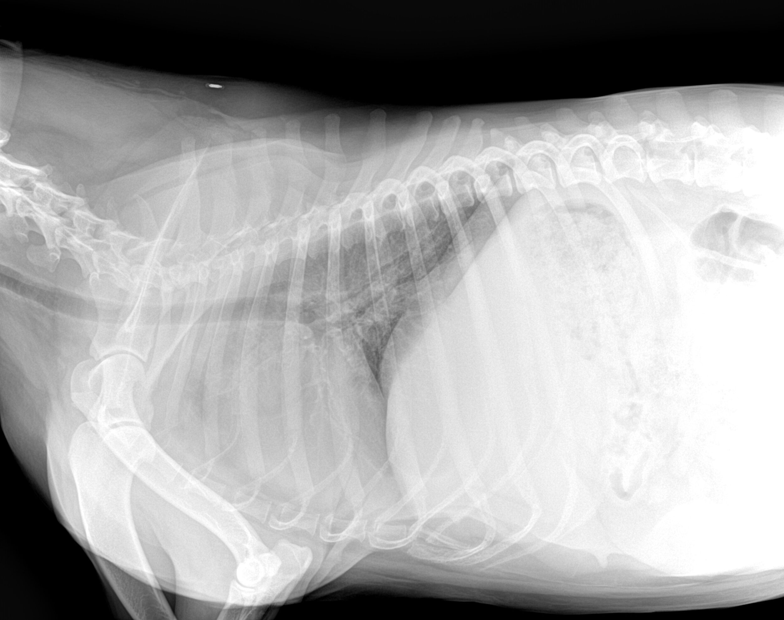
The short answer is because your images will look very light and might not show enough detail or contrast for you to make a diagnosis. An under-exposed radiographic image could mean…
Increased “noise.” This can show up as cloudiness, mottled areas, or even stripes on the image. In the areas outside of the patient, that’s not necessarily a problem—but extra noise or grainy patterns overlying the area you’re looking at could create artifacts or obscure details.
White or light radiographs that are difficult to read. An under-exposed radiograph means there was less penetration of the x-ray beam through the patient’s tissues. This results in an x-ray image that looks overly white or light compared to a properly exposed radiograph. That “whitewash” effect can make it more difficult to see certain lesions or abnormalities.
In other words, under-exposure could mean artifacts, non-diagnostic results, or other confusion when it comes to interpreting your films or digital images.
Reasons why your radiographs might be under-exposed
Here are some possible culprits…
An error in technique (kVp or mAs settings).
A machine or equipment error.
Using grids with a non-grid technique.
Having two films on the same cassette.
Variations in screens.
A long-distance between the x-ray source and the film or plate.
This isn’t an exhaustive list, but it represents good areas to explore for getting better images.
How to get it right the first time
Using the least amount of radiation necessary to get your images is a good practice. However, if your beam isn’t strong enough to produce a diagnostic image, then you’ll have to retake the shots, which produces more potential exposure to patients and staff.
So, it’s best to get it right the first time. Try these tips…
Use technique charts. Charts will give you a good starting point for exposure settings, and take out much of the guesswork. Usually, you’ll have to increase kVp or mAs to improve an underexposed image.
Keep your equipment in good repair, including routine maintenance as necessary.
Use appropriate patient restraint, whether physical or chemical. This can minimize movement artifact, as well as any changes to tissue density that could happen if a patient moves their body in a way that’s different from when you measured them.
Consider using digital radiography. This will minimize wait times between views (allowing you to adjust settings prior to taking more shots).
Digital radiography also prevents errors that come from the film development process. That’s not the same thing as under-exposure… But, taking film development out of the equation does give you one less thing to worry about and makes it faster to troubleshoot if you ever do come across problems with your images.

Have an overexposed radiograph? Check here for more information and tips.
Disclaimer: This article is for general informational purposes only, and not intended as a guide to the medical treatment of any specific animal.
Written by: Dr. Tammy Powell, DVM

Hands-Free X-Rays: The Next Step in Veterinary Safety

Is it time to put restraints on veterinary radiography?
By Julia Bitan, RVT
When Dr. Debrah Berman, a Thornhill veterinarian with over 30 years of experience felt a lump in her tongue, she didn’t think much of it.
Up until that point, her health had been excellent and she was following all current radiology safety regulations such as monitoring her dosimeter values and using all protective equipment.
The diagnosis of Mucoepidermoid Carcinoma of the salivary gland (a type of cancer often associated with excess radiation exposure) came as a shock and made her start questioning the safety of today’s veterinary radiography.
“Since the 1990s, numerous jurisdictions in North America, and worldwide, have strengthened their regulations and best practice guidelines to actively move away from holding patients, in recognition that exposure to scatter radiation poses a risk.”
Outdated regulations
Although there are several provincial and federal laws in place to protect radiation workers in Ontario, the clauses specifically concerning veterinary workers have not been updated since 1990.
According to a veterinary radiography survey conducted in partnership with the Ontario Association of Veterinary
Technicians (OAVT) in April of 2016, 8 out of 10 RVTs still hold their patients during x-rays most of the time, while 7 out of 10 respondents choose to sometimes forgo the use of gloves and other protective equipment simply due to inconvenience.
Our human radiology counterparts in Ontario are rarely present in the room when x-rays are taken because regulations state that no person should regularly perform manual restraining for x-rays.
Technicians commonly request parents hold their own children if x- rays are required.
Since the 1990s, numerous jurisdictions in North America and worldwide have strengthened their regulations and best practice guidelines to actively move away from holding patients, in recognition that every x-ray taken increases the overall risk of cancer.
Because we don’t see regular reports of veterinary workers dying of radiation exposure, we may naively assume that modern science has overcome the dangers with newer machine design and intensive research.
In reality, current occupational exposure limits are derived from decades-old research and we know that the risk of getting cancer increases with every exposure.
Recent findings do show that extended exposure to a low level of radiation increases the risk of developing leukemia, while radiation-induced cataracts are observed at a much lower radiation dose than previously believed. In veterinary medicine, the damage caused by ionizing radiation is simply too low to be felt right away and the ALARA principle (As Low As Reasonably Achievable) is much too ambiguous in the context of our profession.
Change is happening
A small number of veterinary clinics in Ontario, including Toronto Veterinary Emergency Hospital and Referral Center (TVEH), began enforcing a strict no-hold, out-of-the-room x-ray policy to keep their veterinarians and RVTs away from ionizing radiation emitted by the x-ray machines.
TVEH started enforcing the 100% -out-of-the- room radiography six years ago.
RVT Ashley Jenner, head of radiology at TVEH, has developed a number of tools and techniques to make out-of-room x-rays possible.
“Non-manual veterinary radiography is much easier and faster than most people believe,” Jenner says. “All it takes is proper techniques and some extra tools.”
Jenner uses positioning devices alone on 75% of her cases (non-sedated patients), and sedation on the remainder.
Dr. Debrah Berman is now “cancer-free” and back to practicing veterinary medicine. She now endeavors to obtain all her x-rays using positioning/restraining devices or sedation.
“Radiation exposure is cumulative,” says Berman. “You may not realize until 20 or 30 years down the road that you have received too much. It may take a little more effort, but had I known that I would need to have a quarter of my tongue removed because of cancer and that I'd have permanent nerve damage and varying degrees of chronic pain, I wouldn't hesitate. And, I am one of the lucky ones. We need to protect ourselves”.
Transitioning to non-manual radiography is not always an easy task.
Change is rarely welcomed in our profession and as long as the minimum standards are followed, there is little reason or incentive to change.
We do, however, know there are risks involved with radiation, and alternatives do exist.
A program was created to guide clinics through transitioning to non-manual radiography using different tools and techniques.
The Hands-Free X-Rays Initiative aims to promote awareness and encourage change to current veterinary radiography.
For more information about the program, visit our website - www.handsfreexrays.com.
For comments, feedback or help to bring change to your clinic, please contact us at:
email: info@handsfreexrays.com
Phone - +1 (647) 502-4843.
Written by By Julia Bitan, RVT


Your Warranty and Service Coverage for Veterinary X-Ray

Today, every veterinarian recognizes the importance of incorporating digital X-rays into their animal hospitals.
A considerable number of them plan to transition from traditional film and chemical X-rays to digital systems this year.
While the initial concern typically revolves around the cost of acquiring a digital X-ray system, it is equally crucial to inquire about the warranty and its coverage.
Here are some pertinent questions to ask the prospective vendor when considering the purchase of a veterinary digital X-ray system:
1. What is the duration of the hardware warranty for my veterinary digital X-ray plate?
2. How long does the software warranty for my veterinary digital X-ray software last?
3. For how long will I receive software upgrades for the X-ray system used in my animal hospital?
4. How long will my veterinarian technicians have access to tech support, enabling them to seek answers to any queries that may arise during the daily implementation of digital X-rays in my animal practice?
5. What are the costs associated with continued phone support, software upgrades, and technical assistance after the warranty period ends?
6. Which entity is responsible for providing software and technical support? Who developed and owns the software?
7. Does the warranty cover labor and shipping expenses, as needed?
8. In the event that my X-ray plate requires repair, will a temporary replacement plate be provided?
It is common for vendors operating in the animal health sector to offer a minimum of a 1-year warranty.

Not all vendors possess ownership of the software that operates their veterinary digital X-ray plate, which holds significant importance. While a digital X-ray plate can remain functional for many years, the software it relies on may become outdated within a short span of time.
An example of this can be observed with Windows XP platforms. Unless companies continually update their software, it becomes incompatible with new computing platforms.
When veterinarians transition from computed radiography (CR) to direct radiography (DR), the predominant issue they encounter is not the failure of the CR system itself but rather the poor performance or lack of support for the software.
This issue is not limited to small companies alone. Renowned brands like Idexx and Sound have discontinued support for their older product lines by ceasing software upgrades.
Our aim is to offer you transparent service and support. Our animal health software has been developed in-house by JPI which has been manufacturing x-ray equipment, writing software, and providing telephone support for these products since 1994.
Here are the responses to the eight frequently asked questions:
1. What is the duration of the hardware warranty for my veterinary digital x-ray plate?
- The hardware warranty lasts for 5 years.
2. How long is the software warranty for my veterinary digital x-ray software?
- The software warranty is also valid for 5 years.
3. For how long will I receive software upgrades for my x-ray system to be used in my animal hospital?
- You will receive software upgrades for 5 years.
4. How long can my veterinarian technicians receive phone support for any questions they may have regarding the daily implementation of digital X-rays in my animal practice?
- Our tech support helpline will be available for your veterinarian technicians for 5 years.
5. What is the cost of continued phone support and software upgrades after the warranty period?
- The cost for continued phone support and software upgrades after the warranty period is $750.00 per year.
6. Who provides the software and tech support? Who developed and owns the software?
- Our Examvue Veterinary software is developed, supported, and owned by JPI. They have been offering industry support since 1994. For technical support, please call 516-513-1330, and select option 2. Our support team is based in New York.
7. Are labor and shipping costs covered under the warranty if needed?
- Yes, labor and shipping costs are included in the warranty as required.
8. Will a loaner plate be provided in case my X-ray plate needs repair?
- Yes, we will provide a loaner plate in the event that your X-ray plate requires repair.

Is the New Digital X-ray System Compatible with Innovet?

Many veterinarians invested in X-ray systems in the ’90s. Most purchased an Innovet Summit table and generator.
They used the table and generator along with cassettes enclosing film to capture X-rays and provide life-saving next steps for their pet patients.
Processing the film in the chemical rinse and developing the images in the darkroom enabled the practicing veterinarian to review the X-ray.
If the X-ray proved complicating or just interesting then the veterinarian would mail the X-ray to a specialist for a review or save the X-ray case for the next ‘radiology rounds’ in upcoming weeks and months.
During these radiology rounds, X-ray cases would be reviewed amongst veterinarian piers and sometimes even veterinary radiologist.
Even if the patient was deceased by the time of the review huge advances in pet medicine were and still are to this day applied.
If you're reading this you use to film with your old X-ray generator and table. You want to impact the neighborhood and village who depend on precise and timely diagnosis to treat their pets.
Digital X-rays can speed up the process not only of image acquisition but also provide clear diagnostic radiology rounds.
Most old tables and generators still can produce excellent X-rays.
Often times these old systems have new light bulb collimators and replaced fuses that enable the old workhorse to continue to provide the power for an X-ray.
Yes, you can use your old table and generator with two types of digital X-rays.
The first is computed radiography or CR which will work today with any generator as the image is captured on an erasable film cassette then scanned into the software where the digital image can now be interrupted, adjusted or emailed to pet owners.
The next is an AED cesium plate. Automatic exposure detection.
This allows the plate to prepare itself for the exposure to capture the image and send in second the inmate to the display monitor.
This is the newest way to capture X-rays.
Not having to wire the old X-ray system to the new digital X-ray system is a confinement advantage.
It removes an element of future failure in hardware by eliminating the interference box!
We can help answer more questions surrounding your old X-ray table and generator.
Reach out today and we would be happy to help.