


How Veterinary Dentistry Improves Staff Engagement and Productivity
Veterinary Dental Care Supports Team Growth and Retention
Ask most veterinary professionals why dentistry matters, and you'll hear plenty about periodontal disease, oral pain, and improved patient health. What you may hear less often is this:
A strong dental program can also strengthen the veterinary team.
I know how that sounds, but stick with me.
In a profession where burnout, staffing shortages, and compassion fatigue continue to challenge practices of every size, dentistry offers something many teams are searching for:
- Meaningful work
- Skill development
- Collaboration
While dentistry certainly benefits patients and clients, it can also become one of the most rewarding areas of practice for veterinarians, technicians, and support staff alike.
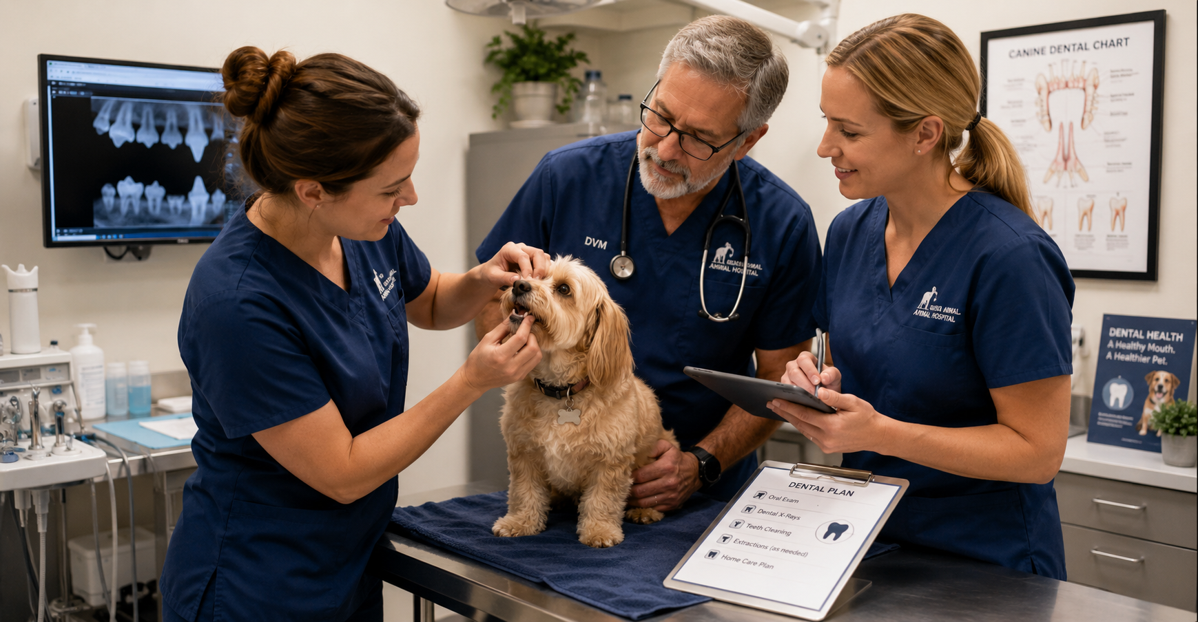
Giving Technicians the Opportunity to Practice at a Higher Level
Veterinary technicians are highly skilled professionals, yet many teams struggle with fully utilizing those skills. Dentistry offers technicians opportunities to become deeply involved in patient care before, during, and after procedures.
Depending on state regulations and practice protocols, technicians often play critical roles in:
- Patient preparation and anesthesia monitoring
- Dental charting and radiography
- Scaling and polishing
- Recovery monitoring
- Client education
Rather than performing the same routine tasks throughout the day, dental technicians can engage in more advanced, hands-on work.
For many team members, that increased responsibility brings greater job satisfaction.
When people are trusted to use their skills, they become more invested in both their work and their patients.
The Moment Things Change: Seeing What’s Hidden
One of the most rewarding moments during a dental procedure often happens when the first radiographs appear on the screen.
A tooth that looked relatively normal externally may reveal severe bone loss. A fractured tooth may show a damaged root. A cat with mild tartar may have advanced resorptive lesions.
These discoveries create valuable learning opportunities for the entire team.
Dental radiography turns procedures into diagnostic investigations rather than routine cleanings. Team members begin asking questions:
- What are we seeing?
- Why is this happening?
- What treatment options are available?
- How can we improve this patient's comfort?
Those moments build clinical confidence and encourage continued learning.
Over time, your team becomes better at recognizing pathology, discussing findings, and contributing to patient care decisions.
Immediate Results Create Meaningful Work
Veterinary medicine can sometimes feel emotionally challenging because progress isn't always obvious.
Chronic disease management takes time. Behavioral improvements may take months. Long-term treatment plans often unfold gradually.
Dentistry is different.
A patient may arrive with severe tartar, inflammation, infection, or oral pain and leave significantly more comfortable the very same day.
The team can see the difference. Clients often notice the difference quickly. And perhaps most importantly, staff members know they helped relieve pain that may have gone unnoticed for months or years.
Those experiences matter.
They remind your team why they entered the field in the first place.
Creating More Predictable Days
Anyone who has worked in veterinary medicine knows that unpredictability is part of the job.
Emergencies happen. Schedules change. Appointments run long.
While no service completely eliminates those challenges, well-organized dental programs can add welcome structure to the workday. Dedicated dental appointments often create:
- Predictable procedure blocks
- Clear team assignments
- Consistent equipment setups
- Efficient patient flow
Your team knows their role. Supplies are prepared. Expectations are clear.
Instead of constantly reacting, your team spends more time working together with purpose.
Investing in Growth Keeps Teams Engaged
Few things improve retention as much as professional growth. Dental programs often encourage continuing education opportunities, such as:
- Dental radiography training
- Anesthesia education
- Dentistry certifications
- Wet labs and hands-on courses
When you invest in these areas, employees often feel valued and supported.
A technician who develops expertise in dentistry may become a mentor, trainer, or internal resource for the rest of the team. That sense of ownership can significantly improve engagement and long-term retention.
In an industry facing staffing challenges, opportunities for growth matter.
Building a Culture Around Better Care
The most successful dental programs become more than a service line. They become part of the practice culture.
Technicians become advocates for dental education. Doctors collaborate on treatment planning. Clients receive more consistent recommendations.
That sense of purpose can strengthen teamwork and create a culture centered around delivering better care.
Dentistry Is About More Than Procedures
It's easy to measure dentistry by production numbers, procedure counts, or equipment investments. But those metrics only tell part of the story. Every dental procedure is also an opportunity:
- To help a pet live more comfortably.
- To strengthen a technician's skills.
- To build confidence within the team.
- To create meaningful work.
- To support professional growth.
Dentistry isn't simply a revenue center. It's a service that allows your team to practice at a higher level while making a real difference in their patients' lives.
And when your team feels challenged, supported, and connected to their work, everyone benefits—including the pets they care for every day.


Building a Sustainable Veterinary Dental Program: The Benefits to Pets, Clients, and Practice Growth
Build a Stronger Veterinary Dental Program
How Better Dental Care Benefits Pets, Clients, Veterinary Teams, and Practice Growth
When veterinarians talk about dentistry, the conversation usually centers on patient health—and for good reason.
Dental disease can cause chronic pain, infection, tooth loss, and a significant decline in a pet’s comfort and quality of life. Helping pets live healthier, more comfortable lives is the primary reason veterinary dentistry matters.
But there is another side of the story that also deserves attention.
A well-developed dental program can strengthen client relationships, help more pet owners follow through with recommended care, create opportunities for team development, and provide a predictable source of revenue that supports the long-term health of the practice.
Veterinary dentistry is one of the rare areas where everyone can benefit: pets, clients, veterinary teams, and the clinic.
Dental Disease Is Often Underdiagnosed
One of the biggest challenges in veterinary dentistry is that many pets do not show obvious signs of discomfort.
Unlike people, dogs and cats often continue eating and following their normal routines even when they are experiencing significant dental pain. They quietly adapt, which can make oral disease difficult for pet owners to recognize.
As a result, serious dental conditions may go unnoticed until they become advanced. Veterinarians commonly discover:
- Advanced periodontal disease
- Tooth-root infections
- Fractured teeth
- Tooth resorption
- Significant bone loss
Many of these conditions can only be fully identified through a comprehensive oral examination and dental imaging.
Because pets are so skilled at hiding oral pain, proactive dental care is especially important.
A strong dental program helps practices identify disease earlier and intervene sooner, before smaller problems become more painful, complicated, and expensive to treat.
Dentistry Creates Opportunities to Improve Patient Health
Unlike procedures that happen only once during a pet’s lifetime, dental care often becomes part of an ongoing healthcare plan.
Oral health requires regular monitoring, preventive care, and treatment throughout a patient’s life. This creates opportunities for:
- Annual dental evaluations
- Professional cleanings
- Dental imaging
- Follow-up procedures
- Preventive care recommendations
- Ongoing client education
Every dental conversation is another opportunity to improve a pet’s quality of life while strengthening the relationship between the veterinary team and the client.
The goal is not simply to perform more procedures. It is to create a proactive approach to oral health that supports patients throughout their lives.
Existing Clients Are Often the Best Opportunity for Growth
Many veterinary practices spend significant time and money attracting new clients.
While new client acquisition is important, existing clients often represent one of the greatest opportunities for sustainable practice growth.
Why?
Because trust has already been established.
When a veterinarian recommends dental care based on examination findings, clients are often more receptive than they would be to an unfamiliar service they have never discussed before.
Dental recommendations are a natural extension of preventive healthcare. The practice is not trying to sell an unnecessary service. It is helping the client understand and address a health problem that already exists.
“Here is what we are seeing.”
“Here is why it matters.”
“Here is how we can help.”
Clear communication helps clients understand the value of treatment and make more confident decisions about their pet’s care.
Dentistry Can Create Predictable, Recurring Revenue
Every veterinary practice needs revenue to operate, invest, and continue providing high-quality care.
One advantage of dentistry is that it can become a relatively predictable service line.
Because dental disease is common and often ongoing, practices can build consistent revenue through:
- Annual dental procedures
- Dental imaging
- Follow-up treatment
- Extractions when medically necessary
- Preventive dental programs
Unlike services that may fluctuate significantly from month to month, dentistry can become a dependable part of practice operations.
That stability can help a practice plan for staffing, equipment, training, and future growth with greater confidence.
Profitable Dentistry Supports Better Veterinary Medicine
Profitability should never take priority over patient care.
However, it gives veterinary practices the resources they need to continue providing excellent medicine.
A profitable dental program can help fund:
- Staff training and continuing education
- New diagnostic equipment
- Facility improvements
- Additional support staff
- Expanded treatment capabilities
- Improved patient-care technology
In many cases, the investments made possible through dental revenue benefit the entire practice, not just the dental department.
The result is a stronger clinic that is better equipped to serve patients, support its team, and help clients make informed healthcare decisions.
Building a Dental Program That Lasts
The most successful dental programs are not built around production goals. They are built around patient care.
Practices that focus on education, early detection, quality diagnostics, and comprehensive treatment recommendations often find that growth follows naturally.
When pets receive better care, clients understand the value of treatment.
When clients understand the value, they are more likely to follow through with recommended care.
That, in turn, gives the practice the resources it needs to invest in its team, equipment, and services.
It becomes a healthy cycle that benefits everyone.
The Real Value of Veterinary Dentistry
At its core, veterinary dentistry is about identifying hidden disease, relieving pain, and improving quality of life.
The business benefits are real, but they are a result of providing valuable care—not the primary objective.
A healthy dental program is not about maximizing revenue. It is about building a sustainable practice that can continue serving pets, supporting veterinary teams, and helping clients make confident decisions about their animals’ healthcare.
Better Dentistry, Better Business: The Value of Digital Dental X-Rays

The Hidden ROI of Veterinary Dentistry: How Digital Dental X-Rays Pay for Themselves
When veterinary practices consider investing in dental equipment, the conversation often starts with patient care…and rightfully so.
Dental disease is one of the most common health issues affecting companion animals, and dental radiographs have become an essential part of providing high-quality care.
But there is another side of the conversation that is equally important: the business case for veterinary dentistry.
Running a successful practice requires more than compassion and clinical expertise. You need to generate enough revenue to pay your team, invest in technology, maintain facilities, and continue delivering exceptional care.
Fortunately, veterinary dentistry is one of the few service areas where improved patient outcomes and financial sustainability often go hand in hand.
The key is making smart equipment investments that support both goals.
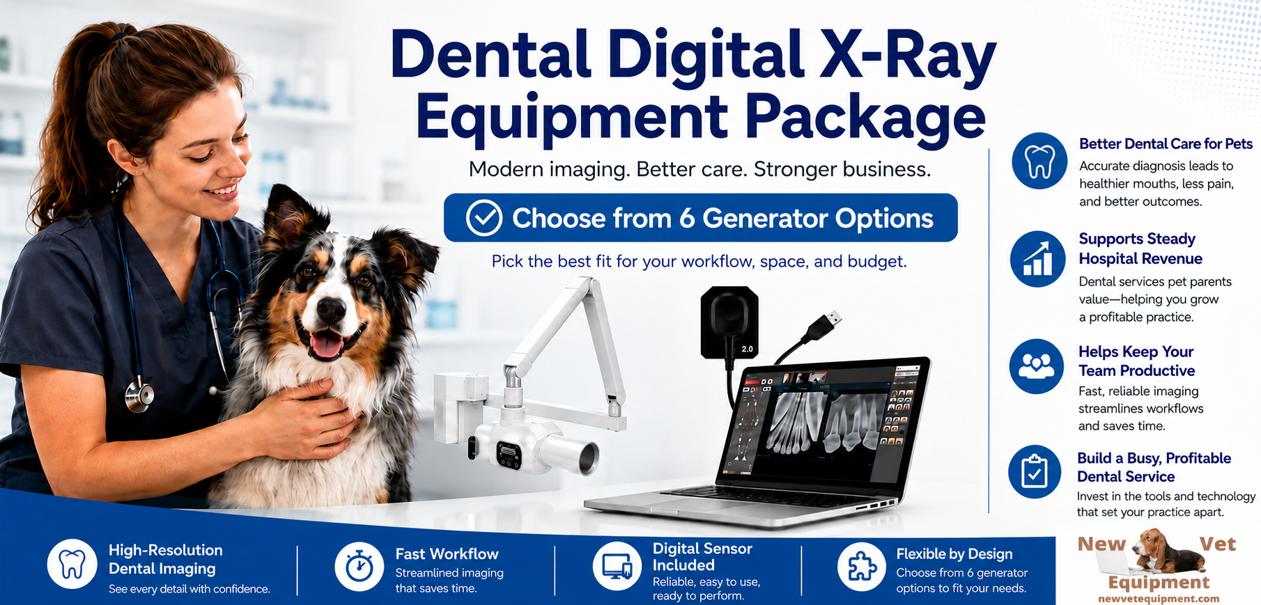
Why Dental Radiographs Are the Standard of Care
Many pet owners are surprised to learn that much of a tooth lies below the gumline. In fact, studies have shown that a significant percentage of dental pathology cannot be identified through a visual examination alone.
Without dental radiographs, veterinarians may miss:
- Tooth root abscesses
- Resorptive lesions
- Periodontal bone loss
- Retained roots
- Fractured teeth
- Endodontic disease
Digital dental X-rays allow you to identify problems that would otherwise remain hidden, helping you provide more accurate diagnoses and treatment recommendations.
Today, many veterinarians consider dental radiography a fundamental component of a complete dental procedure rather than an optional add-on.
Looking Beyond the Price Tag
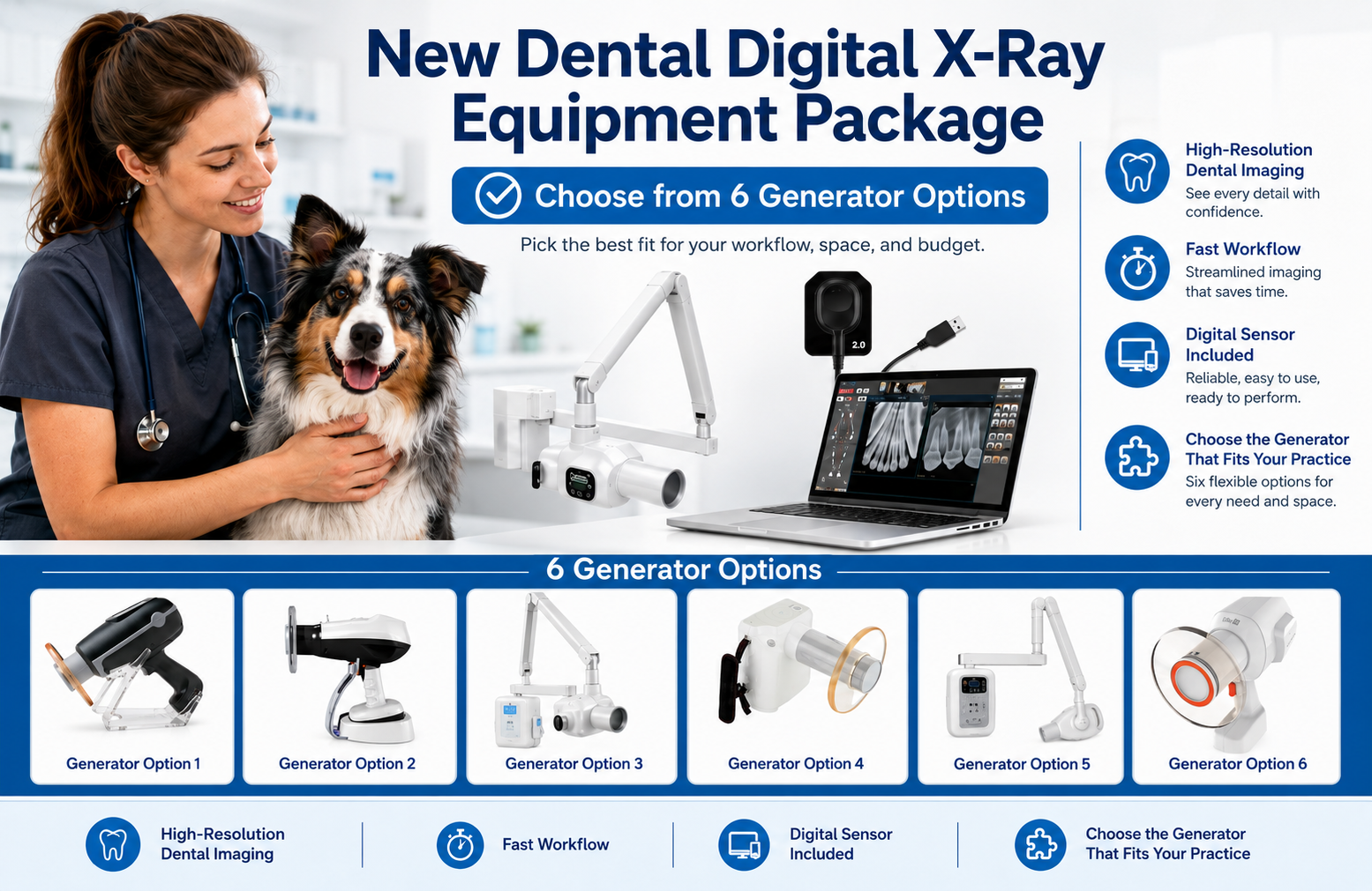
One of the biggest misconceptions about dental equipment is that practices need to spend top dollar to build a successful dental program.
Some premium dental imaging setups can cost $35,000 or more once sensors, generators, software, and accessories are included.
While those systems may offer additional features, you can achieve excellent clinical results with quality digital dental radiography systems in the $12,000-$15,000 range.
The difference matters.
A lower initial investment can significantly shorten the time required to achieve a positive return while still providing the diagnostic capabilities veterinarians need to deliver quality care.
A Simple ROI Example
Let’s look at a realistic scenario:
Assume a practice purchases a digital dental radiography system for $15,000. Now consider a conservative estimate:
Average dental procedure revenue: $600
Dental procedures performed per month: 10
Monthly dental revenue: $6,000
Of course, equipment cost is only one piece of the equation. There are labor, anesthesia, supplies, and overhead expenses to consider. However, even when accounting for those costs, dental procedures often represent a valuable service line for many practices.
If the practice attributes only a portion of that revenue toward recovering the equipment investment, the payback period can still be surprisingly short.
For example:
Equipment investment: $15,000
Contribution toward equipment recovery per procedure: $150
Procedures needed to recover investment: 100
At just 10 dental procedures per month, the equipment could effectively pay for itself in less than a year.
Now compare that to a $35,000 investment.
Using the same assumptions, the practice would need approximately 233 procedures to recoup the equipment cost, significantly extending the payback timeline.
The takeaway is not that higher-priced systems are inherently bad investments. The purchase price directly affects how quickly a clinic can realize a return.
Improving Workflow Efficiency
The ROI of digital dental radiography is not limited to revenue generation. Digital systems can also improve workflow efficiency throughout the practice.
Digital dental X-rays eliminate many time-consuming steps and can help reduce the need for unnecessary retakes.
Benefits often include:
- Immediate image review
- Improved image storage and retrieval
- Easier integration with practice management software
When procedures move more efficiently, you can often complete appointments more predictably and utilize staff time more effectively.
Helping Clients Say “Yes” to Treatment
Client education is one of the most overlooked benefits of digital imaging. When pet owners can actually see pathology on a screen, treatment recommendations become much easier to understand.
A fractured tooth root, severe bone loss, or a resorptive lesion is often far more compelling when accompanied by an image rather than a verbal explanation alone.
The result is better patient care and greater confidence in the veterinary team’s recommendations.
What to Look for When Purchasing Dental Equipment
Not all dental radiography systems are created equal, but the most expensive option is not always the best choice. When evaluating equipment, consider:
- Image Quality: The system should consistently provide diagnostic-quality images with excellent detail.
- Ease of Use: A user-friendly interface can reduce training time and improve adoption among team members.
- Sensor Durability: Dental sensors represent a significant investment. Durability and warranty coverage matter.
- Software Integration: Look for systems that work seamlessly with your existing workflow and practice management software.
- Service and Support: Reliable technical support can make a major difference when issues arise.
- Total Cost of Ownership: Consider not only the purchase price but also future maintenance, replacement costs, and warranty protection.
Better Dentistry, Better Business
Veterinary dentistry should never be viewed solely as a revenue generator. At its core, dental care is about identifying pain, treating disease, and improving quality of life for pets.
But it is also fair, and necessary, to acknowledge the business benefits.
Profitable service lines help you retain talented staff, invest in better technology, expand patient care capabilities, and continue serving your community for years to come.

Pet Dental Care: Why Cleanings and X-rays Matter Most

Pet Dental Care 101: Why Cleaning and X-rays is a Win-Win for Practices and Clients
Pet dental care is often overshadowed by more visibly urgent veterinary services, like surgery or emergency treatments. Yet, we know it’s one of the most valuables most valuable (and misunderstood) aspects.
Offering thorough dental services can be a game-changer for patient outcomes, client satisfaction, and financial stability.
Let’s explain why dental care deserves a prominent place in your practice, and how it benefits everyone involved.
An Overlooked Epidemic: Dental Disease in Pets
Despite the prevalence of periodontal disease in pets, it’s often underdiagnosed and undertreated because its symptoms—bad breath, tartar buildup, or minor gum inflammation—are easy for pet parents to dismiss.
Clients (and sometimes veterinary teams) don’t realize that dental disease doesn’t stay confined to the mouth. Left unchecked, it can lead to:
Chronic Pain: pets often mask discomfort, but dental pain can affect their appetite, behavior, and energy levels.
Systemic Health Issues: bacteria from periodontal disease can enter the bloodstream, impacting organs like the heart, liver, and kidneys. Plaque and tartar are made up of harmful bacteria, and left untreated it can have a more significant impact than just bad breath.
Shortened Lifespan: studies have shown a correlation between untreated dental disease and reduced life expectancy in pets.
Routine cleanings and dental X-rays address these issues early, avoiding unnecessary pain and preventing costly complications.
Why Digital X-rays Are a Game-Changer
The real magic of veterinary dental care happens below the gumline. Roughly 60% of dental disease lurks out of sight, so visual exams alone are insufficient for a proper diagnosis.
Scaling and polishing are a great start, but our patients’ needs go beyond the surface.
This is where digital X-rays come in – they often reveal:
Root Abscesses: these can cause significant pain and require immediate treatment.
Bone Loss: essential for evaluating periodontal disease severity and determining whether teeth can be saved.
Resorptive lesions are widespread in cats, and these painful conditions are impossible to spot without imaging.
Retained Roots: fragments left behind from previous extractions can cause painful infections if not addressed.
Digital X-rays aren’t just a diagnostic tool; they’re an educational one that shows clients images of their pet’s dental issues.
This helps them visualize the problem and understand the urgency of treatment. It transforms what could feel like an optional procedure into a clear medical necessity.
A Financial and Medical Win-Win for Practices and Clients
Let’s look at some of the financial wins for practices:
Increased Revenue Streams: dental services are consistently among the most profitable offerings for veterinary practices. The initial investment in equipment like digital X-rays and dental cleaning stations quickly pays for itself as demand grows.
Recurring Services: unlike one-off emergencies, dental cleanings, and x-rays are part of an ongoing care routine, bringing clients back for regular visits.
Efficiency Gains: up-to-date tools like digital X-rays speed up procedures, allowing you to treat more patients in less time.
Team Empowerment: expanding dental services creates opportunities for staff training and skill development, boosting morale and efficiency.
Now let’s peek at some of the medical wins for pets and pet parents:
Improved Pet Health: regular cleanings and x-rays prevent costly emergency procedures and improve quality of life.
Predictable Costs: preventive care is less expensive than addressing advanced dental disease. When clients understand this, they will commit to regular checkups.
Stronger Relationships: practices emphasizing dental health build deeper connections with clients by showing they care about every aspect of their pet’s well-being.
Industry Insights: The Rising Value of Dental Care
According to the AVMA and other industry reports, practices prioritizing dentistry see significant benefits like revenue growth, client retention, and improved patient outcomes.
Comprehensive dental services are an easy way to increase client satisfaction and improve patient longevity. This often resonates deeply with today’s pet owners, who increasingly view their pets as family members.
Additionally, digital dental x-rays have become a standard of care, and practices that adopt them often set themselves apart as leaders in preventive medicine.
Pet dental care is an opportunity to elevate the standard of care in your practice and investing in the right dental tools ensures pets are healthier, clients are happier, and your clinic thrives.

Hidden Profits: The Value of Regular Pet Dental X-rays

Beneath the Surface: How Routine Dental X-rays and Cleanings Improve Pet Health and Your Bottom Line
In the busy life of a veterinary practice, we know it can be easy to place dental health on the back burner.
After all, pets coming in for vaccinations, surgical procedures, or more obvious pain management often seem more urgent.
Prioritizing dental health through regular teeth cleaning and routine dental X-rays can benefit your patients' well-being and your practice’s financial health.
Let’s talk about why this is a worthwhile investment and why these procedures are essential to our standard of care.
The foundation of good health starts with routine dental cleanings.
Periodontal disease is among the most common health issues in dogs and cats, affecting around 80% of pets over the age of three.
Yet pet owners often underestimate the impact it can have.
Poor dental hygiene doesn’t just affect the mouth, it affects the entire body, leading to issues such as:
Chronic pain
Behavioral change
Weight loss
Systemic infections
Regular cleanings under anesthesia and using a well-equipped dental cleaning station allow us to remove tartar and plaque before they cause these more significant health issues.
They also allow us to discuss pet dental care with clients, which helps improve long-term compliance with at-home care.
Regular cleanings can extend pets’ lives and improve their overall quality of life.
After seeing their pet healthier and more comfortable, our clients often feel reinforced trust in our recommendations and the value of our services.
By encouraging routine dental care, our patients can experience benefits like:
Pain-free eating and play
Prevention of systemic disease
Fresher breath
Enhanced energy and comfort
Lower risk of dental complications
Dental X-rays are an underestimated diagnostic tool and offer an indispensable view of dental health.
Around 60% of dental disease is hidden below the gumline, making it impossible to assess a pet’s oral health without imaging fully.
A tooth may look healthy on the outside, but dental X-rays often reveal hidden problems like:
Root abscesses
Bone loss
Retained roots or resorptive lesions
When we skip these X-rays, we risk missing pain-causing issues that erode the pet's health and comfort over time.
If we avoid routine dental X-rays due to time, cost, or anesthesia concerns, our patients are the ones who ultimately suffer.
Undetected dental disease can progress until it’s obvious enough to require a more aggressive intervention, such as multiple extractions or management of a full-blown abscess.
At that point, the patient has already experienced unnecessary pain and discomfort, and the client faces a larger treatment bill than if the issue had been caught early.
Without early intervention, periodontal disease can lead to chronic infections that eventually spread to other organs, adding further complications to the pet’s health.
For older pets, untreated dental disease may contribute to a shortened lifespan.
Investing in dental health and digital imaging is an opportunity to enhance the health of your practice.
Clients who understand the long-term benefits of preventive dental care and regular checkups will likely invest in these services.
This is especially true when they recognize the care your practice offers—thanks in part to digital imaging technology.
In many cases, veterinary practices that establish clear dental protocols, including routine X-rays, report increased client retention and an expanded service portfolio.
When clients see the advanced diagnostics and appreciate that you’re doing everything possible to prevent disease, they tend to return and spread positive word-of-mouth referrals!
If your practice is ready to integrate or expand digital dental imaging, here are a few tips to ease the transition:
Demonstrate the Importance: educate clients by using examples of cases where X-rays uncovered unseen issues. This helps to give a real-world understanding of what’s at stake.
Offer Dental Packages: Consider bundling dental cleaning and x-ray services for a comprehensive approach. This encourages pet owners to embrace both as part of a complete care package.
Train Your Team: Ensure your entire staff knows the value of digital X-rays and dental cleanings to discuss these services with clients confidently.
Incorporating regular dental care isn’t just about filling appointment slots. It’s about providing your patients with the highest level of care and adding a consistent revenue stream that reflects the value you bring to patient care.

Essential Parts of a Veterinary Dental Machine Explained

Key Components of a Veterinary Dental Machine to Know
Understanding all the parts of a veterinary dental machine will help keep the equipment in good shape to provide optimal patient care. This knowledge can also come in handy when it’s time to buy a new system or for troubleshooting when your dental unit malfunctions.
Here are the key veterinary dental machine parts with which veterinary team members should be familiar, and a few tips for maintaining the equipment in good working order…
Power Equipment of a Veterinary Dental Machine
Dental equipment used during cleaning, polishing, and procedures like extractions include…
Ultrasonic scaler. An ultrasonic scaler is used to quickly and efficiently remove large amounts of dental calculus. They’re powered by electricity and convert sound waves into physical vibrations.
These scalers are especially valuable for supragingival use but can also be used subgingivally with a periodontal tip.
The most popular types of ultrasonic scalers are probably piezoelectric and magnetostrictive. The former pairs with tips, while the latter requires an insert. They must have water flow to function properly and reduce heat injuries, and some include built-in LED lighting.
High-speed handpiece. A high-speed handpiece offers rapid rotations for use with dental burs (also known as “drill bits”). Uses include surgical extractions (removing alveolar bone, exposing tooth roots, and sectioning teeth) as well as softening sharp bone edges prior to closing a surgical extraction site.
There are many different kinds of burs available for different purposes, not to mention different sizes. Water flow is necessary to prevent overheating.
Low-speed handpiece. A low-speed handpiece is used with a prophy angle for polishing the teeth after scaling.
Air/Water syringe. The water aspect of this tool is used for irrigating any areas being worked on (such as an extraction site) or flushing debris in general. The air component can be used for drying as needed, although it’s recommended to avoid using it in open surgical sites (to prevent air embolism).
Suction tool. Some dental units offer a suction component. Weak suction can be a convenient way to remove fluids such as water, blood, and saliva.
Air compressor. A compressor pressurizes air for use with handpieces or other components that are air-powered.
Some compressors require oil (which must be changed at regular intervals), while others are air-cooled. Either way, it’s good to drain moisture out of the system each day it’s used, and to check for air leaks from time to time.
Water bottles. A reservoir is required for the distilled water that flows through some of the tools, such as the ultrasonic scaler, the drill/high-speed handpiece, and the water/air syringe. Have refills available during procedures. And allow the unit to dry when not in use.
Additional Veterinary Dental Equipment
In addition to the dental unit components mentioned above, the following equipment is important for dental procedures…
Hand tools. While ultrasonic scalers are great, hand scaling is still part of any dental cleaning. This includes hand scalers for reaching calculus in tight spots or anything the ultrasonic scaler missed and curettes for subgingival work. Also think of mirrors, probes, and anything else that might be needed.
Have several sizes available, based on the types of patients your clinic sees.
Suture kit. A suture or minor surgery kit is helpful for dental extractions, especially when a gingival flap must be created. This could include things like forceps, gauze, scissors, etc.
Extraction tools. Think of elevators, luxators, extraction forceps, and burs in various sizes (and shapes, in the case of dental burs). Autoclave in packs when it makes sense to do so, or separately for items that are used less often.
Replacement parts. A quick online search reveals a wealth of replacement parts available for veterinary dental machines: everything from turbines for high-speed handpieces, to hoses and connections, to replacement switches, gaskets, and o-rings.
Depending on the knowledge and comfort level of the veterinary team, it might make sense to keep some of these replacement parts on hand in case an urgent replacement is needed (so dental procedures don’t need to be canceled in case of a mechanical malfunction that can be easily corrected).
Manual. It’s probably best to locate the instruction manual for your practice’s dental unit (and all its many parts) BEFORE anything happens. That can allow for a better overall understanding of the machine, including how to properly clean and maintain everything so they’ll last longer and perform better.
If the manual is lost, contact the manufacturer or search online—there are “libraries” of manuals for all sorts of equipment available online.
Keeping Your Veterinary Dental Machine Happy and Healthy
Get to know your machine and all its components. Each component might come with its own unique instructions for maintenance and optimal operation. For example, this might include specific cleaning instructions, knowing when to replace worn tips and burs, and knowing how to sharpen hand tools after each use. Keeping a log can help.
See if there’s a technically-minded team member who is comfortable doing minor repairs or parts replacements. Or, if technical support from the manufacturer isn’t available (or is subpar), see if you can find a local repair person to help.

Becoming familiar with all the components of a veterinary dental machine can help prevent frustrations as much as possible while maximizing efficiency and delivering excellent patient care.
Written by: Dr. Tammy Powell, DVM

Veterinary Dental Cleaning Techniques in Dogs

Dogs come in many shapes and sizes, but all of them can be affected by dental and periodontal disease.
Here are some important things to consider for dental cleanings in dogs…
What’s the Normal Dentition for a Dog?
While it’s important to recognize what’s abnormal, it’s just as important to know what’s NORMAL when it comes to a dog’s teeth.
For example, many clients ask questions like ‘how many teeth does a dog have?’. A quick and confident answer can help instill confidence in the veterinarian’s knowledge.
Also, during a dental procedure, knowing the expected number of teeth will help a vet know if any teeth are missing—an abnormal finding that warrants further exploration and dental radiographs.
It also helps to know how many roots each tooth has, as well as the approximate length and direction of each root. That way, a vet can plan for the most efficient way to extract a tooth, and it will be less likely that any root tips are left behind.
What Are the Most Common Dental Issues in Dogs—and in Specific Breeds?
Periodontal disease is, of course, very common, affecting more than two-thirds of dogs by three years of age.
But while any breed can be affected, certain breeds are more prone to developing tartar and periodontal disease quickly. Often, this happens to small or brachycephalic breeds due to tooth crowding.
Big dogs, on the other hand, maybe more likely to suffer traumatic tooth fractures.
While it’s important to keep a lookout for anything that could be part of a disease process, knowing the common issues in each size and breed of dog could help a vet know where to look for issues and pick up on subtle or early changes.
When Should Dental Radiographs be Taken?
Dental radiographs are considered the standard of care for dental procedures. Some estimates state that dental x-rays can reveal about 40% more pathology than can a visual oral examination alone.
This leads to better patient care. Also, the x-ray images can help clients visualize their dog’s dental health status and therefore better understand the importance of the treatments a veterinarian recommends.
So, when and how should dental radiographs be performed?
Many experts recommend taking a full set of dental radiographs immediately after anesthesia induction. Usually, this task is performed by a skilled vet tech or nurse, along with anesthesia monitoring.
Performing radiographs prior to the cleaning gives the vet a chance to review the radiographs. These findings, along with a visual assessment of the patient’s mouth (including charting), gives a lot of information to support a call to the owner, if needed for additional treatments.
Some veterinarians prefer to do radiographs after the dental cleaning is performed, so they can first remove large chunks of calculus that could interfere with radiographic interpretation. There’s nothing wrong with this, and every practice should do what works best for them.
The most important thing is probably to have a consistent protocol, to improve efficiency.
It’s worthwhile to invest in training for this important skill. Consider sending team members to a conference or course, or asking the company that sold the dental x-ray equipment if they offer training.
Since dogs differ greatly in size, it helps to have more than one size of film/plate/sensor available. In order to be diagnostic, x-ray images must show 2-3 mm of bone from the apex of the root. The crown of the tooth doesn’t necessarily need to be included.
For routine cleanings, one set of radiographs may be all that is needed. But for patients who are receiving extractions or more advanced procedures, it’s beneficial to perform post-op dental x-rays. This confirms that everything was done properly and safely, in case any new concerns arise later.
Tips for Cleaning a Dog’s Teeth During a Dental Procedure
After radiographs and charting are finished, the typical dental procedure consists of scaling and polishing.
Here are a few tips for the dental cleaning:
Scaling of the enamel, above the gumline, can be done with an ultrasonic/and or hand scaler.
It’s recommended not to spend more than 10-15 seconds on a tooth with the ultrasonic scaler, to avoid overheating and damaging the tooth.
Subgingival cleaning may be performed by hand, or via a special “subgingival tip” on the ultrasonic scaler that is designed to be used on a lower setting and is less prone to overheating the tooth. For simple dentals without significant gum recession, this may be all that is needed.
If using ultrasonic scalers, many experts recommend following up with hand scaling for more detailed work or hard to reach places, to ensure no tartar is left behind.
For periodontal pockets ranging from 3-5 mm with no other pathology (mobility, etc.), closed root planing and subgingival curettage may help to reestablish the health of the pocket and soft tissue attachments to the tooth.
For pockets deeper than 5 mm where the tooth is being treated (rather than extracted), open root planing is recommended. Referral to a specialist may be best.
It’s important to stock tools in different sizes to accommodate different breeds of dogs. Having the right tools can make a veterinary team member’s life much easier and allow them to more easily reach and clean crevices or tight spots.
Due to the prevalence of periodontal disease, there’s no doubt that dental health is important for a dog’s wellbeing.

Client expectations are evolving. Many devoted pet owners research dental procedures online and expect a high level of care for their dog’s oral health needs.
By investing in dentistry—and then communicating the value of each step to their clients—a veterinary practice can bring in income while providing excellent care to their canine patients.
Written by: Dr. Tammy Powell, DVM