

Is Advanced CT Imaging Worth It? A Practical ROI for Veterinary Hospitals

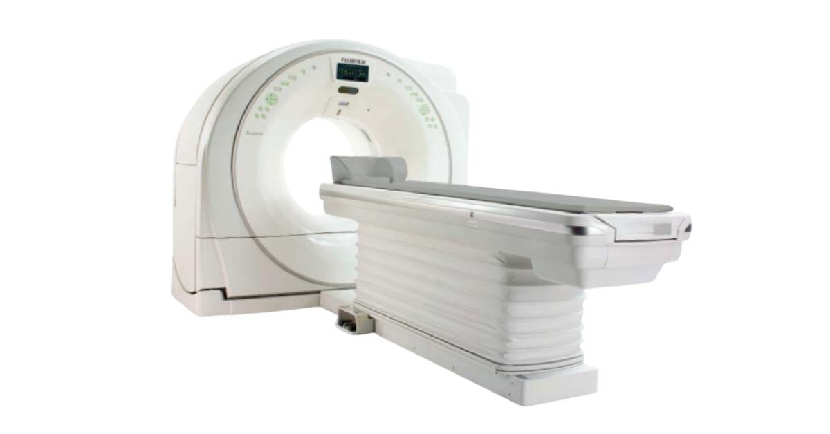
Fujifilm Supria Plus 32-Slice CT ROI for Veterinarians Now
Ever wish you had more diagnostic clarity at your fingertips? Whether it’s a tricky nasal case, a subtle spinal lesion, or a client who needs answers today, in-house CT can change your standard of care—and your bottom line.
With CT becoming more accessible, many practices are asking: is now the time to bring it in-house? Below, we look at the Fujifilm Supria Plus 32-Slice CT to help you decide if it fits your hospital’s clinical goals and financial plan.
What Makes the Supria Plus Different
- Room for everyone: 75 cm gantry and 500 lb table capacity—comfortable for giant-breed dogs.
- Lower dose by design: Automatic, size-based dose reduction helps maintain image quality while minimizing exposure.
- Faster, more detailed scans: Up to 32-slice reconstruction reduces anesthesia time and increases throughput.
- Practical footprint: Compact and user-friendly, easing integration without major renovations or weeks of retraining.
At a glance:
- Excellent for neuro, nasal, dental/maxillofacial, thoracic, abdominal, and orthopedic studies.
- Improved scheduling control and client satisfaction with same-day answers.
- Retention of cases that might otherwise be referred out.
Understanding the Costs
- Acquisition: Approx. $299,000 (many practices finance at roughly $3,995/month).
- Warranty & PM: 2-year warranty; preventative maintenance every six months by Fuji-certified technicians.
- Per-scan costs: Radiologist read ~$250/scan; contrast for injected studies ~$100/scan (estimate).
- After warranty: Set aside a monthly service reserve to cover ongoing parts and service (example below uses $3,000/mo).
How the Numbers Can Work in Your Favor
Here’s a simple, conservative example to sanity-check your local demand:
| Assumption | Example Value |
|---|---|
| Monthly volume | 20 scans (≈5 per week) |
| Average fee per scan | $1,500 |
| Gross monthly revenue | $30,000 |
| Financing payment | $3,995 / month |
| Radiologist reads | 20 × $250 = $5,000 |
| Contrast | 20 × $100 = $2,000 |
| Service reserve (post-warranty planning) | $3,000 / month |
| Total monthly costs | $13,995 |
| Estimated net margin | $30,000 − $13,995 = $16,005 / month |
Break-even snapshot: With revenue of $1,500/scan and variable costs of $350/scan (radiologist + contrast), your contribution margin is ~$1,150/scan. Using the fixed cost proxy above (~$6,995 = payment + reserve), break-even is ≈ 7 scans/month (6.1 rounded up). Your accounting may vary—use your exact fees and costs.
A Few Things to Keep in Mind
- Review cases you currently refer out—this is your most realistic starting volume.
- Plan for a ramp-up period; consistency often lands after a few months.
- Confirm room size, power, shielding, and any site prep requirements in advance.
- Invest in team training; even user-friendly systems have a learning curve.
- Keep a service reserve once the warranty ends to avoid cash-flow surprises.
Taking the Leap
Choosing CT isn’t about chasing the newest gadget—it’s about transforming how you practice medicine. The Supria Plus 32-Slice CT can deliver sharper images, faster answers, and stronger client trust while keeping more care in-house. Run your numbers, map your space and training, and decide if now is the right time.
Looking for specs, workflow, and installation guidance? See the product page: Fujifilm Supria Plus 32-Slice CT Scanner.


Is It Time To Add CT To Your Veterinary Diagnostics?
Bring 32-Slice CT In-House to Elevate Patient Care for Vets
Veterinary medicine is evolving quickly, and with it comes the growing demand for advanced imaging. If you’ve ever found yourself wishing for more detailed views than radiographs can offer—or if you’ve been sending patients elsewhere for CT scans—you may be wondering if it’s time to bring CT technology into your practice. Sound familiar?
Many veterinarians already utilize some form of advanced imaging; however, older CT systems can be limited by factors such as speed, slice count, or patient comfort. A 32-slice system, like the Supria Plus, represents a significant step forward. More slices mean faster acquisition times, sharper image detail, and less time under anesthesia. That’s not just better for diagnostics—it’s safer for patients and reassuring for clients.
Making the Budget Work
Adding advanced imaging has to make sense for your business. Period. A simple way to look at the numbers is to start with a baseline: performing around 15 CT scans per month at an average fee of $1,500 per case. That puts your gross monthly revenue at about $22,500.
From there, factor in common costs:
- A fixed monthly payment for the unit
- Radiologist interpretations (around $250 per scan)
- Contrast materials
- A set amount you save each month for preventive maintenance and future service needs
When you account for those, the math still leaves a healthy margin. Even at the minimum case volume, many practices find they can cover costs, build a maintenance reserve, and still generate meaningful revenue. And as your caseload grows—which often happens once CT becomes available—the financial picture only gets stronger.
Focusing on What Matters
When you’re looking at different imaging options, it helps to focus on what will truly make a difference day-to-day—for your patients, your team, and your clients. The Supria Plus 32-Slice CT has all three in mind, blending precision, efficiency, and practicality in a way that makes it a strong fit. Here are a few advantages that make it stand out:
- Sharper, faster diagnostics – 32-slice technology delivers high-resolution images quickly, reducing anesthesia time and improving patient safety.
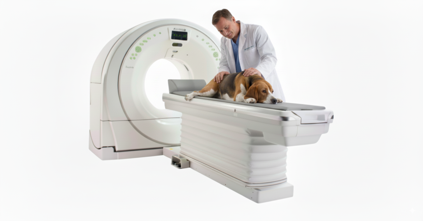
- Built for all patients – A wide-bore gantry accommodates pets of every size and shape.
- Smooth workflow – Compact and intuitive, it makes it easy for your team to integrate CT into your day-to-day.
Designed With Workflow in Mind
The Supria Plus is compact enough to fit comfortably in most veterinary settings, and still spacious where it counts, with high weight capacity to accommodate patients of all sizes. The interface is intuitive, so your team can focus on patient care instead of wrestling with a steep learning curve. And because image acquisition is so fast, your caseload won’t get bottlenecked in the imaging suite.
Looking Ahead
Offering CT services in-house sends a strong message to your clients that your practice is committed to thorough care. For many pet parents, the ability to get same-day results without a referral is invaluable. It strengthens client trust, enhances loyalty, and positions your hospital as a leader in your community.
Whether you’re thinking about adding CT for the first time or considering an upgrade from an older unit, it’s worth looking at how the technology fits into your long-term goals. The right CT doesn’t just expand your diagnostic portfolio—it shapes the kind of medicine you’re able to practice for years to come.

ROI of Veterinary CT Scanners: Profitability Made Simple

Is a CT Scanner Right for Your Veterinary Practice?
Plan the investment, elevate patient care, and see a simple monthly ROI example.
For many veterinary practices, adding advanced imaging is a dream that feels just out of reach. But with the proper planning, CT technology can be both accessible and profitable—while dramatically elevating the level of care you can provide.
Why CT Changes the Conversation
Beyond the financial benefits, adding a CT scanner to your hospital opens the door to faster, more accurate diagnoses for complex cases. CT is beneficial for identifying issues like nasal tumors, thoracic masses, intervertebral disc disease, and complex fractures that can be difficult to assess with traditional radiographs. It’s also great at detecting subtle changes in the lungs, sinuses, and abdomen, giving you and your clients clear answers more quickly. With CT, you can shorten the path from uncertainty to treatment.
Introducing the Fujifilm Supria Plus
The Fujifilm Supria Plus 32-Slice CT Scanner is a traditional CT system designed for practices that want speed, precision, and reliability. It’s designed with both efficiency and patient comfort in mind. Its wide 75 cm gantry and large 500 lb table capacity make positioning easier for veterinary patients of all sizes, while advanced dose-reduction technology ensures high-quality images with lower radiation exposure. Combined with a compact footprint and user-friendly interface, it’s built to deliver powerful diagnostic capabilities without overwhelming your workflow or your facility space.
The Numbers That Matter
When you’re considering a piece of equipment this advanced, it’s natural to wonder: Can my practice really afford this? Here’s a simple way to run the numbers using conservative assumptions:
| Assumption | Value |
|---|---|
| Minimum volume | 15 CT scans per month |
| Average charge per scan | $1,500 |
| Monthly payment plan | $4,000 |
| Radiologist read per scan | $250 |
| Contrast cost per scan | $100 (estimate) |
| Preventive maintenance savings | $3,000/month set aside |
Your Profitability Calculator (Example)
| Line Item | Calculation | Monthly Total |
|---|---|---|
| Revenue | 15 scans × $1,500 | $22,500 |
| CT payment | — | $4,000 |
| Radiologist reads | 15 × $250 | $3,750 |
| Contrast | 15 × $100 | $1,500 |
| Maintenance fund | — | $3,000 |
| Net Monthly Margin | $22,500 – $12,250 | $10,250 |
That’s over $120,000 net margin per year at just 15 cases/month. As awareness grows among clients and referring DVMs, many hospitals see higher volumes—improving ROI even further.
Why Choose the Fuji 32-Slice CT?
- Speed & Accuracy: Detailed imaging in seconds helps reduce anesthesia time and supports precise treatment planning.
- Client Trust: Offering CT communicates clinical excellence and provides peace of mind to pet owners.
- Referral Potential: Nearby clinics without CT are more likely to refer advanced cases to you.
- Longevity: With a disciplined preventive maintenance plan, you’ll get years of reliable performance.
Related: Fujifilm Supria Plus 32-Slice CT Scanner for Veterinary Hospitals

Contrast Enhances Veterinary Cone Beam CT for Pet Care
What is cone beam computed tomography?
Cone Beam Computed Tomography (CBCT) is an imaging technique that is widely utilized in human medicine.
Its unique characteristics make it especially suited to the fields of orthopedics, image-guided radiation therapy, interventional radiology, maxillofacial surgery, and dentistry.
Still, its benefits are only just starting to be realized within the veterinary world. CBCT is capable of producing detailed 3D images that are proving particularly suited to a range of veterinary applications such as maxillofacial surgery (especially trauma), dentistry, imaging of exotic species such as rabbits, and for orthopedics, in particular, imaging of joints and extremities.
CBCT is a specific form of Computed Tomography (CT). As with regular CT imaging, CBCT utilizes a moving beam of X-rays that are detected by a digital X-ray detector.
A series of images are obtained from various angles around the patient which can then be used to generate a 3D image using complex algorithms via specialist computer software.
The main difference between CBCT and regular CT, as its name suggests, is that CBCT utilizes a divergent beam of X-rays which form a cone shape. This allows the X-ray beam to cover a larger area in a single pass.
Regular CT machines in contrast take “slices” through the patient using a flat fan-shaped beam of X-rays that require several passes over the area of interest to acquire sufficient information.
What are its advantages and disadvantages?
The specific characteristics of the CBCT system mean that it has the advantage of being faster than a standard CT scan and exposes the patient to a lower radiation dose due to only a single pass being required to produce an image.
In addition, CBCT machines tend to be smaller and less expensive to purchase, making them more accessible. The images produced by CBCT can also show substantially superior spatial resolution compared with regular CT.
This means that images are clearer and more detailed, allowing better visualization of small structures and abnormalities, particularly within bony tissues.
There are however some disadvantages to the technique. Due to the shape of the beam and the lack of collimation, CBCT scans tend to produce more scatter radiation.
The consequence of this is inferior contrast resolution in soft tissues - meaning that subtle lesions of soft tissues, resulting in only slight differences in contrast, are more likely to be missed.
The scatter also increases the radiation exposure in the surrounding areas and potentially to operating staff. This scatter is amplified when CBCT is used for larger body parts such as the thorax or abdomen.
For these reasons, CBCT tends to be used for smaller body areas such as the head and bones, particularly the extremities. It is beneficial for imaging exotics and their dentistry.
Why is it important to use contrast in CBCT?
The use of contrast is common in many imaging modalities to further enhance and widen their application.
CT typically uses non-ionic contrast media (e.g. iohexol), commonly injected intravenously but can also be injected directly into a body cavity under investigation, such as a joint space.
Contrast is used to highlight specific areas of interest, especially those that may be usually hard to see. In CBCT, iodinated contrast media that are injected intravenously will highlight vessels and soft tissue structures supplied by these.
This has the potential to increase the diagnostic utility of CBCT massively.
The addition of contrast studies in addition to plain CBCT scans is crucial as it will both provide additional information and overcome some of the limitations of the technique.
In orthopedic examinations, the addition of contrast by direct injection into the joint provides high-resolution images of the articular surfaces aiding the assessment of lesions found in degenerative joint disease for example.
In dentistry and maxillofacial surgery, intravenous contrast techniques that highlight blood vessels allow the surgeon to assess vascular structures better and in particular any abnormalities of the vasculature that could be potentially affected by any planned procedures.
Contrast also improves the ability of CBCT to detect and assess soft tissue lesions, for example, neoplasms presenting at an early stage.
Cone beam CT is a relatively new diagnostic modality in the veterinary world.
However, its unique properties and affordability mean that it is likely to be more widely used in the future.
Contrast techniques can greatly increase the range and accuracy of diagnostic procedures possible with these systems and these techniques will likely continue to develop, offering new and exciting opportunities.
References:
[1] Kumar M, Shanavas M, Sidappa A, Kiran M. Cone beam computed tomography - know its secrets. J Int Oral Health. 2015 Feb;7(2):64-8. PMID: 25859112; PMCID: PMC4377156.
[2] Venkatesh E, Elluru SV. Cone beam computed tomography: basics and applications in dentistry. J Istanb Univ Fac Dent. 2017 Dec 2;51(3 Suppl 1):S102-S121. doi: 10.17096/jiufd.00289. PMID: 29354314; PMCID: PMC5750833.
[3] Kim, M., Kim, B., Choi, H., Choi, Y., Oh, S. H., Kang, J., Lee, S., Kang, J., Kim, G. T., Choi, Y., & Hwang, E. (2015). Intravenous contrast media application using cone-beam computed tomography in a rabbit model. Imaging Science in Dentistry, 45(1), 31. https://doi.org/10.5624/isd.2015.45.1.31
[4] Posadzy, M., Desimpel, J., & Vanhoenacker, F. (2018). Cone beam CT of the musculoskeletal system: clinical applications. Insights Into Imaging, 9(1), 35–45. https://doi.org/10.1007/s13244-017-0582-1
[5] Riggs, G. G., Cissell, D. D., Arzi, B., Hatcher, D. C., Kass, P. H., Zhen, A., & Verstraete, F. J. (2017). Clinical Application of Cone Beam Computed Tomography of the Rabbit Head: Part 2-Dental Disease. Frontiers in veterinary science, 4, 5. https://doi.org/10.3389/fvets.2017.00005
[6] Bregger, M. D. K., Koch, C., Zimmermann, R., Sangiorgio, D., & Schweizer-Gorgas, D. (2019). Cone-beam computed tomography of the head in standing equids. BMC Veterinary Research, 15(1), 289. https://doi.org/10.1186/s12917-019-2045-z
[7] Myller, K. A., Turunen, M. J., Honkanen, J. T., Väänänen, S. P., Iivarinen, J. T., Salo, J., Jurvelin, J. S., & Töyräs, J. (2017). In Vivo Contrast-Enhanced Cone Beam CT Provides Quantitative Information on Articular Cartilage and Subchondral Bone. Annals of biomedical engineering, 45(3), 811–818. https://doi.org/10.1007/s10439-016-1730-3
[8] Hamard M, Sans Merce M, Gorican K, Poletti PA, Neroladaki A, Boudabbous S. The Role of Cone-Beam Computed Tomography CT Extremity Arthrography in the Preoperative Assessment of Osteoarthritis. Tomography. 2023 Nov 29;9(6):2134-2147. doi: 10.3390/tomography9060167. PMID: 38133071; PMCID: PMC10747585.
Cone Beam CT and Orthopedic Surgery for Veterinary Hospitals

Cone beam computed tomography (CBCT) has revolutionized diagnostic imaging in the veterinary orthopedic field.
It has enabled delicate structures to be imaged in intricate detail, ensuring no pathologies are missed, diseases can be detected early, and surgery can be planned with the greatest precision.
In veterinary clinics, CBCT is now being used to scan cats and dogs, providing 3D images superior to other imaging modalities.
The benefits of using CBCT for Veterinary
Orthopedic surgeons rely on various imaging techniques for all aspects of their work including diagnostics, surgical planning, intra-surgical imaging, and monitoring post-surgical healing.
The unique properties CBCT has to offer make it the ideal imaging modality for orthopedic work as it greatly reduces a patient’s radiation exposure compared to conventional CT, while still producing high-quality images.
Compared to conventional CT, which uses a continuous beam of radiation, CBCT uses a conical-shaped beam and flat plate receiver, which both rotate 180 to 360° around the patient, while only taking intermittent images at specific intervals. Algorithms are used to convert these 2D images into a reconstructed 3D view that provides orthopedic surgeons with high-resolution images.
The benefits of CBCT:
High-resolution 3D images are produced – The intricate level of detail each scan provides is ideal for assessing the architecture of bones and complex joints.
Patients are only exposed to a low dose of radiation – The use of a focused field of view and intermittent radiation exposure during one scan enables each patient’s radiation dose to be significantly reduced compared to conventional CT.
No issues with superimposition – The 3D images eliminate superimposition, enabling structures to be viewed using CBCT that would otherwise be unable to be assessed with digital radiography.
Scans can be performed rapidly – A full scan takes less than a minute to complete, so it is ideal for both veterinary work and for use during orthopedic surgeries.
The equipment is cost-effective – Compared to the cost of setting up a conventional CT scanner, CBCT is cheaper, enabling veterinary clinics access to CT when conventional CT scanners are out of budget.
CBCT in Orthopedic Veterinary Clinics
CBCT was first used in dentistry where it superseded digital radiographs by providing more accurate information on lesion location while being cost-effective and performing scans rapidly.
More recently, orthopedic surgeons have started using the benefits of CBCT, especially for imaging areas that were previously difficult to assess using digital radiography due to the location of superimposed bones or complex joints.
Why CBCT is superior to digital radiography
Digital radiography is the first-line imaging choice for diagnosing orthopedic conditions in our pets.
However, digital radiography has the disadvantage of only being able to provide limited anatomical detail, restricting its use for diagnosing orthopedic conditions and surgical planning.
Veterinary clinics with access to CBCT can rely on this technique to provide superior detailed scans for all stages of orthopedic work. When used for diagnosing orthopedic conditions, it can assess fractures that would have otherwise been missed and can detect signs of degenerative conditions and bone tumors earlier.
The use of imaging for orthopedic surgery
Orthopedic surgeons are greatly reliant on access to reliable imaging modalities for pre-surgical planning and intra-surgical use.
CBCT is the ideal choice for surgical planning as each scan provides detailed information on lesion location and enables surgical accuracy to be improved while limiting the need for revision surgeries.
Benefits of CBCT for orthopedic surgery:
Ideal for pre-surgical planning – The high-resolution 3D images provide surgeons with maximum information on the location of the lesion, enabling improved surgical accuracy during intricate surgeries while also decreasing anesthetic and surgical time.
Rapid scan times of less than one minute – Each scan may take between 5 to 40 seconds, helping to reduce the surgical and anesthetic time for the patients who require intra-operative scans.
Mobile equipment – Having access to portable CBCT equipment enables scans to be performed during surgery with limited disruption to the patient or the aseptic field.
Lower radiation doses – Compared to conventional CT, CBCT exposes patients to significantly lower radiation doses while not compromising image quality. This enables repeated scans to be performed when necessary.
Ideal for monitoring healing post-surgery – CBCT allows healing to be more accurately assessed, especially when monitoring fracture repair and callus formation. In comparison, digital radiographs can over- or underestimate healing, making it difficult to assess post-operative recovery.
CBCT is currently gaining rapid popularity in veterinary hospitals due to its ability to produce accurate 3D images in a rapid, cost-effective way that is unique to other imaging modalities.
ts ability to overcome superimposition and its suitability for surgical planning and intra-surgical use make it essential for all veterinary orthopedic work.
References
Posadzy M, Desimpel J, Vanhoenacker F. Cone Beam CT of the musculoskeletal system: clinical applications. Insights into imaging. 2018 Feb;9(1):35-45.
Ricci M, Boldini M, Bonfante E, Sambugaro E, Vecchini E, Schenal G, Magnan B, Montemezzi S. Cone-beam computed tomography compared to X-ray in the diagnosis of extremities bone fractures: a study of 198 cases. European Journal of Radiology open. 2019 Jan 1;6:119-21.
Meneses F, Maiolini A, Forterre F, Oevermann A, Schweizer-Gorgas D. Feasibility of a Frameless Brain Biopsy System for Companion Animals Using Cone-Beam CT-Based Automated Registration. Frontiers in veterinary science. 2022 Feb 9;8:779845.
Lee J, Stayman JW, Otake Y, Schafer S, Zbijewski W, Khanna AJ, Prince JL, Siewerdsen JH. Volume-of-change cone-beam CT for image-guided surgery. Physics in Medicine & Biology. 2012 Jul 17;57(15):4969.
Venkatesh E, Elluru SV. Cone beam computed tomography: basics and applications in dentistry. Journal of Istanbul University Faculty of Dentistry. 2017 Dec 2;51(3 Suppl 1):102-21.
Fotouhi J, Fuerst B, Unberath M, Reichenstein S, Lee SC, Johnson AA, Osgood GM, Armand M, Navab N. Automatic intraoperative stitching of nonoverlapping cone‐beam CT acquisitions. Medical physics. 2018 Jun;45(6):2463-75.

Cone Beam CT: A Powerful Tool for Veterinary Surgeons

What is Cone beam CT (CBCT)
Cone beam CT (CBCT) is a type of 3D imaging that uses X-rays, similar to regular CT scans. But instead of a fan-shaped beam, CBCT uses a cone-shaped beam, making it smaller, faster, and lower in radiation.
Why use Cone beam CT (CBCT)
Veterinary surgeons are finding CBCT especially useful for:
Maxillofacial disease: CBCT excels at imaging the complex structures of the jaws, teeth, and sinuses. This helps diagnose and treat dental problems, trauma, and tumors more accurately. Studies show it provides more detailed information than traditional X-rays.
Small and exotic animals: CBCT's compact size and lower radiation make it ideal for examining the heads and teeth of small pets and even exotic species like rabbits.
Joint disease: CBCT can reveal subtle changes in joints, helping diagnose fractures, dislocations, and arthritis, especially in areas with overlapping bones.
When not to use it?
CBCT isn't perfect for everything. Its uncollimated beam means it's not ideal for:
Soft tissue, thorax, or abdomen: These areas require more focused imaging, best achieved with regular CT scans.
Large animals: Scattered radiation makes images less clear in larger animals like big dogs.
The future of CBCT?
As vets see the benefits, CBCT is likely to become more common in veterinary clinics. Its ability to provide detailed 3D images can lead to better diagnoses, treatment plans, and ultimately, improved patient care for a wider range of animals.
Key points:
CBCT uses a cone-shaped beam for faster, lower radiation 3D imaging.
It excels at imaging the jaws, teeth, and joints in small animals.
Not ideal for soft tissue, thorax, or abdomen, or large animals.
Holds promise for improved diagnosis and treatment in veterinary medicine.

3 Key Reasons Veterinary Surgeons Should Use Cone Beam CT
Three reasons why a veterinary surgeon would want to use a cone beam CT
Cone beam computed tomography (CBCT) has been available in human dentistry for years, but it is now also entering veterinary medicine.
Some specialist vets who work with maxillofacial and dentistry cases have already started to use this technology. I
t provides detailed 3D images which can be helpful for oncology or trauma cases, as well as those suffering from dental disease. Here we are going to explore this imaging modality in more detail, including some scenarios in which you might choose to use it.
What is cone beam CT?
CBCT uses radiation, in the same way that normal computed tomography (CT) does. However, the shape of the x-ray beam is different. In CBCT the beam is in the shape of a cone, whereas normal CT has a fan-shaped beam.
The cone-shaped beam covers the area of interest, and the X-ray source only needs to pass once around the animal, being picked up by a sensor on the opposite side of the patient to create an image.
This means the average dose of radiation a patient receives is more similar to that of one thoracic radiograph than a conventional helical CT.
Due to the low amounts of radiation it emits, its lower purchase cost and the machine’s compact design, CBCT is becoming easier to access than normal CT. Its design makes it particularly ideal for imaging the maxillofacial area, which is a complicated area of anatomy to visualize.
Three reasons why a veterinary surgeon would want to use cone beam CT
Maxillofacial disease
As previously discussed, CBCT is commonly used for maxillofacial disease.
This imaging modality works well in small anatomical areas with high tissue density. This fits the bill for the maxillofacial area, which has several complicated structures including the teeth, upper and lower jaws, the temporomandibular joint, and the sinuses filled with turbinates. All of these structures can overlap and confuse diagnostic imaging, particularly in brachycephalic breeds.
CBCT can create a highly detailed, superimposition-free image that shows the area of interest in 3D. This gives a clear advantage over traditional, 2D dental x-rays.
A recent study into dental pathology in cats found that ‘CBCT provided more clinically relevant detailed information as compared to dental radiography’.
This was also the case in a 2018 paper on CBCT vs dental radiography in small to medium-sized brachycephalic dogs. These studies indicate that practitioners could be missing clinically relevant pathology when using dental X-rays alone.
As well as endodontic disease, other examples where CBCT may be superior to X-rays include –
Evaluating maxillofacial trauma
Assessment of temporomandibular joint disease, dislocation, luxation, or fracture
Viewing bone integrity associated with cystic lesions
Assessing the severity of oronasal communications and cleft palates
Evaluating the extent of oral tumors in hard tissue of the maxilla and mandible
Small and exotic animals
CBCTs can be used in examinations of the head and dentition of small and exotic species, as well as canine and feline patients.
A 2017 paper holds ‘CBCT to be superior to conventional CT for evaluation of normal dentition in rabbits’. Subtle changes in the periodontal ligament space can be detected more easily in rabbits, which can be an indication of emerging disease.
This could otherwise go unnoticed and be missed in conventional CT scans.
Whole body exams of many small, exotic animals may be possible with this technology too, further widening its appeal and usage.
Joint disease
CBCT can be used in extremities like the limbs to evaluate joint disease.
There are indications for its use in the musculoskeletal system including evaluation of fractures and dislocations in small bones and joints, and it can be used in conjunction with other imaging techniques to assess cartilage. It is easier to evaluate degenerative joint disease (like osteoarthritis) with CBCT than conventional radiography, particularly in areas where there is superposition of other bony structures.
This paper on CBCT in people with joint disease further demonstrates this point.
In veterinary medicine, a 2021 study in standing horses demonstrated that CBCT provided diagnostic imaging in a timely fashion. No doubt this modality will start to become more commonly used for joint disorders in a variety of species as veterinary practitioners start to utilize it further.
When would you not want to use cone beam CT?
The X-rays from the CBCT are not collimated.
They make a divergent, uncollimated cone meaning more scattered radiation is produced. This means there is a reduction in soft tissue contrast, and the resolution in areas like the thorax and abdomen is poorer, especially in large dogs.
Larger body parts cause greater scatter. Using CBCT in these areas will mean contrast is lower than if it is used in smaller areas, like the jaw or a limb.
This is different from normal CT scanners which have a collimated beam that runs as straight as possible, with little scattered radiation.
So normal CT should be chosen for soft tissue, thorax, and abdomen examinations (especially in larger cats and dogs) and CBCT would not be advised.
Equally, good radiation shielding – especially when a horizontal beam is likely – is necessary for imaging facilities utilizing this technique.
Summary
Cone beam CT shows great promise across multiple areas of veterinary medicine, but in particular for maxillofacial disease.
It provides highly detailed 3D images of areas that are traditionally hard to assess with conventional dental radiography. This helps veterinary surgeons to plan their treatment accordingly, which can only lead to better patient care and outcomes.
CBCT will probably be seen more and more frequently in hospitals, as its potential gains recognition by general practitioners.

Veterinary CT Terminology and Technology, when buying

In recent years, veterinarians have had a lot to choose from in terms of CT system sizes, technology, combinations, and price points.
While it’s good to have options, it can also feel like “information overload” trying to choose the best machine for an individual practice. The following information may help with this important decision…
Conventional CT
Traditional, fan-beam-style CT scanners were the norm for a long time. And they still serve a valuable purpose. A fan-beam model is a good all-around option, especially for hospitals that see all different sizes of patients including large dogs.
Fan-beam CT machines work by taking image “slices” (cross-sectional images) that are picked up by an array of detectors. Then the patient is advanced further into the gantry (entryway) and another slice is taken—and so on until the area of interest has been fully imaged.
Slice Counts
Advances in fan-beam technology include systems that image multiple slices at once. Instead of a single slice, this could mean 4, 8, 16, 32, or even 64 slices at a time. These multidetector CT scanners have the advantage of being faster than a single-slice machine.
Although slice thickness factors in, as you can imagine, shooting a larger area (i.e., multiple slices) at once means the study takes less time. In some cases, this can reduce anesthesia or sedation requirements. But chemical restraint may still be needed for areas in which motion artifact is a bigger confounding factor, such as respiratory movements during lung studies.
As you can probably also imagine, the price of the machine goes up as the slice count increases.
Slice Thickness
There are pros and cons to both thinner and thicker image slices. Thinner slices allow for the acquisition of more details. Thicker slices allow for faster study times.
Helical or Spiral Scanners
Originally, the patient would be advanced into the machine, then stopped while the first image slice is obtained, then advanced a little further, and so on…
With helical models, the patient is continuously advanced without stopping. This results in the x-ray tube head moving in a spiral motion around the patient.
This can mean shorter study times. Helical models can also have great image quality, including 3D renderings.
Cone-beam Technology
Cone-beam CT is a newer, increasingly popular option. Instead of the images being captured on a narrow array of detectors like fan-beam technology, they are captured on a wider flat panel detector similar to those used for regular X-ray machines.
Veterinary cone-beam CT is a nice option because of the smaller footprint and lower price point. They’re also ideal for certain studies, like the skull, extremities, and some musculoskeletal views.
However, these small machines don’t accommodate larger patients or offer as much contrast and detail for soft tissue studies. And they may be more susceptible to motion artifact.
Portable CT Machines
Battery-powered, portable CT scanners are also available. They might be advantageous for busy hospitals with limited space, or for any practitioners who need to move their machine to the patient.
Many of these machines use standard electrical outlets for power or for battery charging, which is convenient compared to higher-powered machines. Some also have built-in shielding, which can decrease the requirement for lead-lined walls.
A practice should check the specifications for the specific machine they are purchasing.
Combo Machines
Some models offer combinations of CT, fluoroscopy, and even standard digital x-rays all in one. But are they worth it?
It depends. If there is a significant price advantage—and the image quality is great for ALL modalities—a combo could be a practical, space-saving option.
If one or both of these requirements aren’t met, a veterinary practice might find it’s better for them to focus on one modality at a time rather than purchasing a combination machine. It’s also important to consider whether there is a potential “bottleneck” in scheduling—for example, team members waiting to do a CT scan while someone else finishes radiographs.
Veterinary Radiologist Consultations
It’s exciting that CT technology is becoming more widely available and even used by some general practitioners. However, there is a large learning curve for any new imaging modality a practice adopts. Veterinary CT is no exception.
Specialists exist for a reason. Veterinary radiologists have significantly more knowledge and experience with advanced imaging modalities. A consultation with a radiologist can help with everything from pre-purchase planning (which model to purchase, requirements to set it up at a clinic, maintenance cost considerations, etc.) to doing teleradiology consultations on the images obtained from a CT scanner.
For teleradiology consultations, ask ahead of time if the radiologist has any specific requirements for the type of machine used, the settings, etc.
Investing In the Right Machine
As time goes on and more veterinary practices show an interest in CT scanners, there’s no doubt that new technology and options will be developed.

Decision makers at each practice must perform due diligence and talk to experts (veterinary radiologists, business/financial consultants, and state regulations for radiation safety) prior to purchasing and using a CT system, to ensure the machine they purchase is a good fit for them.
With that in mind, it’s an exciting time in veterinary medicine, as practitioners have yet another option for providing advanced patient care at their practice.
Written by: Dr. Tammy Powell, DVM

What Is the Cost of a Veterinary CT System?

Computed tomography (CT) seems to be gaining in popularity in veterinary medicine, since the diagnostic scans are beneficial in the workup and care of many patients. As the technology continues to advance, with more affordable options being marketed to veterinarians, CT scanners are even becoming available to non-referral or general practices.
Although veterinarians now have more CT models to choose from, is the price tag worth it? How much do CT systems actually cost, and can a veterinary practice make a profit by offering this service?
How Much Do Veterinary CT Scanners Cost?
As with any big-ticket equipment purchase, costs can vary greatly depending on the type of CT scanner, the model and manufacturer, deals from the vendor, whether it’s new or used or refurbished, and other factors. CT investments tend to cost more than a standard veterinary digital x-ray system. But the good news is, some newer models are certainly less expensive than previous ones.
According to Sound a new standard (fan-beam) CT machine can cost upwards of $500,000 or more, depending on the number of slices the machine produces. Manufacturer-refurbished models can run in the $100,000-200,000 range, or possibly significantly less if refurbished by a third party.
Portable models are listed from the high $100,000s to $500,000 or more—again, depending on the number of slices and whether the model is new or refurbished.
Cone-beam CT, on average, tends to be less of a financial investment than standard CT. That’s one of the reasons it has become popular, in addition to other factors such as taking up less space and producing better images for certain studies (particularly for skull/head studies and certain musculoskeletal conditions). According to Sound, a new cone-beam CT system can cost between $180,000-$250,000, whereas refurbished ones might start around $150,000.
While this smaller price tag is inviting, cone-beam does have its limitations. A veterinary practice must consider the pros and cons of each type of CT scanner and decide what they need for the patients they see and the types of studies they plan to perform.
What Additional Costs Should Be Expected?
Although the purchase price of a veterinary CT system is important, it’s certainly not the only number that factors into a purchase decision. Here are a few more to think about…
Shipping and installation. Check to see if these costs are included. However, keep in mind that some installation costs and logistics can’t be covered solely by the manufacturer or vendor.
Specifically, think about lead lining or shielding that the machine requires, as well as electrical requirements. For certain CT models, consultations with licensed physicists/radiation experts and electricians may be required, as well as remodeling of the CT suite, to accommodate the machine and meet local radiation safety regulations.
Ongoing maintenance and repairs. See if a warranty is included. Service and maintenance plans can also be a great option for many types of veterinary equipment.
However, service plans do tend to be on the pricier side for CT machines. Consider the cost of the plan, as well as the average cost of repairs, to see whether it makes more sense to pay for the plan or simply set aside some cash for unexpected repairs.
Consulting with a veterinary radiologist, or even the radiology department of a human hospital, may be helpful to determine which repairs are most common and how much they tend to cost.
Software. All digital imaging technology requires appropriate software to process, view, share, and store images. Since CT scanners take a lot of images per study, the file storage can be quite large. Make sure hardware and software can accommodate these files.
Explore which financing option makes the most sense for your practice. Consult a tax professional to determine which tax benefits you may receive from the purchase. And perhaps most importantly, contact a boarded veterinary radiologist for advice.
Think about the return on your investment. How much would your practice charge for a CT study? Are clients likely to say ‘yes’ to this price, including anesthesia costs? And how many studies could the practice realistically expect to perform in a week or a month? As much as a veterinary practice would love to provide every possible service to their patients, the business must make sure the purchase fits into their financial plan.

Veterinary CT systems represent an amazing and useful technology that can benefit many patients. Knowing the numbers, talking to experts, and having a plan in place can help a veterinary practice profit financially while also providing excellent patient care.
Written by: Dr. Tammy Powell, DVM

What to Know About Veterinary CT Unit Installation

When purchasing a CT scanner, veterinary practices should look at not only the purchase price but also the costs of installing and maintaining the unit.
Planning for the following factors can help a practice avoid surprises and smoothly purchase and integrate a veterinary CT machine…
Space Requirements
CT systems vary greatly in terms of size. Traditional, fan-beam CT scanners can be very large and require their own dedicated room. Some veterinary practices just might not have the space for these units, or they might need to do some extensive remodeling to accommodate them.
Other CT scanners, such as cone-beam or portable units, can be significantly smaller in size. This can make them attractive to veterinary practices that are limited on space.
Electrical Requirements
Some units can operate on standard electrical outlets. Others require a higher current—which might mean consultation with an electrician and rewiring are necessary.
As you might expect, the former is more common with newer, smaller CT units, while the latter is more common with large, traditional units that take a lot of power to operate. Portable units can even be battery-powered. But it’s crucial to check the specific electrical requirements for the unit you are purchasing.
Lead Shielding Requirements
Radiation safety is crucial with any x-ray modality. And CT is certainly no exception, especially since the amount of radiation produced with multiple slices can be large. In addition to personal protective equipment, shielding might include other requirements, such as lead-lined walls in the room in which the unit is housed.
As might be expected, a larger CT scanner generally has more lead shielding requirements than a smaller one. However, it again comes down to the requirements of the specific model of CT unit in which you are interested.
Also, while the specs of the individual machine are important, they’re not the only consideration. Every jurisdiction has its own radiation safety laws and regulations. Do your research and hire a radiation consultant prior to installing a new machine. They can instruct your practice on shielding requirements based on the radiation doses and expected usage, occupancy in the spaces outside the CT room, etc.
This often includes speaking to a veterinary radiologist, as well as a radiation physics consultant or licensed physicist who understands local legalities and how to keep your veterinary practice in compliance. Your local veterinary medical board may be able to point you in the right direction, too, in terms of regulations and finding licensed consultants to work with.
Doing this step prior to purchase can also help a veterinary practice understand what costs are involved in installing various machines and shielding, which might factor into the purchase decision.
Cooling Requirements and Additional Considerations
Some CT units, especially larger ones, can get quite warm during use. It’s important to ensure there is adequate cooling and ventilation in the room in which the machine will be used.
The flooring should be sturdy enough to support a heavy CT unit.
Also, in addition to the up-front costs (purchase and installation), it’s crucial to factor in long-term operating and maintenance costs. Some warranties and service plans are very pricy and designed for machines with heavy use. Other service plans (or even paying out of pocket for repairs) might be more practical for typical, light veterinary use.
Also, a veterinary practice team should always consider whether the CT system they are purchasing meets their expected usage requirements on the types of patients they see. For example, maybe a practice wants a smaller CT machine due to space limitations, but they also see a lot of large dogs that need abdominal scans, for which a larger CT unit would be ideal. It’s a matter of balancing these interests and finding the best solution.
Although the price tag plus additional costs can seem daunting, it helps to break down each consideration one at a time.
Then compare everything to the expected income from your new veterinary CT system to determine what are the most that your practice can or should invest.

This will greatly help with determining if CT is a good fit for your practice, and then selecting the right machine for your needs.
Written by: Dr. Tammy Powell, DVM

Vet CT Technology Comparison Traditional vs. Cone-Beam
As CT (computed tomography) technology advances, veterinarians have more options than ever to choose from for their clinic—including the increasingly popular cone-beam CT scanner.
So, is cone-beam technology superior to traditional CT units? It all depends on what a veterinary practice needs. Here are some factors that can help a veterinarian choose which veterinary CT system is right for them.
What’s the Difference Between Traditional and Cone-beam CT?
With traditional CT, images are created as a series of fan-shaped slices, which are picked up by a narrow array of detectors. Cone-beam CT units use a wider, cone-shaped beam with a flat panel detector or plate.
The simplicity of the plate means a cone-beam machine can be smaller and might have lower maintenance costs. However, cone-beam technology takes longer to acquire the images.
Both types of technology create a series of images that give a veterinarian a deeper look at a patient’s anatomy compared to standard radiographs. The total series of images essentially allows a 3D view of the area being studied, thanks to CT’s ability to eliminate the problem of superimposition. And newer cone-beam technology can create some impressive 3D renderings.
When Is Traditional CT Most Beneficial?
Here are a few situations in which a veterinarian might prefer a conventional veterinary CT unit…
Larger patients. Cone-beam, which were originally designed for studies of the head in humans, have a gantry or entry point that is relatively small. Thus, cone-beam CT might only be practical for cats, small dogs, exotics, or a larger patient’s head or extremities. Traditional CT, on the other hand, could potentially be used for full scans on larger patients.
Soft tissue differentiation. One big advantage of conventional CT is better soft tissue differentiation compared to traditional radiographs and even compared to cone-beam CT. Examples of uses could include visualizing individual muscle bodies and blood vessels, metastasis checks of the lungs, characterizing a soft tissue mass within an organ such as the liver, and detailed surgical planning.
Abdominal or thoracic studies. Due to a combination of soft tissue definition and accommodating larger anatomy, standard CT is usually the better choice for abdominal and thoracic studies.
Limiting motion artifact. Cone-beam units have slower revolutions, meaning motion artifact can be more pronounced. General anesthesia can help prevent this. However, when motion is a concern (such as when detailed thoracic studies are needed), traditional CT might provide an advantage. This is yet another reason why traditional CT is often preferred for thoracic studies.
When Is Cone-beam CT Advantageous?
Here are some situations when veterinary cone-beam CT might be a better choice…
Smaller footprint. Cone-beam tends to be much smaller than standard CT units. Some are even portable. In veterinary hospitals where space is at a premium, a cone-beam unit might be the only practical option. Additionally, some cone-beam models can be plugged into a standard wall outlet, which is very convenient.
Lower price point. On average, cone-beam CT units cost less to purchase than traditional CT units. It’s also important to look at ongoing maintenance costs regardless of which type of technology is being purchased. Cone-beam might come out ahead for saving on maintenance costs, too, especially since it’s often easier to find replacement parts or repair services for newer equipment models.
Skull and dental images. Cone-beam CT is ideal for studies of the head, since that’s the purpose for which it was originally developed. This could also include studies of the inner or middle ears, pharyngeal area, nasal passages, etc. Some have even suggested that it may replace dental radiographs in the future.
Small patients and small anatomy. Whereas traditional CT is ideal for thoracic and abdominal studies, cone-beam technology might take the lead with smaller anatomy, especially if it’s an area with inherent tissue contrast. Orthopedic issues of the limbs and paws are one possibility. Cone-beam CT can also be an option for small exotic patients who fit into the machine.
Every case is different, so a veterinarian should always use their own clinical judgment. Plus, new technology is being developed all the time, so there is even a third option—a “hybrid” CT model—available, giving veterinarians plenty of choices.

When in doubt, a vet can also consult with a radiologist for their recommendation. Some services even provide teleradiology consultations for CT scans.
Cone-beam is an exciting option that could be affordable and practical for a lot of veterinary clinics. However, each practice should consider all factors to decide which type of technology—and which individual model—is the best fit for their needs.
Written by: Dr. Tammy Powell, DVM

Enhancing Veterinary Care with CT Imaging Technology

While CT imaging was initially reserved for veterinary universities and referral hospitals, the technology is making its way into some general practices.
Like any technology, it becomes more compact, affordable, and practical to own over time, so a CT scanner might be a reasonable option for practices in some locations.
Here is an overview of CT in veterinary medicine, which will be followed by a deeper dive into the technology and its uses in the next few articles.
What Is CT?
CT stands for computed tomography, and it’s a specialty type of imaging modality. It uses x-ray technology, but instead of a single 2D image, the machine circles around the body to produce “slices” or cross-sectional images of the area being evaluated. Added up, these cross-sections allow the veterinarian to get more of a 3D look inside the body.
Depending on the technology being used, this could be performed by scanning a thin slice of the patient, then advancing them a little further into the machine (just a few millimeters at a time) and obtaining another slice, then repeating until the entire area under study has been imaged in this manner.
New technology features “multi-slice” imaging in which many cross-sectional images can be obtained simultaneously. As you might expect, newer technology can perform the task much faster.
Advantages of CT Scans
Some of the “pros” of CT technology, compared to other imaging modalities, include…
Overcoming superimposition. The cross-sectional nature of the images allows a veterinarian to see much more than they could with just a few traditional x-ray views. It reduces or eliminates the problem of superimposition. This is especially valuable for structures such as the skull, which is notoriously difficult to radiograph due to superimposition of the many structures inside the head, nose, etc. Another example might be a soft tissue mass in the abdomen that overlaps with structures such as the liver. CT imaging helps a vet see deep or superimposed structures more clearly.
Better tissue differentiation. Compared to conventional radiographs, CT allows better differentiation of tissues and more precise detection of x-ray attenuation, especially for neighboring soft tissue structures. Examples might include visualizing individual muscles, differentiating vasculature from surrounding tissues, or being able to see a mass in the brain or within an abdominal organ such as the liver or spleen. For these reasons, CT is often a great choice for evaluating musculoskeletal lesions and looking for/describing the extent of lung metastasis.
Contrast studies. These may be performed when more information or differentiation is needed. Although CT generally provides excellent detail, contrast studies may help further characterize a lesion.
Surgical planning. CT can help surgeons plan for a complicated mass removal—such as with nasal tumors, intrathoracic neoplasias, and more—by delineating and characterizing the full extent of the abnormal growth.
Disadvantages of Using CT
While CT is a great modality that is superior to conventional radiographs in many ways, every technology has its pros and cons. Here are some challenges to consider for using CT…
Anesthesia. Dogs and cats must usually be anesthetized for their CT scan. This allows proper positioning and prevents motion artefact. Even something as simple as normal respiration might create motion artefact. Ventilation may be controlled during the shot while the patient is anesthetized, to prevent motion as well as lung atelectasis that could be mistaken for a lesion.
Costs. Although more affordable than it once was, there’s no doubt that CT machines can be a significant financial investment—in terms of both the initial purchase and the upkeep. Given the cost of the technology, as well as the added costs of general anesthesia, some clients might be deterred by the price tag of a CT study. It may still be more affordable than MRI, though, in cases in which a CT study can be used in place of MRI (there are many).
Size constraints. This applies to both the patient and the hospital. Some CT models, especially smaller machines like cone beam CT, only fit patients of a certain size. Although they might still be used for the head or extremities of larger patients, this would eliminate some potential studies altogether on large dogs or other big patients. It’s also important to consider the footprint of the machine itself and where it will fit in the hospital.
Learning curve. CT images look a bit different than traditional x-ray images. Not to mention, most vets aren’t familiar with the evaluation of cross-sectional images. As with any new skill, it can take time and training to feel up to speed and confidently interpret CT studies. Plus, the staff must learn how to use the machine. Fortunately, there are many resources (texts, courses/training, etc.) to help. Also, teleradiology consultations are available for added support.

With all the potential uses of CT, it’s no surprise that it seems to be gaining in popularity and is available at some general veterinary practices. It will be exciting to see how the technology continues to evolve over time to advance veterinary medical knowledge and patient care.
Written by: Dr. Tammy Powell, DVM