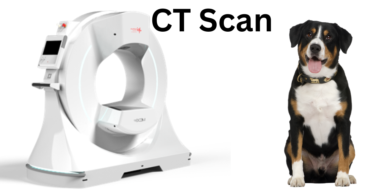
Vet CT Technology Comparison Traditional vs. Cone-Beam
As CT (computed tomography) technology advances, veterinarians have more options than ever to choose from for their clinic—including the increasingly popular cone-beam CT scanner.
So, is cone-beam technology superior to traditional CT units? It all depends on what a veterinary practice needs. Here are some factors that can help a veterinarian choose which veterinary CT system is right for them.
What’s the Difference Between Traditional and Cone-beam CT?
With traditional CT, images are created as a series of fan-shaped slices, which are picked up by a narrow array of detectors. Cone-beam CT units use a wider, cone-shaped beam with a flat panel detector or plate.
The simplicity of the plate means a cone-beam machine can be smaller and might have lower maintenance costs. However, cone-beam technology takes longer to acquire the images.
Both types of technology create a series of images that give a veterinarian a deeper look at a patient’s anatomy compared to standard radiographs. The total series of images essentially allows a 3D view of the area being studied, thanks to CT’s ability to eliminate the problem of superimposition. And newer cone-beam technology can create some impressive 3D renderings.
When Is Traditional CT Most Beneficial?
Here are a few situations in which a veterinarian might prefer a conventional veterinary CT unit…
Larger patients. Cone-beam, which were originally designed for studies of the head in humans, have a gantry or entry point that is relatively small. Thus, cone-beam CT might only be practical for cats, small dogs, exotics, or a larger patient’s head or extremities. Traditional CT, on the other hand, could potentially be used for full scans on larger patients.
Soft tissue differentiation. One big advantage of conventional CT is better soft tissue differentiation compared to traditional radiographs and even compared to cone-beam CT. Examples of uses could include visualizing individual muscle bodies and blood vessels, metastasis checks of the lungs, characterizing a soft tissue mass within an organ such as the liver, and detailed surgical planning.
Abdominal or thoracic studies. Due to a combination of soft tissue definition and accommodating larger anatomy, standard CT is usually the better choice for abdominal and thoracic studies.
Limiting motion artifact. Cone-beam units have slower revolutions, meaning motion artifact can be more pronounced. General anesthesia can help prevent this. However, when motion is a concern (such as when detailed thoracic studies are needed), traditional CT might provide an advantage. This is yet another reason why traditional CT is often preferred for thoracic studies.
When Is Cone-beam CT Advantageous?
Here are some situations when veterinary cone-beam CT might be a better choice…
Smaller footprint. Cone-beam tends to be much smaller than standard CT units. Some are even portable. In veterinary hospitals where space is at a premium, a cone-beam unit might be the only practical option. Additionally, some cone-beam models can be plugged into a standard wall outlet, which is very convenient.
Lower price point. On average, cone-beam CT units cost less to purchase than traditional CT units. It’s also important to look at ongoing maintenance costs regardless of which type of technology is being purchased. Cone-beam might come out ahead for saving on maintenance costs, too, especially since it’s often easier to find replacement parts or repair services for newer equipment models.
Skull and dental images. Cone-beam CT is ideal for studies of the head, since that’s the purpose for which it was originally developed. This could also include studies of the inner or middle ears, pharyngeal area, nasal passages, etc. Some have even suggested that it may replace dental radiographs in the future.
Small patients and small anatomy. Whereas traditional CT is ideal for thoracic and abdominal studies, cone-beam technology might take the lead with smaller anatomy, especially if it’s an area with inherent tissue contrast. Orthopedic issues of the limbs and paws are one possibility. Cone-beam CT can also be an option for small exotic patients who fit into the machine.
Every case is different, so a veterinarian should always use their own clinical judgment. Plus, new technology is being developed all the time, so there is even a third option—a “hybrid” CT model—available, giving veterinarians plenty of choices.
When in doubt, a vet can also consult with a radiologist for their recommendation. Some services even provide teleradiology consultations for CT scans.
Cone-beam is an exciting option that could be affordable and practical for a lot of veterinary clinics. However, each practice should consider all factors to decide which type of technology—and which individual model—is the best fit for their needs.
Written by: Dr. Tammy Powell, DVM


