
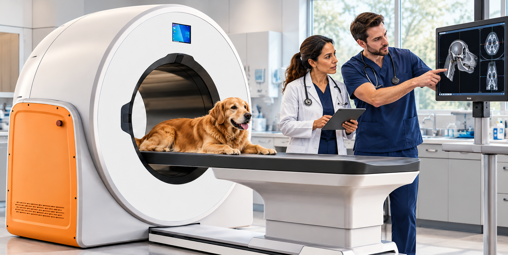
Beyond X-Rays: 7 Ways Veterinary CT Can Improve Diagnostic Workups
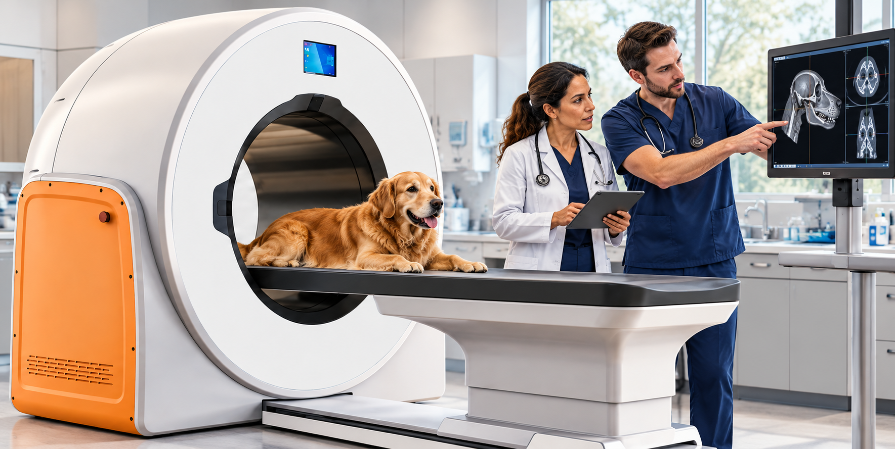
7 Veterinary CT Applications That Can Improve Diagnosis and Treatment Planning
Veterinary CT is often thought of as an advanced imaging option reserved for referral hospitals or complicated cases. But as more practices bring CT in-house, it's worth considering how many everyday diagnostic challenges could benefit from a closer look.
Unlike traditional radiographs, which produce two-dimensional images of three-dimensional anatomy, CT creates cross-sectional images that can be reconstructed into detailed views of bones, organs, and other structures. This can help veterinarians better understand a condition's location, extent, and complexity before deciding on the next step.
Of course, CT isn't the answer for every patient. Ultrasound, radiographs, and MRI each have important roles in veterinary diagnostics. The key is knowing when CT can provide information that changes the diagnostic or treatment plan.
Let's look at seven applications worth knowing.
1. Head and Nasal Disease: Seeing Beyond the Surface
Chronic nasal discharge, sneezing, facial swelling, or unexplained nosebleeds can be difficult to investigate with traditional radiographs alone.
The complex anatomy of the nasal passages, sinuses, and skull creates overlapping structures that can make subtle abnormalities difficult to identify.
CT provides detailed cross-sectional images that help veterinarians evaluate conditions such as:
- Nasal masses
- Changes associated with fungal infections
- Turbinate destruction
- Sinus disease and changes involving surrounding bone
CT can also help determine whether a mass has extended into adjacent structures, making it particularly valuable when planning surgery or obtaining diagnostic samples.
2. Dental Conditions: A Closer Look at Oral Health
Veterinary dental radiographs remain essential for evaluating individual teeth and their roots, but CT can provide additional information when disease involves more complex anatomy.
It can help identify and evaluate:
- Jaw lesions
- Tooth root abnormalities
- Bone loss
- Fractures
- Changes involving the temporomandibular joints
For complicated oral surgery, trauma, or suspected pathology involving surrounding structures, CT can give veterinarians a more complete picture before treatment begins.
3. Complex Fractures: Better Visualization for Surgical Planning
Not all fractures are straightforward. Injuries involving joints, small bones, or multiple fracture fragments can be difficult to fully evaluate using traditional radiographs.
CT provides detailed images from multiple angles, along with 3D reconstructions that help surgeons understand fracture alignment, bone displacement, and joint involvement.
This supports surgical planning, implant selection, and decisions about the best approach to repair.
4. Orthopedic Planning: Investigating What's Behind the Limp
When a patient presents with persistent lameness or an orthopedic condition that isn't fully explained by radiographs, CT may help uncover the underlying problem.
It's particularly useful for evaluating:
- Elbow dysplasia
- Fragmented coronoid processes
- Subtle bone abnormalities
- Complex joint conditions
CT can also help with measuring bone angles and alignment when planning corrective orthopedic procedures.
Ultrasound and MRI may be more appropriate for certain soft tissue injuries, but CT offers valuable detail when bone structure is the primary concern.
5. Thoracic Imaging: A More Detailed Look at the Chest
Thoracic digital radiographs are often the first step when investigating respiratory concerns or screening for metastatic disease.
However, conventional multidetector CT can reveal smaller pulmonary nodules and provide more detailed information about:
- Lung tissue
- Blood vessels
- Mediastinal structures
- Chest wall abnormalities
CT can be especially valuable when staging cancer, investigating suspected pulmonary disease, or determining the extent of a mass before surgery.
For certain vascular conditions, CT angiography can provide additional diagnostic information.
6. Abdominal Investigations: Understanding What's Happening Inside
Abdominal ultrasound remains a go-to imaging tool for evaluating many soft tissue organs, but CT can offer advantages when veterinarians need a broader view of anatomy, vascular structures, or the extent of a mass.
CT can help assess tumor size, location, involvement of surrounding tissues, and relationships with major blood vessels.
CT angiography is also useful when investigating vascular abnormalities, including portosystemic shunts.
These details can be particularly helpful when determining whether a mass is surgically removable and planning the procedure.
7. Selected Neurological Cases: Evaluating the Skull and Spine
CT can play an important role in investigating certain neurological conditions, particularly those involving bone, the skull, and spinal structures.
It can help identify vertebral abnormalities, fractures, bone remodeling, and some forms of intervertebral disc disease.
However, when the primary concern involves the brain, spinal cord, or other soft tissues of the central nervous system, MRI generally provides better detail.
Choosing between CT and MRI depends on the suspected condition and the information needed to guide treatment.
Frequently Asked Questions About Veterinary CT
What is veterinary CT used for?
Veterinary CT is used to evaluate complex anatomical structures and conditions involving the head, nasal passages, teeth and jaws, bones, joints, chest, abdomen, and spine. It can be especially helpful when traditional imaging doesn't provide enough detail for diagnosis or treatment planning.
Is CT better than regular veterinary X-rays?
Not necessarily. Radiographs remain an important first-line imaging tool for many conditions. CT provides cross-sectional views that eliminate much of the anatomical overlap seen on standard X-rays, making it especially useful for complex structures and selected surgical cases.
When should a veterinarian consider CT instead of ultrasound?
Ultrasound is valuable for examining soft tissue organs and evaluating many abdominal conditions. CT may be more useful when a broader anatomical overview is needed, especially for complex masses, bone involvement, or vascular anatomy.
What is the difference between veterinary CT and MRI?
CT is particularly useful for evaluating bone, lung anatomy, fractures, and certain complex anatomical structures. MRI generally provides better soft tissue detail for many conditions involving the brain, spinal cord, and central nervous system.
Can veterinary CT help with surgical planning?
Yes. CT can provide detailed cross-sectional images and 3D reconstructions that help veterinarians evaluate fractures, masses, joint abnormalities, and other conditions before surgery. The information may assist with treatment decisions and surgical planning.
Knowing When Veterinary CT Makes Sense
CT isn't intended to replace radiographs, ultrasound, or MRI. Each imaging modality has a purpose, and the best choice depends on the patient, clinical presentation, and diagnostic question.
For veterinary practices, having access to CT can expand diagnostic capabilities, provide more detailed information for surgical planning, and help identify conditions that may be difficult to fully evaluate with conventional imaging.
There are also different types of CT systems. Conventional multidetector CT and cone-beam CT have different strengths, particularly when evaluating soft tissues, the chest, and vascular structures. Practices should confirm that a specific system supports the examinations they intend to perform.
The real value isn't simply having another piece of equipment. It's having another way to get answers when the clinical picture calls for a closer look.
Considering CT for Your Veterinary Practice?
Choosing advanced imaging equipment is an important decision. Understanding the clinical applications, equipment capabilities, and practical requirements can help you determine whether CT is the right fit for your hospital.
Learn more about the VetiST 3 Veterinary CT Imaging System, or contact Brad Haven, Jr. to discuss your questions and equipment options.
Questions? Call 877-545-1202
Stop Paying Too Much for Veterinary Equipment.


Is Advanced CT Imaging Worth It? A Practical ROI for Veterinary Hospitals

Fujifilm Supria Plus 32-Slice CT ROI for Veterinarians Now
Ever wish you had more diagnostic clarity at your fingertips? Whether it’s a tricky nasal case, a subtle spinal lesion, or a client who needs answers today, in-house CT can change your standard of care—and your bottom line.
With CT becoming more accessible, many practices are asking: is now the time to bring it in-house? Below, we look at the Fujifilm Supria Plus 32-Slice CT to help you decide if it fits your hospital’s clinical goals and financial plan.
What Makes the Supria Plus Different
- Room for everyone: 75 cm gantry and 500 lb table capacity—comfortable for giant-breed dogs.
- Lower dose by design: Automatic, size-based dose reduction helps maintain image quality while minimizing exposure.
- Faster, more detailed scans: Up to 32-slice reconstruction reduces anesthesia time and increases throughput.
- Practical footprint: Compact and user-friendly, easing integration without major renovations or weeks of retraining.
At a glance:
- Excellent for neuro, nasal, dental/maxillofacial, thoracic, abdominal, and orthopedic studies.
- Improved scheduling control and client satisfaction with same-day answers.
- Retention of cases that might otherwise be referred out.
Understanding the Costs
- Acquisition: Approx. $299,000 (many practices finance at roughly $3,995/month).
- Warranty & PM: 2-year warranty; preventative maintenance every six months by Fuji-certified technicians.
- Per-scan costs: Radiologist read ~$250/scan; contrast for injected studies ~$100/scan (estimate).
- After warranty: Set aside a monthly service reserve to cover ongoing parts and service (example below uses $3,000/mo).
How the Numbers Can Work in Your Favor
Here’s a simple, conservative example to sanity-check your local demand:
| Assumption | Example Value |
|---|---|
| Monthly volume | 20 scans (≈5 per week) |
| Average fee per scan | $1,500 |
| Gross monthly revenue | $30,000 |
| Financing payment | $3,995 / month |
| Radiologist reads | 20 × $250 = $5,000 |
| Contrast | 20 × $100 = $2,000 |
| Service reserve (post-warranty planning) | $3,000 / month |
| Total monthly costs | $13,995 |
| Estimated net margin | $30,000 − $13,995 = $16,005 / month |
Break-even snapshot: With revenue of $1,500/scan and variable costs of $350/scan (radiologist + contrast), your contribution margin is ~$1,150/scan. Using the fixed cost proxy above (~$6,995 = payment + reserve), break-even is ≈ 7 scans/month (6.1 rounded up). Your accounting may vary—use your exact fees and costs.
A Few Things to Keep in Mind
- Review cases you currently refer out—this is your most realistic starting volume.
- Plan for a ramp-up period; consistency often lands after a few months.
- Confirm room size, power, shielding, and any site prep requirements in advance.
- Invest in team training; even user-friendly systems have a learning curve.
- Keep a service reserve once the warranty ends to avoid cash-flow surprises.
Taking the Leap
Choosing CT isn’t about chasing the newest gadget—it’s about transforming how you practice medicine. The Supria Plus 32-Slice CT can deliver sharper images, faster answers, and stronger client trust while keeping more care in-house. Run your numbers, map your space and training, and decide if now is the right time.
Looking for specs, workflow, and installation guidance? See the product page: Fujifilm Supria Plus 32-Slice CT Scanner.

ROI of Veterinary CT Scanners: Profitability Made Simple

Is a CT Scanner Right for Your Veterinary Practice?
Plan the investment, elevate patient care, and see a simple monthly ROI example.
For many veterinary practices, adding advanced imaging is a dream that feels just out of reach. But with the proper planning, CT technology can be both accessible and profitable—while dramatically elevating the level of care you can provide.
Why CT Changes the Conversation
Beyond the financial benefits, adding a CT scanner to your hospital opens the door to faster, more accurate diagnoses for complex cases. CT is beneficial for identifying issues like nasal tumors, thoracic masses, intervertebral disc disease, and complex fractures that can be difficult to assess with traditional radiographs. It’s also great at detecting subtle changes in the lungs, sinuses, and abdomen, giving you and your clients clear answers more quickly. With CT, you can shorten the path from uncertainty to treatment.
Introducing the Fujifilm Supria Plus
The Fujifilm Supria Plus 32-Slice CT Scanner is a traditional CT system designed for practices that want speed, precision, and reliability. It’s designed with both efficiency and patient comfort in mind. Its wide 75 cm gantry and large 500 lb table capacity make positioning easier for veterinary patients of all sizes, while advanced dose-reduction technology ensures high-quality images with lower radiation exposure. Combined with a compact footprint and user-friendly interface, it’s built to deliver powerful diagnostic capabilities without overwhelming your workflow or your facility space.
The Numbers That Matter
When you’re considering a piece of equipment this advanced, it’s natural to wonder: Can my practice really afford this? Here’s a simple way to run the numbers using conservative assumptions:
| Assumption | Value |
|---|---|
| Minimum volume | 15 CT scans per month |
| Average charge per scan | $1,500 |
| Monthly payment plan | $4,000 |
| Radiologist read per scan | $250 |
| Contrast cost per scan | $100 (estimate) |
| Preventive maintenance savings | $3,000/month set aside |
Your Profitability Calculator (Example)
| Line Item | Calculation | Monthly Total |
|---|---|---|
| Revenue | 15 scans × $1,500 | $22,500 |
| CT payment | — | $4,000 |
| Radiologist reads | 15 × $250 | $3,750 |
| Contrast | 15 × $100 | $1,500 |
| Maintenance fund | — | $3,000 |
| Net Monthly Margin | $22,500 – $12,250 | $10,250 |
That’s over $120,000 net margin per year at just 15 cases/month. As awareness grows among clients and referring DVMs, many hospitals see higher volumes—improving ROI even further.
Why Choose the Fuji 32-Slice CT?
- Speed & Accuracy: Detailed imaging in seconds helps reduce anesthesia time and supports precise treatment planning.
- Client Trust: Offering CT communicates clinical excellence and provides peace of mind to pet owners.
- Referral Potential: Nearby clinics without CT are more likely to refer advanced cases to you.
- Longevity: With a disciplined preventive maintenance plan, you’ll get years of reliable performance.
Related: Fujifilm Supria Plus 32-Slice CT Scanner for Veterinary Hospitals