


Cone-Beam CT vs. Conventional CT: What's the Difference for Veterinary Practices?
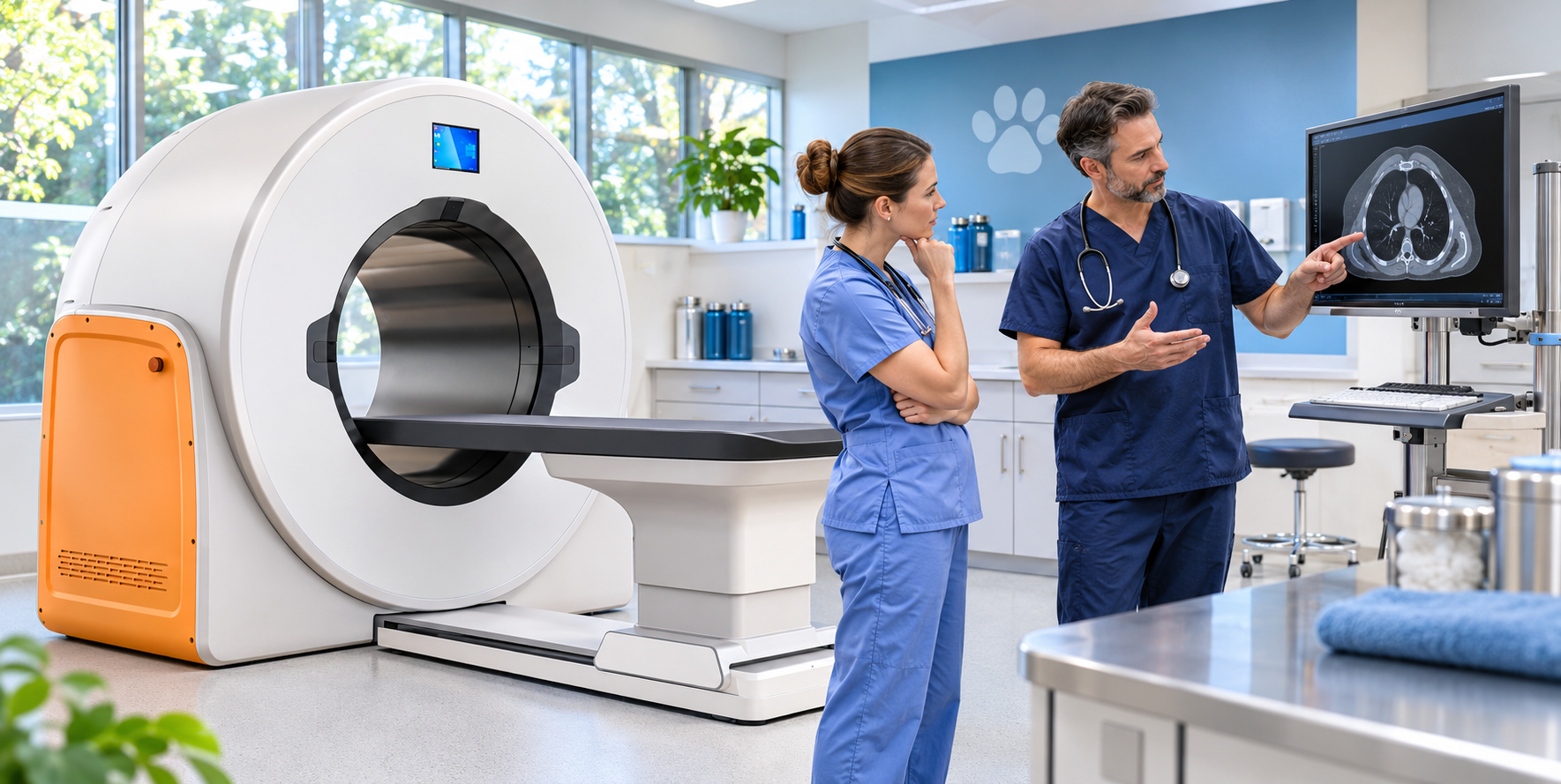
If your veterinary practice is considering investing in a CT scanner, one of the first decisions you'll encounter is whether to choose cone-beam CT (CBCT) or conventional CT.
Both technologies produce detailed images that can help veterinarians diagnose medical conditions, evaluate injuries, and plan treatment. However, they use different imaging methods and offer different advantages depending on the patients and conditions your practice treats.
So, is one technology better than the other?
Not necessarily. The right choice depends on what you need to see, the examinations you perform most often, and the capabilities you want to bring into your practice.
Understanding how each system works will help you make a more informed equipment decision.
Let's take a closer look.
How Do Cone-Beam and Conventional CT Work?
The biggest difference between these two technologies begins with how they capture X-ray information and reconstruct images.
Conventional CT (Multi-Slice CT)
Conventional CT, often called multi-slice CT or multi-detector CT (MDCT), uses an X-ray source and detector system that rotate around the patient.
- Uses a fan-shaped X-ray beam, or a relatively narrow cone-shaped beam in some modern systems.
- Detectors collect X-ray information from multiple angles as the system rotates.
- Creates cross-sectional images, often using continuous helical scanning to acquire data through a region of the body.
- Software reconstructs the collected information into detailed images that can be viewed in multiple planes or as 3D images.
- Multi-slice systems use multiple detector rows to acquire several image sections during each rotation.
Conventional CT is widely used for evaluating both bone and soft-tissue structures, including complex examinations involving the chest, abdomen, and internal organs.
Cone-Beam CT (CBCT)
Cone-beam CT uses a different approach to collect imaging information.
- Uses a cone-shaped X-ray beam and a flat-panel detector.
- The scanner rotates around the patient, capturing a series of 2D projection images from different angles.
- Software combines the collected projections to reconstruct a three-dimensional imaging volume.
- Images can then be reviewed as cross-sectional views or 3D reconstructions.
- Unlike conventional multi-slice CT, CBCT reconstructs its imaging volume from the acquired projection data rather than collecting narrow sections in the same manner.
These differences in image acquisition influence spatial resolution, soft-tissue contrast, scan coverage, motion sensitivity, and the types of examinations each system may be best suited for.
Important distinction: A scanner's advertised slice count does not tell the entire story. Cone-beam reconstruction slices and conventional multi-slice CT detector configurations are not directly interchangeable measurements.
Image Quality: What Are You Actually Getting?
When comparing veterinary CT scanners, image quality is one of the most important considerations.
However, choosing the scanner with the highest slice count doesn't necessarily mean you're getting the best system for your practice.
The more important question is whether the scanner produces the type of diagnostic images your veterinarians need.
Conventional CT: Strong Soft-Tissue Imaging
Conventional CT generally offers better soft-tissue contrast resolution, making it particularly useful when evaluating structures such as:
- Internal organs
- Muscle tissue
- Blood vessels
- Thoracic and abdominal structures
- Other internal soft tissues
Conventional CT is widely used for thoracic and abdominal imaging, cancer staging, surgical planning, and complex diagnostic investigations.
Depending on the examination, contrast agents and specialized acquisition protocols may provide additional diagnostic information.
Cone-Beam CT: Excellent Detail for Bone and High-Contrast Structures
CBCT systems are particularly well suited for evaluating bone and other high-contrast structures.
Depending on the system and imaging protocol, CBCT can provide excellent spatial detail for:
- Skull anatomy
- Nasal passages and surrounding bony structures
- Teeth and dental roots
- Jaw and maxillofacial structures
- Selected orthopedic examinations
This makes cone-beam CT especially attractive for practices that perform a substantial amount of dental, oral, maxillofacial, or bone-focused imaging.
However, CBCT generally has more limited soft-tissue contrast than conventional multi-slice CT. Scatter and certain image artifacts can also affect diagnostic image quality.
Some modern CBCT scanners include specialized reconstruction algorithms and imaging protocols designed to improve visualization of additional anatomical structures.
Their actual clinical performance should be evaluated for the specific examinations your practice intends to perform.
Field of View and Patient Positioning
Field of view (FOV) determines how much anatomy can be captured within an imaging examination.
This is particularly important in veterinary medicine, where a practice may treat everything from small cats to large-breed dogs.
Conventional CT Field of View
Conventional CT scanners offer different imaging field sizes and scan lengths depending on their configuration.
Many systems accommodate examinations ranging from small anatomical regions to larger portions of the body.
Cone-Beam CT Field of View
CBCT systems can also offer generous fields of view, although coverage varies considerably among manufacturers and models.
Some CBCT systems are designed primarily for dental and head imaging, while others accommodate larger anatomical regions.
For example, the VetiST 3 Veterinary CT System features:
- Gantry aperture: 80 cm
- Maximum field of view: 72 × 42 cm
- Imaging technology: Cone-beam CT
- Additional imaging modes: Digital radiography, fluoroscopy, and panoramic imaging
These specifications are worth considering when evaluating patient positioning, anatomical coverage, and the variety of imaging examinations your practice may want to perform.
Don't confuse gantry size with imaging coverage. A large scanner opening does not automatically mean the system can capture every anatomical region in a single scan.
Always compare the gantry aperture, actual field of view, scan length, patient table capacity, and positioning requirements.
Scan Protocols and Motion Sensitivity
Patient movement is another important consideration, especially in veterinary medicine.
Unlike human patients, animals generally cannot be instructed to remain perfectly still throughout an examination.
Sedation or general anesthesia is often necessary to obtain diagnostic-quality images, depending on the patient and the examination.
Conventional CT Scan Speed
Conventional multi-slice CT systems can acquire images quickly, depending on the scanner, acquisition protocol, and anatomical region.
Faster acquisitions can be useful when performing examinations where respiratory motion or patient movement is a concern.
Cone-Beam CT and Motion Artifacts
CBCT systems typically acquire a series of projections during a rotational scan.
Since image reconstruction uses the collected projections, patient movement during acquisition can introduce motion artifacts and affect the reconstructed imaging volume.
This does not mean all CBCT examinations are slow, nor does it mean conventional CT is immune to motion-related problems.
Both technologies require appropriate patient preparation, positioning, and imaging protocols.
When comparing specific scanners, ask about actual acquisition times, motion-management features, anesthesia requirements, and how the system performs during the examinations you expect to perform most frequently.
Cone-Beam vs. Conventional CT: Side-by-Side Comparison
The following comparison summarizes the general differences between the two technologies. Individual scanner capabilities vary by model, configuration, and imaging protocol.
| Feature | Cone-Beam CT | Conventional CT |
|---|---|---|
| X-ray beam | Cone-shaped beam | Fan-shaped or relatively narrow cone-shaped beam |
| Detector | Typically flat-panel detector | Typically multi-row detector array |
| Bone detail | Often excellent spatial detail | Strong diagnostic capability |
| Soft-tissue contrast | Generally more limited | Generally stronger |
| Dental and skull imaging | Particularly well suited | Also capable |
| Thoracic and abdominal imaging | Depends on system and clinical task; generally more limited for soft tissues | Well established for these examinations |
| Motion sensitivity | Movement during projection acquisition may affect the reconstructed volume | Motion can affect acquired image sections |
| Field of view | Varies significantly by model | Varies by model and configuration |
| Additional imaging modes | Available on certain integrated systems | Depends on system configuration |
Which CT Technology Is Right for Your Veterinary Practice?
The best choice depends on your clinical priorities, patient population, and equipment budget.
Cone-Beam CT May Be a Good Fit If Your Practice:
- Performs a high volume of dental, oral, and maxillofacial imaging.
- Frequently evaluates bone, skull, and selected orthopedic conditions.
- Needs detailed visualization of high-contrast anatomical structures.
- Wants access to multiple imaging modalities in one system, depending on the model.
- Is looking for a system that fits its facility, workflow, and investment requirements.
For practices evaluating an integrated CBCT platform, the VetiST 3 is one example worth investigating because it combines cone-beam CT, digital radiography, fluoroscopy, and panoramic imaging.
Conventional CT May Be a Better Fit If Your Practice:
- Routinely performs thoracic and abdominal imaging.
- Needs strong soft-tissue contrast for a wide range of diagnostic investigations.
- Performs complex cancer staging and surgical planning.
- Requires particular acquisition protocols or advanced imaging capabilities.
- Needs a system designed for broader anatomical coverage or specific clinical applications.
For example, the Fujifilm Supria Plus CT Scanner is a conventional CT system available for veterinary practices evaluating multi-slice imaging technology.
Comparing actual scanner specifications and representative clinical images is more useful than relying on technology labels alone.
What Else Should You Consider Before Buying a Veterinary CT Scanner?
Imaging performance is essential, but it is not the only factor that determines whether a CT purchase makes sense for your practice.
Before making a decision, consider the following practical questions.
1. What Examinations Will You Perform Most Often?
Start with your current caseload.
Are you primarily evaluating dental and bone conditions, or do you need advanced imaging for abdominal, thoracic, and other soft-tissue examinations?
Your answer should guide which technology and scanner capabilities you prioritize.
2. Will the Equipment Accommodate Your Patients?
Compare patient size, table capacity, gantry aperture, imaging field of view, and positioning requirements.
A system should accommodate the patients and anatomical regions you routinely need to examine.
3. What Are the Facility Requirements?
CT installation involves more than finding enough floor space for the scanner.
Electrical service, room dimensions, equipment access, and radiation protection requirements should be evaluated before purchasing.
If you're planning a CT room, a veterinary X-ray or CT room shielding report can help identify appropriate radiation protection requirements for your facility.
4. What Support Is Included?
Ask about warranty coverage, installation, training, software updates, service response, and long-term technical support.
These details matter because your team needs reliable imaging equipment and access to assistance when questions arise.
5. Does the Investment Make Financial Sense?
Consider the total cost of ownership, not simply the purchase price.
Include installation, facility preparation, service, maintenance, staffing, and financing when evaluating the investment.
The goal is to purchase equipment that supports the diagnostic services you expect to provide without paying for capabilities your practice may rarely use.
Frequently Asked Questions About Veterinary CT Scanners
1. Is cone-beam CT better than conventional CT?
Neither technology is automatically better for every veterinary practice. CBCT is particularly useful for bone, dental, and other high-contrast structures. Conventional CT generally offers better soft-tissue contrast and is widely used for thoracic, abdominal, and complex diagnostic examinations. The best choice depends on your clinical needs.
2. Can cone-beam CT be used for soft-tissue imaging?
Some CBCT systems offer specialized modes and reconstruction algorithms for additional imaging applications. However, CBCT generally provides less soft-tissue contrast than conventional CT. The suitability of a particular CBCT scanner for a soft-tissue examination should be confirmed using system-specific clinical evidence.
3. Does a higher CT slice count mean better image quality?
Not necessarily. Image quality also depends on detector technology, spatial resolution, contrast resolution, acquisition protocols, and reconstruction software. CBCT reconstruction slice counts should not be directly compared with conventional CT detector-row counts.
4. Do veterinary patients need anesthesia for CT scans?
Many veterinary CT examinations require sedation or general anesthesia to minimize patient movement. The specific approach depends on the patient, examination, scanner, and veterinary team's clinical assessment. Both CBCT and conventional CT can be affected by motion artifacts.
5. Can one veterinary imaging system provide CT, digital X-ray, and fluoroscopy?
Yes. Certain integrated systems offer multiple imaging modes. For example, the VetiST 3 combines cone-beam CT, digital radiography, fluoroscopy, and panoramic imaging. The capabilities and limitations of each mode should be reviewed before purchasing.
6. What should I compare before purchasing a veterinary CT scanner?
Start with the types of examinations your practice performs. Then compare image quality, field of view, patient capacity, acquisition speed, facility requirements, warranty, technical support, and total ownership costs. Ask to review representative images for your intended clinical applications.
Choosing the Right Veterinary CT Scanner
Ultimately, the best veterinary CT scanner isn't necessarily the most advanced or most expensive option.
It's the scanner that provides the imaging capabilities your caseload requires, supports your clinical goals, and makes financial sense for your practice.
If you're comparing cone-beam CT and conventional CT, Brad Haven, Jr. at New Vet Equipment can help you review equipment options, specifications, installation considerations, and pricing.
The goal is to help you understand your options so you can make a confident, informed equipment decision.
Stop Paying Too Much for Veterinary Equipment.
Questions? Call 877-545-1202

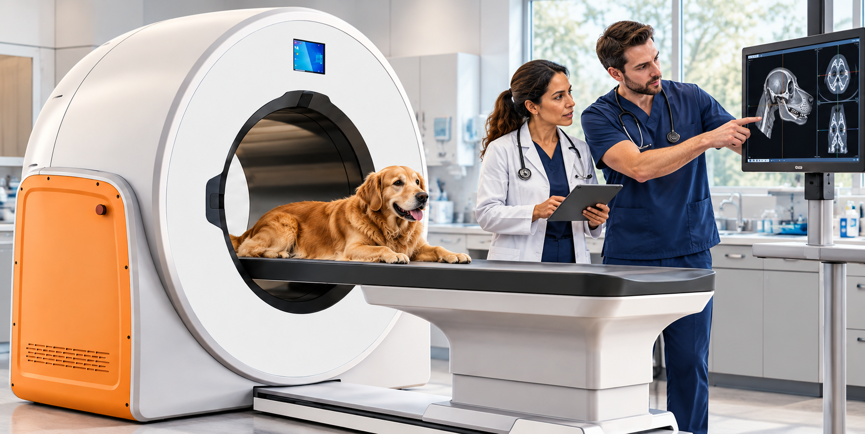
Beyond X-Rays: 7 Ways Veterinary CT Can Improve Diagnostic Workups
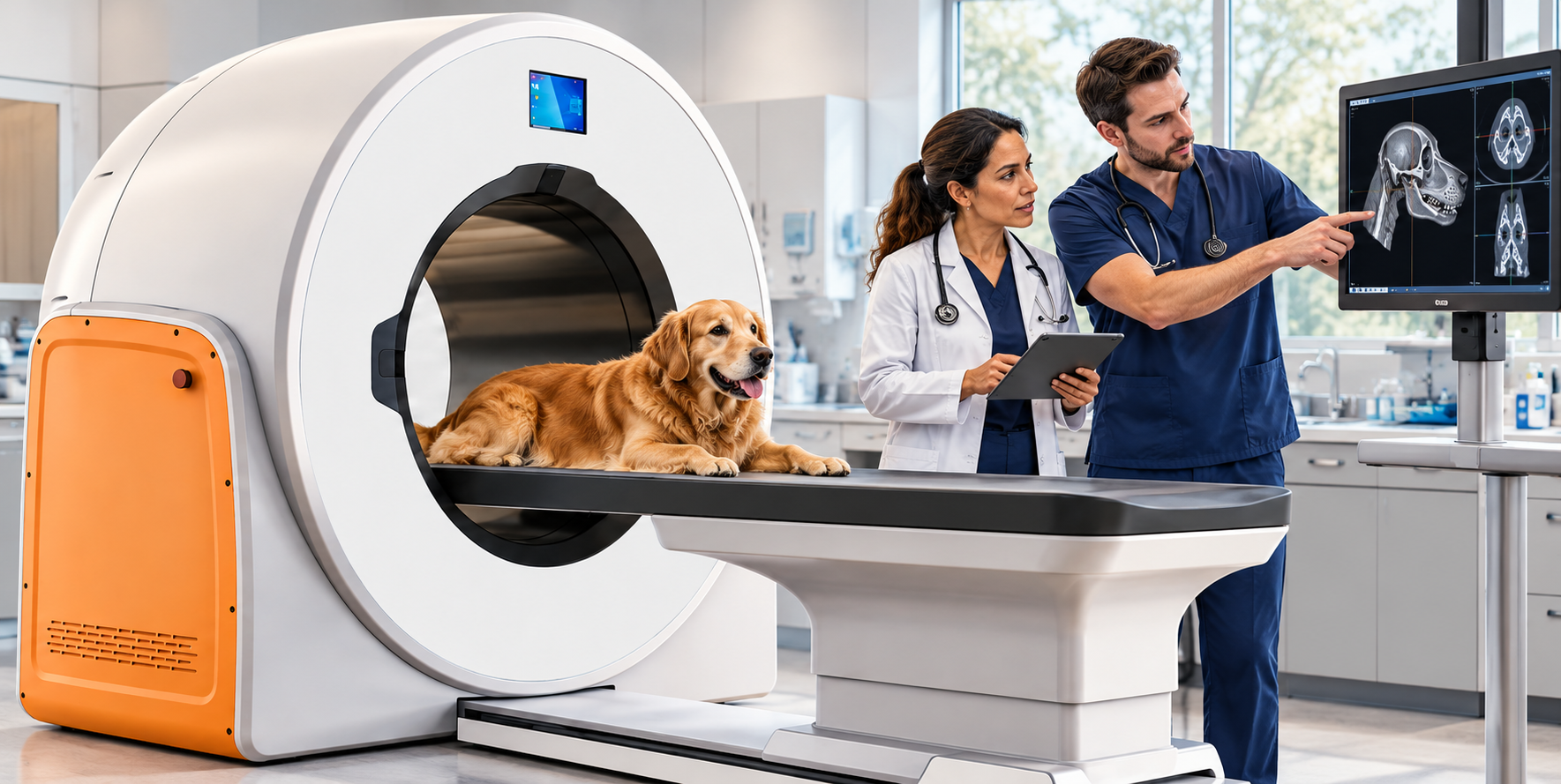
7 Veterinary CT Applications That Can Improve Diagnosis and Treatment Planning
Veterinary CT is often thought of as an advanced imaging option reserved for referral hospitals or complicated cases. But as more practices bring CT in-house, it's worth considering how many everyday diagnostic challenges could benefit from a closer look.
Unlike traditional radiographs, which produce two-dimensional images of three-dimensional anatomy, CT creates cross-sectional images that can be reconstructed into detailed views of bones, organs, and other structures. This can help veterinarians better understand a condition's location, extent, and complexity before deciding on the next step.
Of course, CT isn't the answer for every patient. Ultrasound, radiographs, and MRI each have important roles in veterinary diagnostics. The key is knowing when CT can provide information that changes the diagnostic or treatment plan.
Let's look at seven applications worth knowing.
1. Head and Nasal Disease: Seeing Beyond the Surface
Chronic nasal discharge, sneezing, facial swelling, or unexplained nosebleeds can be difficult to investigate with traditional radiographs alone.
The complex anatomy of the nasal passages, sinuses, and skull creates overlapping structures that can make subtle abnormalities difficult to identify.
CT provides detailed cross-sectional images that help veterinarians evaluate conditions such as:
- Nasal masses
- Changes associated with fungal infections
- Turbinate destruction
- Sinus disease and changes involving surrounding bone
CT can also help determine whether a mass has extended into adjacent structures, making it particularly valuable when planning surgery or obtaining diagnostic samples.
2. Dental Conditions: A Closer Look at Oral Health
Veterinary dental radiographs remain essential for evaluating individual teeth and their roots, but CT can provide additional information when disease involves more complex anatomy.
It can help identify and evaluate:
- Jaw lesions
- Tooth root abnormalities
- Bone loss
- Fractures
- Changes involving the temporomandibular joints
For complicated oral surgery, trauma, or suspected pathology involving surrounding structures, CT can give veterinarians a more complete picture before treatment begins.
3. Complex Fractures: Better Visualization for Surgical Planning
Not all fractures are straightforward. Injuries involving joints, small bones, or multiple fracture fragments can be difficult to fully evaluate using traditional radiographs.
CT provides detailed images from multiple angles, along with 3D reconstructions that help surgeons understand fracture alignment, bone displacement, and joint involvement.
This supports surgical planning, implant selection, and decisions about the best approach to repair.
4. Orthopedic Planning: Investigating What's Behind the Limp
When a patient presents with persistent lameness or an orthopedic condition that isn't fully explained by radiographs, CT may help uncover the underlying problem.
It's particularly useful for evaluating:
- Elbow dysplasia
- Fragmented coronoid processes
- Subtle bone abnormalities
- Complex joint conditions
CT can also help with measuring bone angles and alignment when planning corrective orthopedic procedures.
Ultrasound and MRI may be more appropriate for certain soft tissue injuries, but CT offers valuable detail when bone structure is the primary concern.
5. Thoracic Imaging: A More Detailed Look at the Chest
Thoracic digital radiographs are often the first step when investigating respiratory concerns or screening for metastatic disease.
However, conventional multidetector CT can reveal smaller pulmonary nodules and provide more detailed information about:
- Lung tissue
- Blood vessels
- Mediastinal structures
- Chest wall abnormalities
CT can be especially valuable when staging cancer, investigating suspected pulmonary disease, or determining the extent of a mass before surgery.
For certain vascular conditions, CT angiography can provide additional diagnostic information.
6. Abdominal Investigations: Understanding What's Happening Inside
Abdominal ultrasound remains a go-to imaging tool for evaluating many soft tissue organs, but CT can offer advantages when veterinarians need a broader view of anatomy, vascular structures, or the extent of a mass.
CT can help assess tumor size, location, involvement of surrounding tissues, and relationships with major blood vessels.
CT angiography is also useful when investigating vascular abnormalities, including portosystemic shunts.
These details can be particularly helpful when determining whether a mass is surgically removable and planning the procedure.
7. Selected Neurological Cases: Evaluating the Skull and Spine
CT can play an important role in investigating certain neurological conditions, particularly those involving bone, the skull, and spinal structures.
It can help identify vertebral abnormalities, fractures, bone remodeling, and some forms of intervertebral disc disease.
However, when the primary concern involves the brain, spinal cord, or other soft tissues of the central nervous system, MRI generally provides better detail.
Choosing between CT and MRI depends on the suspected condition and the information needed to guide treatment.
Frequently Asked Questions About Veterinary CT
What is veterinary CT used for?
Veterinary CT is used to evaluate complex anatomical structures and conditions involving the head, nasal passages, teeth and jaws, bones, joints, chest, abdomen, and spine. It can be especially helpful when traditional imaging doesn't provide enough detail for diagnosis or treatment planning.
Is CT better than regular veterinary X-rays?
Not necessarily. Radiographs remain an important first-line imaging tool for many conditions. CT provides cross-sectional views that eliminate much of the anatomical overlap seen on standard X-rays, making it especially useful for complex structures and selected surgical cases.
When should a veterinarian consider CT instead of ultrasound?
Ultrasound is valuable for examining soft tissue organs and evaluating many abdominal conditions. CT may be more useful when a broader anatomical overview is needed, especially for complex masses, bone involvement, or vascular anatomy.
What is the difference between veterinary CT and MRI?
CT is particularly useful for evaluating bone, lung anatomy, fractures, and certain complex anatomical structures. MRI generally provides better soft tissue detail for many conditions involving the brain, spinal cord, and central nervous system.
Can veterinary CT help with surgical planning?
Yes. CT can provide detailed cross-sectional images and 3D reconstructions that help veterinarians evaluate fractures, masses, joint abnormalities, and other conditions before surgery. The information may assist with treatment decisions and surgical planning.
Knowing When Veterinary CT Makes Sense
CT isn't intended to replace radiographs, ultrasound, or MRI. Each imaging modality has a purpose, and the best choice depends on the patient, clinical presentation, and diagnostic question.
For veterinary practices, having access to CT can expand diagnostic capabilities, provide more detailed information for surgical planning, and help identify conditions that may be difficult to fully evaluate with conventional imaging.
There are also different types of CT systems. Conventional multidetector CT and cone-beam CT have different strengths, particularly when evaluating soft tissues, the chest, and vascular structures. Practices should confirm that a specific system supports the examinations they intend to perform.
The real value isn't simply having another piece of equipment. It's having another way to get answers when the clinical picture calls for a closer look.
Considering CT for Your Veterinary Practice?
Choosing advanced imaging equipment is an important decision. Understanding the clinical applications, equipment capabilities, and practical requirements can help you determine whether CT is the right fit for your hospital.
Learn more about the VetiST 3 Veterinary CT Imaging System, or contact Brad Haven, Jr. to discuss your questions and equipment options.
Questions? Call 877-545-1202
Stop Paying Too Much for Veterinary Equipment.
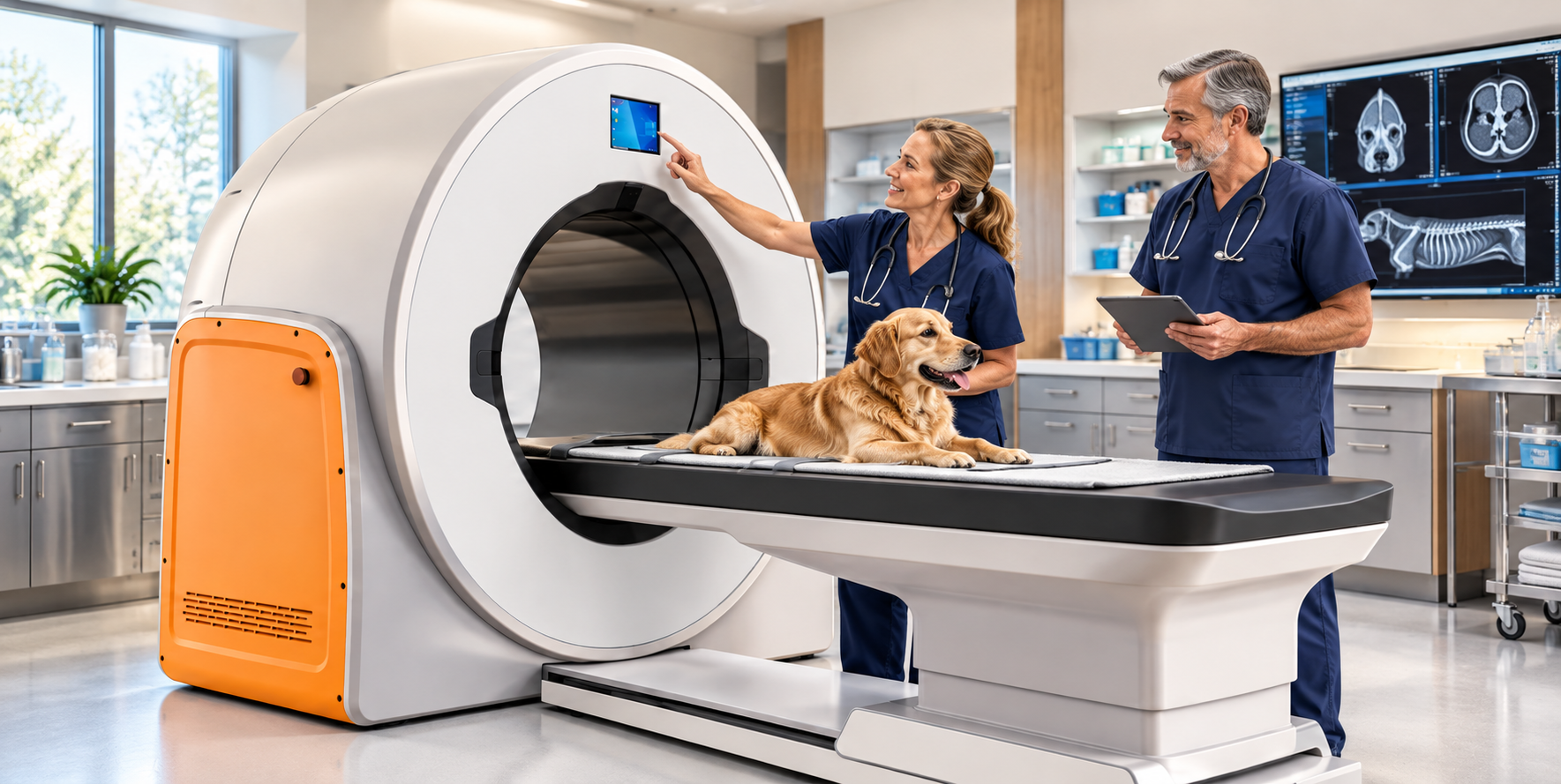
Veterinary CT Buyers Guide: How Does the VetiST 3 Stack Up Against Alternatives?
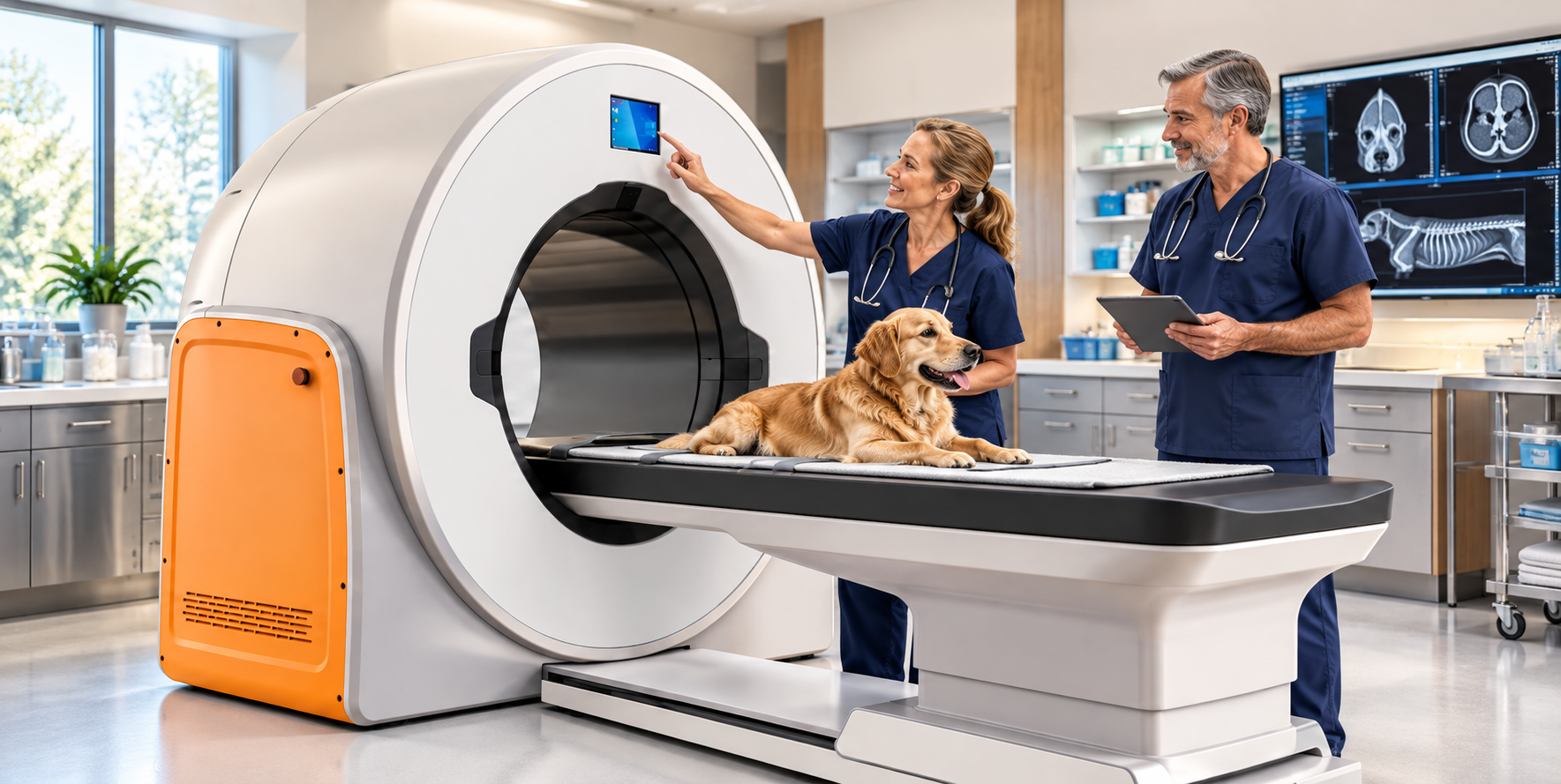
Comparing Veterinary CT Scanners: Features, Costs, and Patient Capacity
Choosing a CT scanner is a big investment for any veterinary practice. There's a lot to consider before bringing advanced imaging in-house.
- Purchase price
- Installation requirements
- Maintenance and support
- Patient size and capacity
- Imaging capabilities
The good news? Today's veterinary CT market offers options for different budgets, caseloads, and clinical needs.
The challenge is understanding what you're actually getting for your money.
Here's the key: Don't compare scanners by price alone. Compare the entire package, including installation, warranties, service, and the types of patients you can scan.
What to Consider Before Buying a Veterinary CT Scanner
Choosing the right CT scanner goes beyond the initial purchase price. Before committing to a system, consider how its capabilities, ongoing costs, and support align with your practice's needs.
- Imaging capabilities: Consider slice count, image resolution, scan speed, and whether cone-beam or conventional CT best suits your clinical caseload.
- Patient capacity: Evaluate gantry aperture, table weight limits, and scan coverage to make sure the system accommodates the dogs and cats you typically treat.
- Total cost of ownership: Include purchase price, installation, room modifications, radiation shielding, maintenance, software, and service agreements.
- Warranty and technical support: Review warranty duration, exclusions, response times, replacement-parts availability, and post-warranty service costs.
- Space and installation requirements: Confirm room dimensions, electrical requirements, facility modifications, and necessary regulatory approvals.
- Return on investment: Estimate scan volume, pricing, referral opportunities, and operating expenses. Determine whether bringing CT in-house makes financial sense.
Meet the Contenders: Six Veterinary CT Options
Let's compare the VetiST 3 Veterinary CT System with five refurbished conventional CT alternatives.
These systems have different technologies, capabilities, and price points. Understanding those differences will help you identify which features matter most for your practice.
VetiST 3 Veterinary CT System
The VetiST 3 is a cone-beam CT (CBCT) system that combines four imaging modalities in one system.
- CT imaging
- Digital radiography
- Fluoroscopy
- Panoramic imaging
Financing is available.
The package includes:
- 3-year warranty
- Technical support
- Software updates
- Shielding plan
- Shipping and installation
Patient specifications: 80 cm gantry aperture, 72 × 42 cm maximum field of view, and 135 kg (298 lb) table capacity.
View the VetiST 3 CT scanner for additional product details.
Five Refurbished CT Alternatives Worth Investigating
Refurbished conventional CT systems can provide another route to advanced imaging. However, pricing and included services may vary considerably between sellers.
GE BrightSpeed 16 Vet
A refurbished 16-slice conventional CT option for veterinary practices.
Approx. $110,000–$185,000
Pricing depends on condition, configuration, and included installation or service.
GE LightSpeed 16 Vet
An established conventional CT platform available refurbished.
Approx. $95,000–$160,000
Final pricing depends on the specific scanner and package.
GE LightSpeed VCT 64 Vet
A 64-slice conventional CT system for practices evaluating higher-throughput imaging capabilities.
Approx. $175,000–$325,000
Pricing varies by model year, configuration, and refurbishment level.
Siemens SOMATOM Emotion 16 Vet
A refurbished conventional CT option worth evaluating for veterinary use.
Approx. $95,000–$150,000
Availability and configuration can affect pricing.
GE Optima 660 Vet
A refurbished conventional CT system to consider when evaluating advanced imaging capabilities.
Approx. $225,000–$375,000
Compare the complete refurbishment, warranty, installation, and service package.
Note: Refurbished prices are approximate planning estimates, not verified current seller quotes. Actual pricing, condition, availability, and package inclusions must be confirmed.
Why Does CT Slice Count Matter?
Slice count can influence how a conventional CT scanner captures images, especially how quickly it can cover a region of the body.
Higher-slice systems can acquire more image sections per rotation, which may benefit larger anatomical studies and faster scanning.
However, slice count alone does not determine image quality or clinical usefulness. Detector technology, image resolution, scan protocols, and reconstruction software also play important roles.
Important distinction: Cone-beam CT and conventional multi-slice CT use different imaging technologies. Their slice-count specifications are not directly interchangeable.
The better question is whether the system can produce the diagnostic images your veterinarians need for the exams they perform most often.
What About the Size of Dogs and Cats?
Patient capacity can be a deciding factor, especially for practices treating everything from small cats to giant-breed dogs.
Two important specifications are:
- Gantry aperture: The opening available to position a patient for imaging.
- Patient table capacity: The maximum patient weight the table can safely support.
Scan field of view and coverage also matter. A table may support a large dog, but the scanner must still be able to capture the required anatomy.
Veterinary CT Patient Capacity Comparison
Here's a side-by-side look at reported gantry openings and table capacities.
| CT System | Gantry Aperture | Patient Table Capacity |
|---|---|---|
| VetiST 3 | 80 cm | 135 kg (298 lb) |
| GE BrightSpeed 16 | 70 cm | Approx. 205 kg (452 lb) |
| GE LightSpeed VCT 64 | 70 cm | Approx. 227 kg (500 lb) |
| Siemens SOMATOM Emotion 16 | 70 cm | Approx. 200 kg (440 lb) |
| GE Optima 660 | Confirm with seller | Confirm with seller |
| GE LightSpeed 16 | Confirm with seller | Confirm with seller |
Table capacities and apertures are comparison specifications supplied for this article. Confirm the exact specifications for each individual system and configuration before making a purchase. The VetiST 3 also has a reported maximum field of view of 72 × 42 cm.
When comparing patient capacity, evaluate the complete picture:
- Gantry opening
- Patient table capacity
- Maximum field of view
- Scan coverage
- Types of examinations performed
These details help determine whether a system can handle your everyday veterinary caseload.
Don't Overlook the Total Cost of Ownership
The initial purchase price is only part of the equation. A less expensive scanner upfront may not be the least expensive system to own over time.
For example, the VetiST 3 has a $175,000 package price that includes shipping, installation, and a three-year warranty.
When comparing different systems, pay close attention to:
- Warranty exclusions
- Preventive maintenance requirements
- Replacement parts
- Software and update costs
- Technical support and service pricing
Additional Questions for Refurbished CT Systems
If you're considering refurbished conventional CT equipment, request a detailed written breakdown covering:
- Installation: Is delivery, rigging, and installation included?
- Tube condition: What is the tube's operating history, and what would replacement cost?
- Parts availability: Are replacement components readily available?
- Service coverage: Who provides repairs, and what are the response times and costs?
- Facility requirements: What electrical, cooling, structural, and radiation shielding modifications will your building need?
A smart buying decision: Request an itemized quote showing the complete installed price, warranty coverage, recurring expenses, and anticipated service costs.
This gives you a much more meaningful comparison than purchase price alone.
Which Veterinary CT Scanner Is Right for Your Practice?
There isn't one perfect CT system for every veterinary hospital.
The right choice depends on your caseload, diagnostic goals, available space, and budget.
Before making a final decision, ask for written documentation covering:
- Complete purchase and installation pricing
- Warranty terms and exclusions
- Technical support and service expenses
- Patient weight and size capacity
- Clinical imaging capabilities
- Facility and installation requirements
These details can help you make a more informed investment in your practice and your patients.
Have Questions About the VetiST 3 Veterinary CT?
Advanced imaging is an important investment. You deserve clear answers before making that decision.
If you're considering the VetiST 3 CT scanner, I'd be happy to help you review its features, patient capacity, pricing, financing, and installation requirements.
My goal is simple: Help you make a confident, informed equipment purchasing decision.
Brad Haven, Jr.
NewVetEquipment.com
877-545-1202
Veterinary CT Systems: Side-by-Side Comparison
Compare six veterinary CT systems by imaging technology, patient capacity, warranty, installation, and facility requirements. Be sure to confirm specifications and included services before purchasing.
← Swipe or scroll horizontally to compare all six systems →
| Feature | VetiST 3 | GE BrightSpeed 16 | GE LightSpeed 16 | GE LightSpeed VCT 64 | Siemens SOMATOM Emotion 16 | GE Optima 660 |
|---|---|---|---|---|---|---|
| CT Technology | Cone-beam CT | Conventional CT | Conventional CT | Conventional CT | Conventional CT | Conventional CT |
| Slice Configuration | Up to 600 slices advertised | 16-slice | 16-slice | 64-slice | 16-slice | 128-slice configuration |
| Additional Imaging | Digital radiography (DR), fluoroscopy, panoramic imaging | Depends on configuration | Depends on configuration | Depends on configuration | Depends on configuration | Depends on configuration |
| Gantry Aperture | 80 cm | 70 cm | Verify unit | 70 cm | 70 cm | Approx. 70 cm |
| Maximum Table Load | 135 kg / 298 lb | 205 kg / 452 lb | Verify unit | 227 kg / 500 lb | Approx. 200–220 kg / 441–485 lb | Approx. 227 kg / 500 lb |
| Advertised Purchase Price | $175,000 | Quote required | Quote required | Quote required | Quote required | Quote required |
| Warranty | 3 years advertised | 1 year parts and labor advertised | Confirm with seller | 1 year parts and labor advertised | Confirm with seller | Confirm with seller |
| Installation | Included in advertised package | Typical installation included | Confirm with seller | Typical installation included | Confirm with seller | Confirm with seller |
| Room Size | Confirm with vendor | Approx. 18.2 m² | Confirm with vendor | Approx. 34.2 m² | Approx. 18 m² | Confirm with vendor |
Important: Advertised features and specifications may vary by configuration, model year, equipment condition, and seller. Room sizes are approximate and may not include all required support space. Confirm current pricing, clinical capabilities, warranty terms, facility requirements, and installation costs directly with each equipment supplier.
Understanding slice counts: Cone-beam CT and conventional multi-slice CT use different acquisition methods. The VetiST 3's advertised 600-slice figure should not be interpreted as equivalent to the slice configuration of a conventional 16-, 64-, or 128-slice CT scanner.