
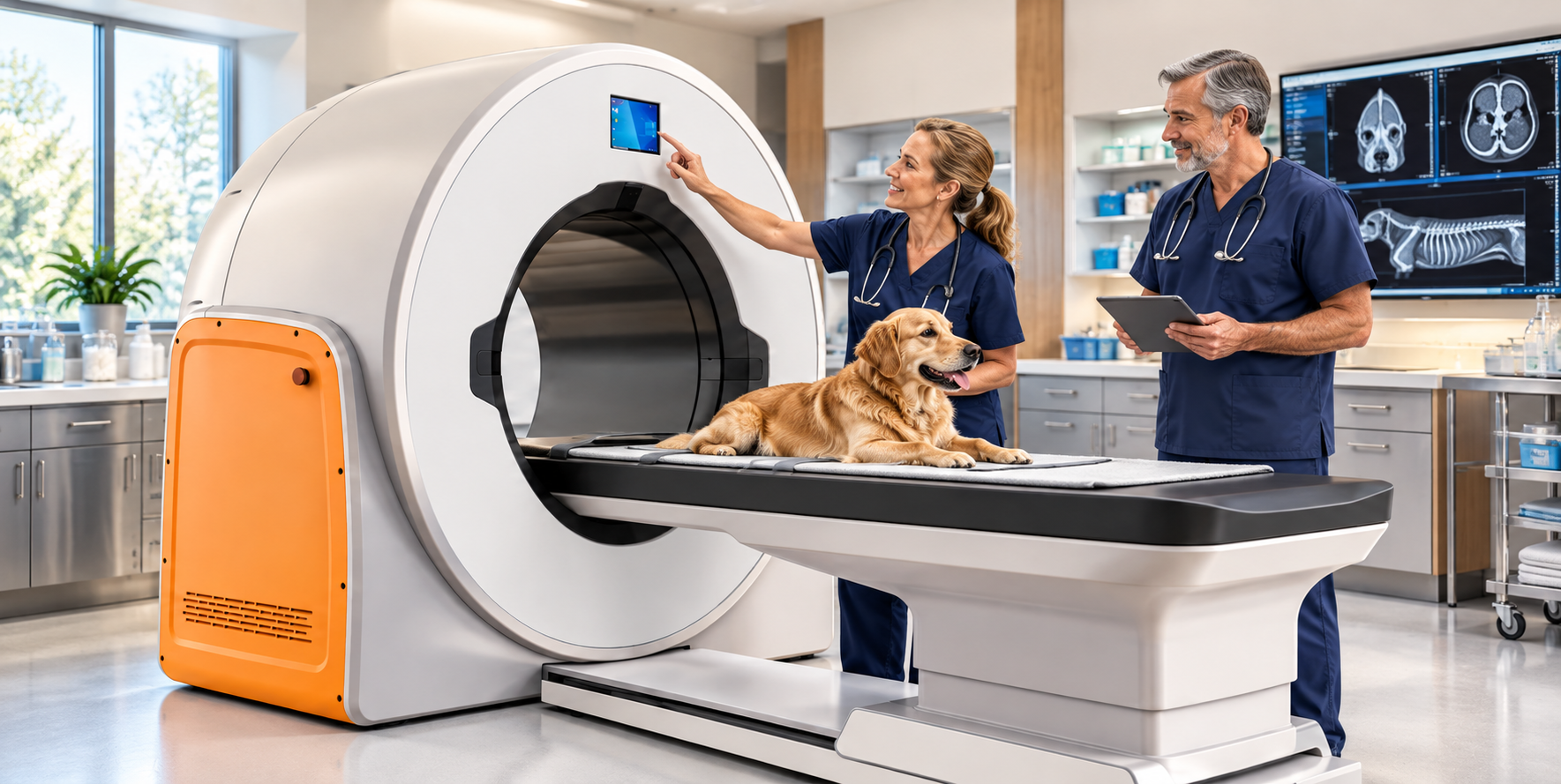
Veterinary CT Buyers Guide: How Does the VetiST 3 Stack Up Against Alternatives?
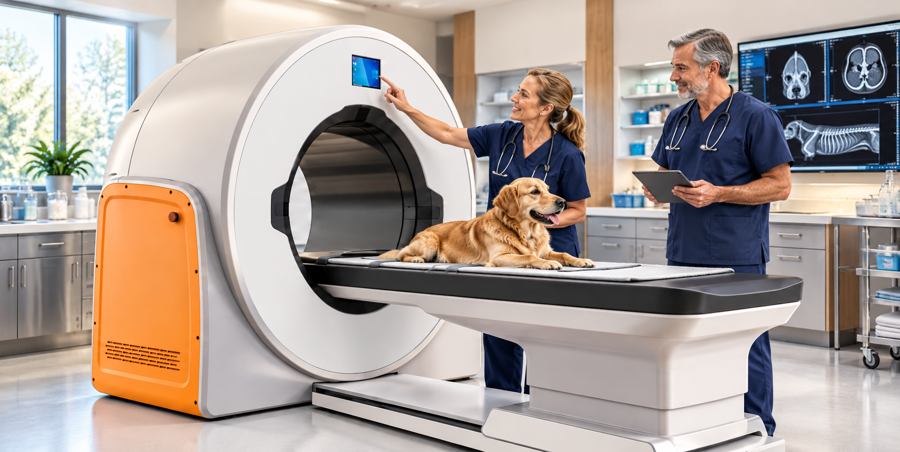
Comparing Veterinary CT Scanners: Features, Costs, and Patient Capacity
Choosing a CT scanner is a big investment for any veterinary practice. There's a lot to consider before bringing advanced imaging in-house.
- Purchase price
- Installation requirements
- Maintenance and support
- Patient size and capacity
- Imaging capabilities
The good news? Today's veterinary CT market offers options for different budgets, caseloads, and clinical needs.
The challenge is understanding what you're actually getting for your money.
Here's the key: Don't compare scanners by price alone. Compare the entire package, including installation, warranties, service, and the types of patients you can scan.
What to Consider Before Buying a Veterinary CT Scanner
Choosing the right CT scanner goes beyond the initial purchase price. Before committing to a system, consider how its capabilities, ongoing costs, and support align with your practice's needs.
- Imaging capabilities: Consider slice count, image resolution, scan speed, and whether cone-beam or conventional CT best suits your clinical caseload.
- Patient capacity: Evaluate gantry aperture, table weight limits, and scan coverage to make sure the system accommodates the dogs and cats you typically treat.
- Total cost of ownership: Include purchase price, installation, room modifications, radiation shielding, maintenance, software, and service agreements.
- Warranty and technical support: Review warranty duration, exclusions, response times, replacement-parts availability, and post-warranty service costs.
- Space and installation requirements: Confirm room dimensions, electrical requirements, facility modifications, and necessary regulatory approvals.
- Return on investment: Estimate scan volume, pricing, referral opportunities, and operating expenses. Determine whether bringing CT in-house makes financial sense.
Meet the Contenders: Six Veterinary CT Options
Let's compare the VetiST 3 Veterinary CT System with five refurbished conventional CT alternatives.
These systems have different technologies, capabilities, and price points. Understanding those differences will help you identify which features matter most for your practice.
VetiST 3 Veterinary CT System
The VetiST 3 is a cone-beam CT (CBCT) system that combines four imaging modalities in one system.
- CT imaging
- Digital radiography
- Fluoroscopy
- Panoramic imaging
Financing is available.
The package includes:
- 3-year warranty
- Technical support
- Software updates
- Shielding plan
- Shipping and installation
Patient specifications: 80 cm gantry aperture, 72 × 42 cm maximum field of view, and 135 kg (298 lb) table capacity.
View the VetiST 3 CT scanner for additional product details.
Five Refurbished CT Alternatives Worth Investigating
Refurbished conventional CT systems can provide another route to advanced imaging. However, pricing and included services may vary considerably between sellers.
GE BrightSpeed 16 Vet
A refurbished 16-slice conventional CT option for veterinary practices.
Approx. $110,000–$185,000
Pricing depends on condition, configuration, and included installation or service.
GE LightSpeed 16 Vet
An established conventional CT platform available refurbished.
Approx. $95,000–$160,000
Final pricing depends on the specific scanner and package.
GE LightSpeed VCT 64 Vet
A 64-slice conventional CT system for practices evaluating higher-throughput imaging capabilities.
Approx. $175,000–$325,000
Pricing varies by model year, configuration, and refurbishment level.
Siemens SOMATOM Emotion 16 Vet
A refurbished conventional CT option worth evaluating for veterinary use.
Approx. $95,000–$150,000
Availability and configuration can affect pricing.
GE Optima 660 Vet
A refurbished conventional CT system to consider when evaluating advanced imaging capabilities.
Approx. $225,000–$375,000
Compare the complete refurbishment, warranty, installation, and service package.
Note: Refurbished prices are approximate planning estimates, not verified current seller quotes. Actual pricing, condition, availability, and package inclusions must be confirmed.
Why Does CT Slice Count Matter?
Slice count can influence how a conventional CT scanner captures images, especially how quickly it can cover a region of the body.
Higher-slice systems can acquire more image sections per rotation, which may benefit larger anatomical studies and faster scanning.
However, slice count alone does not determine image quality or clinical usefulness. Detector technology, image resolution, scan protocols, and reconstruction software also play important roles.
Important distinction: Cone-beam CT and conventional multi-slice CT use different imaging technologies. Their slice-count specifications are not directly interchangeable.
The better question is whether the system can produce the diagnostic images your veterinarians need for the exams they perform most often.
What About the Size of Dogs and Cats?
Patient capacity can be a deciding factor, especially for practices treating everything from small cats to giant-breed dogs.
Two important specifications are:
- Gantry aperture: The opening available to position a patient for imaging.
- Patient table capacity: The maximum patient weight the table can safely support.
Scan field of view and coverage also matter. A table may support a large dog, but the scanner must still be able to capture the required anatomy.
Veterinary CT Patient Capacity Comparison
Here's a side-by-side look at reported gantry openings and table capacities.
| CT System | Gantry Aperture | Patient Table Capacity |
|---|---|---|
| VetiST 3 | 80 cm | 135 kg (298 lb) |
| GE BrightSpeed 16 | 70 cm | Approx. 205 kg (452 lb) |
| GE LightSpeed VCT 64 | 70 cm | Approx. 227 kg (500 lb) |
| Siemens SOMATOM Emotion 16 | 70 cm | Approx. 200 kg (440 lb) |
| GE Optima 660 | Confirm with seller | Confirm with seller |
| GE LightSpeed 16 | Confirm with seller | Confirm with seller |
Table capacities and apertures are comparison specifications supplied for this article. Confirm the exact specifications for each individual system and configuration before making a purchase. The VetiST 3 also has a reported maximum field of view of 72 × 42 cm.
When comparing patient capacity, evaluate the complete picture:
- Gantry opening
- Patient table capacity
- Maximum field of view
- Scan coverage
- Types of examinations performed
These details help determine whether a system can handle your everyday veterinary caseload.
Don't Overlook the Total Cost of Ownership
The initial purchase price is only part of the equation. A less expensive scanner upfront may not be the least expensive system to own over time.
For example, the VetiST 3 has a $175,000 package price that includes shipping, installation, and a three-year warranty.
When comparing different systems, pay close attention to:
- Warranty exclusions
- Preventive maintenance requirements
- Replacement parts
- Software and update costs
- Technical support and service pricing
Additional Questions for Refurbished CT Systems
If you're considering refurbished conventional CT equipment, request a detailed written breakdown covering:
- Installation: Is delivery, rigging, and installation included?
- Tube condition: What is the tube's operating history, and what would replacement cost?
- Parts availability: Are replacement components readily available?
- Service coverage: Who provides repairs, and what are the response times and costs?
- Facility requirements: What electrical, cooling, structural, and radiation shielding modifications will your building need?
A smart buying decision: Request an itemized quote showing the complete installed price, warranty coverage, recurring expenses, and anticipated service costs.
This gives you a much more meaningful comparison than purchase price alone.
Which Veterinary CT Scanner Is Right for Your Practice?
There isn't one perfect CT system for every veterinary hospital.
The right choice depends on your caseload, diagnostic goals, available space, and budget.
Before making a final decision, ask for written documentation covering:
- Complete purchase and installation pricing
- Warranty terms and exclusions
- Technical support and service expenses
- Patient weight and size capacity
- Clinical imaging capabilities
- Facility and installation requirements
These details can help you make a more informed investment in your practice and your patients.
Have Questions About the VetiST 3 Veterinary CT?
Advanced imaging is an important investment. You deserve clear answers before making that decision.
If you're considering the VetiST 3 CT scanner, I'd be happy to help you review its features, patient capacity, pricing, financing, and installation requirements.
My goal is simple: Help you make a confident, informed equipment purchasing decision.
Brad Haven, Jr.
NewVetEquipment.com
877-545-1202
Veterinary CT Systems: Side-by-Side Comparison
Compare six veterinary CT systems by imaging technology, patient capacity, warranty, installation, and facility requirements. Be sure to confirm specifications and included services before purchasing.
← Swipe or scroll horizontally to compare all six systems →
| Feature | VetiST 3 | GE BrightSpeed 16 | GE LightSpeed 16 | GE LightSpeed VCT 64 | Siemens SOMATOM Emotion 16 | GE Optima 660 |
|---|---|---|---|---|---|---|
| CT Technology | Cone-beam CT | Conventional CT | Conventional CT | Conventional CT | Conventional CT | Conventional CT |
| Slice Configuration | Up to 600 slices advertised | 16-slice | 16-slice | 64-slice | 16-slice | 128-slice configuration |
| Additional Imaging | Digital radiography (DR), fluoroscopy, panoramic imaging | Depends on configuration | Depends on configuration | Depends on configuration | Depends on configuration | Depends on configuration |
| Gantry Aperture | 80 cm | 70 cm | Verify unit | 70 cm | 70 cm | Approx. 70 cm |
| Maximum Table Load | 135 kg / 298 lb | 205 kg / 452 lb | Verify unit | 227 kg / 500 lb | Approx. 200–220 kg / 441–485 lb | Approx. 227 kg / 500 lb |
| Advertised Purchase Price | $175,000 | Quote required | Quote required | Quote required | Quote required | Quote required |
| Warranty | 3 years advertised | 1 year parts and labor advertised | Confirm with seller | 1 year parts and labor advertised | Confirm with seller | Confirm with seller |
| Installation | Included in advertised package | Typical installation included | Confirm with seller | Typical installation included | Confirm with seller | Confirm with seller |
| Room Size | Confirm with vendor | Approx. 18.2 m² | Confirm with vendor | Approx. 34.2 m² | Approx. 18 m² | Confirm with vendor |
Important: Advertised features and specifications may vary by configuration, model year, equipment condition, and seller. Room sizes are approximate and may not include all required support space. Confirm current pricing, clinical capabilities, warranty terms, facility requirements, and installation costs directly with each equipment supplier.
Understanding slice counts: Cone-beam CT and conventional multi-slice CT use different acquisition methods. The VetiST 3's advertised 600-slice figure should not be interpreted as equivalent to the slice configuration of a conventional 16-, 64-, or 128-slice CT scanner.


Why Now Is a Smart Time to Buy Veterinary Equipment Before the End of 2026
Your veterinary practice may need new equipment. Section 179 could make this the right time to invest.
As we approach the end of 2026, veterinary practice owners have an opportunity worth considering.
If you've been thinking about upgrading your digital X-ray system, adding dental equipment, buying an ultrasound, or improving your surgery suite, now may be a great time to make that investment.
Why?
Because of a federal tax provision called Section 179, which may allow your veterinary practice to deduct the full cost of qualifying equipment on your 2026 tax return.
At New Vet Equipment, we help veterinarians find quality equipment at competitive prices. And with year-end approaching, we want to make sure you understand this potential tax benefit.
What Is Section 179?
Section 179 is part of the federal tax code that lets businesses, including veterinary practices, deduct the cost of qualifying equipment in the year it is placed in service.
Normally, when a business purchases expensive equipment, it depreciates that equipment over several years.
Section 179 may let you take the full deduction in the first year instead.
For 2026, the IRS allows eligible businesses to deduct up to $2,560,000 in qualifying equipment purchases, subject to applicable limitations.
The deduction begins to phase out when total qualifying equipment purchases exceed $4,090,000.
Most independently owned veterinary practices will never approach those limits.
That makes Section 179 worth discussing with your accountant before the end of the year.
What Could This Mean for Your Veterinary Practice?
Let's use a simple example.
Suppose your veterinary hospital purchases $50,000 in qualifying equipment before the end of 2026.
If your practice qualifies for the full Section 179 deduction, you may be able to deduct the entire $50,000 on your 2026 federal tax return.
Here's how that might look:
Equipment investment$50,000Potential Section 179 deduction$50,000Assumed federal marginal tax rate24%Illustrative federal tax savings$12,000
That's a potential $12,000 reduction in federal income taxes.
Of course, the equipment still costs $50,000. Section 179 is a tax deduction, not a government reimbursement.
But when your practice needs equipment anyway, the potential tax savings can make the investment more attractive.
Actual savings depend on your tax situation, business income, and eligibility. Your accountant can help determine what applies to your practice.
What Veterinary Equipment May Qualify?
The good news is that many common veterinary equipment purchases may qualify for Section 179.
At New Vet Equipment, we offer equipment that can help improve patient care, increase efficiency, and expand your hospital's services.
Some examples include:
Digital X-ray systems: Upgrade your imaging capabilities and help your team work more efficiently.
Dental X-ray and dental cleaning equipment: Strengthen your dental services and improve diagnostic capabilities.
Veterinary ultrasound systems: Bring more diagnostic capabilities into your practice.
Surgery tables and surgical equipment: Improve your operating room's efficiency and functionality.
Anesthesia machines and patient monitoring: Support safer, more efficient veterinary procedures.
Kennels, cages, and treatment tables: Improve everyday patient care and hospital workflow.
CT scanners and advanced imaging: Expand the diagnostic services your practice can provide.
Eligibility depends on the equipment, its business use, and applicable IRS rules.
Explore veterinary equipment and prices at NewVetEquipment.com
Why You Shouldn't Wait Until December
Here's something important that many business owners overlook.
Simply purchasing equipment before December 31 isn’t necessarily enough to qualify for the 2026 deduction.
Generally, the equipment must also be placed in service during the tax year.
That means it must be ready and available for use in your veterinary practice.
Consider what happens when you wait until the last minute:
Equipment may need to be ordered or shipped.
Installation may require scheduling.
Certain systems may need setup, testing, or training.
A holiday shipping delay could push your equipment's availability into January.
If the equipment isn't placed in service until 2027, it generally won't qualify for a 2026 Section 179 deduction.
That's why October and November are good months to start planning.
You have time to compare equipment, evaluate pricing, discuss tax considerations with your accountant, and arrange delivery and installation.
Don't Buy Equipment Just for the Tax Deduction
A tax deduction is valuable, but it shouldn't be the only reason to purchase equipment.
The better question is:
Will this investment improve my veterinary practice?
For example:
Could a new dental X-ray system help your team identify dental disease more effectively?
Could an ultrasound system allow your veterinarians to perform additional diagnostic procedures in-house?
Would a new surgery table make procedures more comfortable and efficient for your team?
Could an upgraded digital X-ray system reduce workflow interruptions and help you serve patients more effectively?
These are the reasons to invest in equipment.
The potential Section 179 deduction is an additional financial advantage.
Why Buy From New Vet Equipment?
Purchasing veterinary equipment is a significant investment, and we believe you should be able to compare your options without unnecessary confusion.
At New Vet Equipment, we help veterinarians find the equipment they need at competitive prices.
We show you our prices.
We also help you understand your equipment options so you can make a confident purchasing decision.
Whether you're opening a new veterinary hospital, expanding your services, or replacing older equipment, we're here to help you evaluate your choices.
Explore our equipment at NewVetEquipment.com or call me directly to discuss your practice's needs.
Three Things to Do Before the End of 2026
1. Review your equipment needs.
Which pieces of equipment are getting older? What equipment would help your practice improve patient care, efficiency, or revenue?
2. Talk with your accountant.
Ask whether Section 179 or other depreciation provisions could benefit your practice this year. Your accountant can help determine the amount you may qualify to deduct.
3. Contact New Vet Equipment now.
Let's look at equipment options, pricing, and realistic delivery or installation schedules so you can plan your purchase before year-end.
Make a Smart Investment in Your Practice
The end of the year is approaching, but there's still time to make a thoughtful equipment purchase.
If your veterinary practice needs new equipment, why not explore your options while there may also be a valuable tax deduction available?
The goal isn't simply to save on taxes. It's to invest in equipment that will serve your patients, your staff, and your practice for years to come.
I'd be happy to help you find the right equipment at a competitive price.
Brad Haven, Jr.
New Vet Equipment
Call 877-545-1202
Visit NewVetEquipment.com — See Our Equipment and Prices
Tax information: Section 179 rules include eligibility requirements, taxable-income limitations, purchase thresholds, and placed-in-service requirements. Tax treatment may differ by state. This article provides general information, not tax advice. Please consult your CPA or tax professional before making equipment purchases based on potential deductions.
Sources: IRS Publication 946 — How to Depreciate Property and Section179.org.

5 Ways to Use a Veterinary Surgical Instrument Table in Your Practice
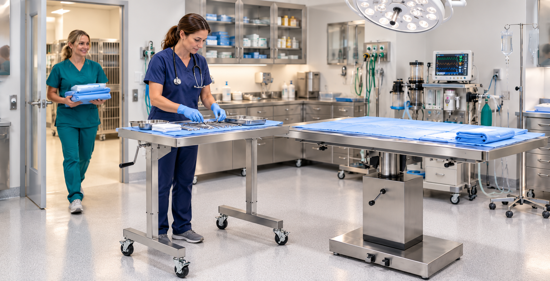
A well-organized surgical suite helps your veterinary team work efficiently and keep essential instruments close at hand.
When you're preparing for surgery, having the right instruments is only part of the equation. Where you place those instruments, how easily your team can reach them, and how well your surgical suite is organized can all influence the flow of a procedure.
A veterinary surgical instrument table offers a practical way to keep essential tools and supplies within reach.
Whether your practice performs routine spays and neuters, soft tissue procedures, or more complex surgeries, a versatile piece of equipment can help you create a more organized workspace.
Featured Equipment: Suburban Surgery Instrument Table
The Suburban Surgery Instrument Table is designed with this in mind.
It has an adjustable-height stainless steel surface, a mobile base, and a raised back rail for easy access to instruments and equipment during procedures.
5 Practical Ways to Use a Veterinary Surgical Instrument Table
Keep Surgical Instruments Organized and Within Reach
During a surgical procedure, the last thing you or your team needs is to waste time searching for an instrument. A dedicated instrument table gives your surgical team a designated place to organize the tools needed for the procedure.
Depending on the surgery, the table can accommodate instrument trays, clamps, scissors, forceps, and other essential supplies.
Arranging instruments in a consistent, easy-to-follow layout helps the team anticipate what the surgeon needs next and keeps the procedure moving smoothly.
Practical Tip: Establish a setup routine that works for your team. And make it repeatable.
Organize instruments by procedure, keep frequently used items accessible, and maintain the sterile field throughout surgery.
Create a More Efficient Surgical Workflow
When instruments and supplies are close to the operating table, veterinary teams can focus more on the patient and the procedure.
The Suburban Surgery Instrument Table can be positioned next to or over part of the surgery table, depending on your setup.
This flexibility makes it easier to arrange your workspace around the surgeon's and surgical team's needs.
For busy practices performing multiple procedures throughout the day, small workflow improvements can really add up.
Of course, the table is just one part of an efficient surgical workflow. Clear communication, appropriate preparation, and established cleaning and sterilization protocols remain essential.
Adjust the Work Surface to Suit Your Team
Veterinary teams come in all shapes and sizes, and not every procedure calls for the same working position. A table that sits too high or too low can make it harder to stay comfortable during a lengthy procedure.
The Suburban Surgery Instrument Table offers a height range of 39 to 62 inches, adjusted using a hand crank accessible from either side.
This lets team members position the work surface to suit their needs and the surgical suite layout.
When planning your setup, consider the height of your primary surgery table, the surgeon's reach, and the space needed for other equipment.
Keep in Mind: The goal is to create a workspace that supports comfortable access without obstructing the surgical team or compromising sterile technique.
Make Better Use of Your Surgical Suite
Space is often at a premium in veterinary practices. Between anesthesia machines, monitoring equipment, patient warming systems, and instrument trays, even a well-designed surgical suite can feel crowded.
A mobile instrument table gives your team more flexibility when arranging the room.
With its 40-inch-wide by 23-inch-deep tabletop, the Suburban Surgery Instrument Table provides room for instruments and supplies without requiring a permanent workstation.
You can reposition the table as needed and move it out of the way when the procedure is complete.
For practice owners planning a surgical suite update, this flexibility is worth considering.
Before You Buy: Measure your surgical area and confirm that the table's dimensions and base clearance work with your existing equipment.
Support a Clean, Professional Surgical Environment
Maintaining a clean surgical environment is fundamental to veterinary patient care.
The Suburban Surgery Instrument Table is constructed entirely from Type 304 stainless steel.
Stainless steel is durable and compatible with many routine cleaning practices when you follow the right products and procedures.
The table also features a raised back rail designed to help prevent equipment from accidentally sliding off the rear of the work surface.
Choosing the Right Surgical Instrument Table for Your Practice
When evaluating a surgical instrument table, think beyond its basic function.
Consider these three important questions:
- How does your surgical team typically perform procedures?
- Which instruments and supplies need to remain accessible during surgery?
- How will the instrument table fit alongside your existing equipment?
A surgical instrument table may not be the most prominent piece of equipment in your surgical suite, but it plays an important supporting role.
Keeping instruments accessible, adapting the workspace to your team, and using available space more effectively can help create a more organized surgical environment.
Looking for a Better Surgical Suite Setup?
The Suburban Surgery Instrument Table may be a practical addition to your veterinary practice.
Explore the equipment details and contact Brad Haven, Jr. with questions about your surgery equipment needs.

America’s Biggest Shelter Projects: 10 Animal Shelters Investing in Future Care
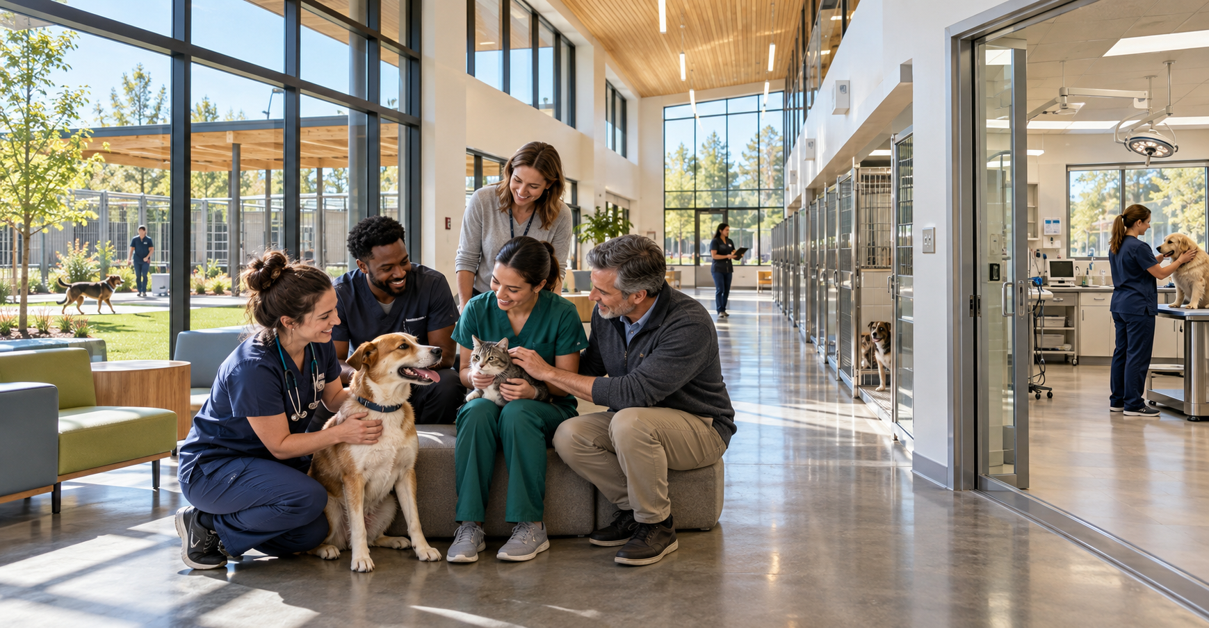
10 Major Animal Shelter Construction Projects Shaping the Future of Shelter Medicine
Animal shelters are changing, and the buildings themselves are becoming a major part of that change.
Across the country, communities are investing millions of dollars in new animal shelters, medical centers, and expanded campuses designed around much more than simply housing dogs and cats.
Modern shelter projects are incorporating:
- Veterinary hospitals and medical clinics
- Isolation areas
- Adoption spaces
- Behavior and enrichment areas
- Community services
As of 2026, here are 10 of the most significant animal shelter construction, replacement, and expansion projects underway across the United States.
1. Maricopa County West Valley Animal Care Center — Glendale, Arizona
One of the largest projects on the list, Maricopa County's new West Valley Animal Care Center carries an estimated $78.4 million price tag. Construction officially broke ground in June 2026, with an opening expected in 2028.
The new campus will include approximately 630 animal housing spaces, a state-of-the-art veterinary clinic, adoption areas, and dedicated spaces for staff and volunteers. Multistudio designed the facility in collaboration with Animal Arts.
2. Orange County Animal Services — Orlando, Florida
At approximately $49 million and 123,419 square feet, Orange County's new animal shelter will be Florida's largest when completed. The two-story facility will house approximately 265 dogs and 200 cats and will feature air conditioning throughout.
Construction broke ground in April 2026, with completion expected in 2027. Borrelli + Partners is part of the design team.
3. Aurora Animal Shelter — Aurora, Colorado
Aurora is replacing its aging 13,000-square-foot shelter with a roughly 42,000-square-foot, two-story facility designed to accommodate more than 250 animals.
The approximately $51.3 million project includes veterinary and medical space, separate intake pathways, adoption areas, and room for future expansion. Funding combines city capital sources, impact fees, federal ARPA funds, and community donations.
Animal Arts designed the facility, with Hausmann serving as the builder. Construction began in June 2026.
4. Bronx Animal Care Center — New York, New York
New York City's Bronx Animal Care Center represents a $92 million investment in a 50,000-square-foot facility. Designed by Marvel, the center will house approximately 70 dogs, 140 cats, 30 rabbits, and 20 additional animals.
The facility will include admissions, adoptions, medical services, and community space. Veterinary services will include a low-cost clinic intended to help financially vulnerable pet owners keep their pets with their families.
Construction is underway, with the opening currently targeted for 2027.
5. Durham County Animal Shelter — Durham, North Carolina
Durham County is taking a different approach by designing its new shelter around population needs, veterinary medicine, and long-term operational efficiency.
The planned $55 million, approximately 37,000-square-foot facility will house 131 dogs and 132 cats. It will also include a fully equipped medical clinic with infrastructure for veterinary surgery.
RND Architects, working with animal-care specialists Animal Arts, is leading the design. Construction is scheduled for completion in 2030.
6. Central Missouri Humane Society Animal Care Campus — Columbia, Missouri
The Central Missouri Humane Society is developing a new $25 million, 30,000-square-foot animal care campus on 17.5 acres.
The project is being funded primarily through a major private fundraising campaign, with $15 million in gifts and pledges secured as of mid-2026.
The campus will expand animal housing, medical care, intake services, and community programming. Construction is targeted to begin in 2027.
7. Olathe Animal Shelter — Olathe, Kansas
Olathe's new shelter illustrates how quickly the cost of modern shelter construction can increase as expectations for animal care evolve.
What began as a roughly $15 million project has increased to approximately $25 million. The new facility will replace a 4,400-square-foot shelter built decades ago that was never designed to meet today's animal-care demands.
SFS Architecture is handling the design, with construction beginning in 2026.
8. Sugar Land Animal Shelter — Sugar Land, Texas
Sugar Land's project combines public investment with private fundraising. The approximately $20 million shelter will include:
- Animal housing
- Veterinary space
- Quarantine areas
- Surgical capabilities
- Adoption areas
- Community classrooms
The project is funded largely through voter-approved general obligation bonds, supplemented by approximately $2.4 million in donations.
PGAL is the design firm, with construction scheduled to begin in late 2026.
9. Jefferson Parish East Bank Animal Welfare & Adoption Center — Louisiana
After Hurricane Ida destroyed the former East Bank facility, Jefferson Parish is building a new 27,775-square-foot animal shelter at an estimated cost of approximately $15.6 million.
The project includes indoor and outdoor animal areas as well as a low-cost veterinary clinic. Construction began following a 2026 groundbreaking, with an opening anticipated in 2027.
Watermark Design Group is involved in the architectural development.
10. Orphans of the Storm — Riverwoods, Illinois
Not every major shelter investment comes from a government capital budget. Orphans of the Storm is replacing its nearly century-old facility with a new 23,120-square-foot animal shelter costing approximately $15 million.
The project is being supported through private fundraising and community donations.
Plans include modern medical and isolation areas, improved air quality, larger kennels with outdoor access, indoor exercise space, and community gathering areas. Construction broke ground in July 2026.
What These Projects Tell Us About the Future of Shelter Medicine
The most interesting thing about these projects isn't simply how much money is being spent. It's where the money is going.
Veterinary medicine is increasingly being designed into animal shelters from the beginning rather than squeezed into whatever space happens to be available. The same is true for animal behavior, enrichment, and welfare.
Staffing and workflow are also becoming important parts of shelter design. Several of these projects are being developed around more efficient workflows so veterinary teams, animal-care staff, volunteers, and adoption personnel can spend less time working around the limitations of an outdated building and more time caring for animals.
Ultimately, these projects point toward a broader shift in animal welfare. The modern animal shelter is becoming a healthcare facility, a community resource, and a lifesaving operation all under one roof.
The building isn't simply where the animals stay. It's becoming an important part of how we care for them.

Diagnosing Bladder Stones in Pets: What You Need to Know
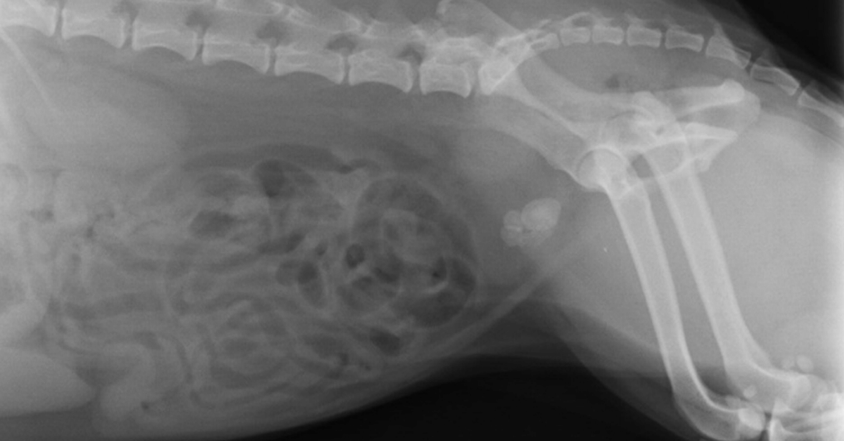
Sometimes, diagnosing urinary bladder stones in dogs and cats is simple: one set of x-rays and the mineral-dense uroliths glow bright white on your viewing screen.
Other times, it’s not so straightforward… especially for small or radiolucent stones.
Here’s how radiographs and ultrasound can be used to help you find pesky, hard-to-view bladder stones.
Are bladder stones on your differential diagnosis list?
Bladder stones may be at the forefront of your mind if you see a dog or cat who’s…
Having blood in their urine.
Urinating more frequently, and in short streams.
Having urinary accidents in the home.
Straining or experiencing pain during urination.
Excessively grooming around their genitals.
Usually, a client will bring their pet into you for these concerns, and your physical exam will help to determine that there’s no urinary obstruction.
With urinary bladder stones, you may notice some discomfort on palpation of the caudal abdomen. On a cat or small dog, you may even feel stones or crepitus in the area of the bladder.
Some patients, on the other hand, may exhibit minimal symptoms and their physical exam may be normal (sometimes bladder stones are an incidental finding).
Either way, most pets will need some type of imaging to confirm that bladder stones are there. Radiographs are a great place to start…
Finding uroliths via radiographs
In addition to any other needed tests—such as a urine analysis or bloodwork—radiographs are often recommended for pets with urinary symptoms, in order to look for uroliths or other abnormalities.
Typically, you’ll start with two simple views of the abdomen—a lateral and a VD.
Take a close look at the urinary bladder to look for radiopaque stones, which should show up as a white opacity relative to soft tissues thanks to their mineral composition.
Stones can range in size from small sand-like grains to more than two inches across. There may be just one or two stones present… or a small group… or even more than 100.
Remember to check the urethra for small stones that could be stuck—especially if the patient is straining or in pain during urination.
For better visualization of the entire urethra in male dogs, consider taking another lateral view with the hindlimbs pulled forward.
Also, check the kidneys and the areas of the ureters. While much less common in pets than in people, occasionally uroliths may be seen higher up in the urinary tract.
If you see stones now—you have your answer.
If you suspect urinary bladder stones but still don’t see them, a contrast study may allow better visualization.
For finding bladder stones, a double-contrast study is ideal.
This includes using both a positive contrast agent (soluble iodinated contrast medium) and a negative contrast agent (room air, or ideally carbon dioxide to reduce the risk of an air embolus) in the bladder together.
Anesthesia or sedation may be needed for the patient’s safety and comfort since the contrast agents are administered via a urinary catheter.
In addition to radiographs, an ultrasound is a useful tool…

Finding uroliths via ultrasound
An ultrasound study is another great option for finding bladder stones, especially radiolucent ones.
The fluid-filled bladder provides contrast for the ultrasound waves so that stones can be found (and often emphasized by acoustic shadowing).
Besides radiolucent stones, you may also see…
Bladder stones (radiopaque or radiolucent) that were too small to visualize radiographically (smaller than 1-3mm).
Other problematic issues in the bladder, such as ‘sludge’ buildup in cats with crystalluria.
Damage to the urinary bladder itself, such as inflammation.
The condition of the upper urinary tract—the kidneys and ureters.
Unexpected findings, such as tumors or anatomical abnormalities of the bladder.
Because of this, ultrasound imaging is a valuable tool for helping you diagnose and treat problems of the bladder, including urinary stones.
Follow-up
Depending on your findings, you may recommend a diet change for dissolvable stones, or a cystotomy to remove the stones.
For dissolution, follow-up imaging can help to track the patient’s progress and see whether or not the stone is dissolving.
When a cystotomy is recommended, remember to use imaging on the day of surgery…
Take pre-op radiographs to confirm the stones are still there, and that your urinary catheter is in place.
Include post-op views to confirm and document that all stones were successfully removed.
Since most stones are radiopaque, standard radiographs are a good option for follow-ups—and typically the imaging choice on the day of surgery.
But ultrasound can also be used in conjunction with other diagnostic tests to monitor the health of the urinary system long-term and to look for early signs of a problem such as a reoccurrence of stones.

Catching stones early, when they’re small, may allow less invasive treatment options such as voiding urohydropropulsion.
With the right combination of imaging modalities, you can help your clients stay on top of treating, monitoring, and preventing urinary bladder stones in their pets.
Written by: Dr. Tammy Powell, DVM
Disclaimer: This article is for general informational purposes only, and not intended as a guide to the medical treatment of any specific animal.

Old X-ray Table and Generator Be Used with a New Digital X-ray System?
Can My Old Universal Easymatic X-ray Table and Generator Be Used with a New Digital X-ray System?

Some of the Universal Easymatic systems look like this
Some veterinarians purchased their X-ray table and generator 25+ years ago, and they still work perfectly!
With proper care and maintenance, they've produced diagnostic images for years using film.
For some veterinarians, 2017 is the year they would like to stop using chemicals and standard film.
However, they don't want to spend 17k-25k on a new table and generator when their old system produces excellent mas/kvp options for each case.
The good news is your existing system can work well with proper installation with your new digital X-ray equipment. For computed radiography (CR), a simple calibration will enable the old table and generator to be ready for use on the day of installation.
The installer should carry out this installation on the day of installation, before the training session with the veterinary staff.
For direct radiography (DR), the plate may be wired into the foot switch as a prep switch. The old universal Easymatic X-ray table and generator send out a 120-volt prep and exposure signal.
Our on-site installers bring a conversion box that uses cesium technology to convert the voltage down to 5 volts (high) and 0 volts (low).
We also include a new foot pedal switch with our system. Oftentimes, the old switch can be worn out and the wiring loose.
A new foot pedal switch ensures a smooth transition from prep to exposure. In some instances, we also include a hand switch, which can be mounted on the wall.
This secondary option is especially useful if the foot switch fails; you always have another way to take a high-quality digital X-ray.
You do not always need to buy a new table and generator to enjoy the speed, safety, and quality of digital X-rays.
Contact us today.
We can help you upgrade your system to digital.
Here is a video showing the conversion from film to digital using the old table and generator.

Buy Veterinary Digital X-ray Equipment with Confidence

How to Buy Veterinary Digital X-ray Equipment without Experiencing Buyer's Remorse
Have you ever bought something and regretted the purchase?
I think I have at some point. However, it’s one thing to regret ordering a cheeseburger and fries, but quite another when I regret buying a $50,000 car or truck.
When I make a big purchase, I want to feel good about my decision. I want to make sure that I made the right choice.
I want you to feel great about buying from me! I understand it is no small thing for a Veterinarian to spend $20,000 or $30,000 on digital X-ray equipment, and I want you to feel great about buying from us.
So, here is my “purchase without buyer's remorse” plan:
The 30-day satisfaction guarantee, or your money-back offer. When you buy a digital X-ray system, I will give you 30 days to feel comfortable and confident you made the right choice.
And if you are not satisfied, return your digital X-ray equipment for a full refund.
Yes, that is correct! A FULL refund!!
I know that after the vet digital X-ray equipment is installed in your clinic, and once you learn how to use the software, you will be very happy with the results you get.
I can offer this guarantee because I'm confident in the quality of the digital X-ray equipment we sell, and I want you to be completely satisfied.
Here is the fine print:
Equipment must be returned in original packaging.
Equipment cannot be returned if the user damages it during the 30-day period.
The buyer pays the cost of shipping/packing/insurance of all returned equipment.
You must give us a reasonable chance to correct any dissatisfaction.
I am offering this “30-day satisfaction guarantee or your money back” to give you peace of mind.
I understand I’m not as big as Idexx, Cuattro, or Sound-Eklin. That may make you think twice about trying us out.
But I am confident that our equipment is as good, if not better, and now we have a “30-day satisfaction guarantee or your money back” - which the big boys do not offer!

The bottom line is simple:
I am offering quality digital X-ray equipment for the veterinary industry at an amazing price, with a great warranty and now a 30-day guarantee.
I want to do the right thing, treat you with respect, and help you be successful in your veterinary practice.

Upgrade to Digital X-Ray with Your Existing Table/Generator

Saying Goodbye to Chemicals and Dark Rooms: The Digital X-Ray Advantage
Advantages of upgrading to digital X-ray:
Faster viewing of X-rays compared to film x-ray.
Elimination of the need for a dark room, saving 20-30 minutes of processing time.
Improved environmental conditions and staff health:
No more purchasing and handling of chemicals.
Elimination of film, storage, toxic chemical fumes, and odors.
Convenient sharing of X-rays:
Sending x-rays to a radiologist via email or PACS.
Easy sharing of X-rays with customers through email.
Enhanced customer service and interaction:
Digital technology allows for easier and more efficient viewing of each X-ray.
More time can be spent with customers, providing better service.
We had one location that had a table/generator which was over 15 years old, and we were able to upgrade to digital, still using his old table and generator.
Money saved!
The Doctor upgraded to a DR system, and we saved him money by using his old table and generator.
We can do the same for you and it doesn't matter if you want a CR or DR digital x-ray system.
TESTIMONY
Here is what the Doctor had to say in an unsolicited email a few days after the installation...
“Image quality is superb! Far better than Sound x-ray we use to have at Bakersfield hospital. We took abdominal X-rays on a 70-pound dog and the x-rays came out great. Thank you! Let me know if anybody wants to visit and see the images...I will be more than happy to show them the images” — Dr. Singh - Family Pet Hospital - Vacaville, CA
Use your current table and generator and go digital - for a CR or a DR system.

Diagnosing Tumors of the Heart in Dogs and Cats

Cancers of the heart are uncommon in dogs and cats. When they do occur, it’s important to differentiate a tumor from other conditions that could cause generalized or focal enlargement of the heart and cardiovascular symptoms.
Which cancers occur in cardiac tissues?
A cancerous lesion based at the heart could be a primary tumor, or due to metastasis.
Primary tumors that occur at the heart include:
Hemangiosarcomas—the most common heart cancer in dogs, usually occurring at the right atrium. These occur most frequently in larger dogs with longer noses, such as Golder Retrievers, Doberman Pinschers, and German Shepherds.
Chemodectomas, also known as aortic body tumors or heart base tumors. Generally thought to be slow-growing, these tumors occur most commonly in brachycephalic breeds like Boxers, Bulldogs, and Boston Terriers.
Myxomas.
Sarcomas.
Ectopic thyroid tumors.
Lymphoma/lymphosarcoma—the most common heart cancer in cats.
Other cancers are possible, but these are the most commonly diagnosed types. Breeds may vary as noted above, but most pets with tumors of the heart are middle-aged or older.
How are heart tumors diagnosed?
If slow-growing, tumors near the heart base are often an incidental finding, seen on thoracic radiographs that are taken for another reason.
Other times, diagnostics are pursued because of clinical symptoms—which are often sudden in onset.
Once a mass is large enough to push on the heart and major blood vessels, many cardiovascular symptoms are possible, such as:
Coughing
Ascites
Lethargy
Weakness
Vomiting
Loss of appetite
Difficulty breathing
Collapse
Sudden death
Note: If a dog presents with some of these symptoms, especially sudden weakness and collapse, a quick ultrasound scan may help to identify pericardial effusion and aid in guiding a needle for emergency pericardiocentesis.
Finding heart masses early
As with most cancers and disease processes, discovering a problem earlier rather than later can allow for more treatment options.
General screening radiographs or ultrasound checks—such as with a senior wellness health check—can be a good opportunity to discover heart base tumors before they cause cardiovascular dysfunction and symptoms.
An echocardiogram performed via ultrasound can help to provide more information on the location, size, and invasiveness of the mass.
In some cases, a presumptive diagnosis may be made based on the appearance and location of the mass on an ultrasound study, along with the patient’s signalment. If possible to perform without undue risk, an ultrasound-guided aspirate of the mass can provide more information about which type of tumor is present.
Treatment options for cancers of the heart
Treatment will be based on the type of neoplasia, how fast the mass is growing, whether metastasis is present, and whether or not the pet is symptomatic.
When a tumor of the heart is diagnosed, a good next step is to screen for metastasis and concurrent conditions via chest x-rays (if not already done), bloodwork, lymph node evaluation, and abdominal ultrasound.
Once a diagnosis is made, treatment options may include:
Periodic monitoring with a cardiologist (especially for slow-growing chemodectomas/heart base tumors) prior to pursuing more invasive treatments.
Surgery to remove the tumor.
Pericardiectomy to remove the pericardium and prevent life-threatening cardiac tamponade or pericardial effusion.
(A pericardiocentesis may be necessary on an emergency basis prior to diagnostics in a pet who presents with acute symptoms. After that, a planned pericardiectomy can help to prevent further emergency episodes of fluid buildup around the heart.)
Chemotherapy, often in conjunction with surgery.
Radiation therapy—either conventional, or via Cyberknife therapy.
Even though cancers of the heart are uncommon in pets, they can be scary to pet owners because of the possibility of sudden onset of serious clinical symptoms (especially with hemangiosarcomas).

By performing diagnostics, referring to specialists as needed, and giving your clients as much information as possible, you can help them make an informed decision for their pet.
Disclaimer: This article is for general informational purposes only, and not intended as a guide to the medical treatment of any specific animal.
Written by: Dr. Tammy Powell, DVM

COVID-19 and Your Veterinary Practice- COVID-19 Safety Tips

What we know right now about pets and COVID-19
Currently, pets are not considered to be involved in the transmission of SARS-CoV-2. A handful of pets around the world tested positive (compared to over 4 million human beings), and it is thought that these dogs and cats contracted the virus from a human.
So, pets may have some risk of being infected from us, but at this time there’s no evidence of zoonosis from pets.
Seeing patients during a pandemic
While veterinary medicine is generally considered an essential service, you’ve probably had to change the number of appointments you see—and the manner in which you see your clients and patients—during the coronavirus pandemic.
The CDC recommends using your professional judgment to provide care to pets while minimizing human-to-human exposure and conserving PPE (personal protective equipment).
This could mean…
Seeing urgent care visits only while postponing routine and elective visits.
Using telehealth consultations in lieu of in-person visits whenever possible.
Requesting that clients call ahead of time to schedule their pet’s visit if possible.
Implementing “curbside” service, where one of your team members picks up the pet from the client’s car in the parking lot. The pet owner stays outside and communicates by phone as you examine and treat their pet.
If clients must enter the facility, insisting they wear a face mask and practice social distancing.
If a client is ill with COVID-19 and their pet must be seen, they should have someone else (a healthy person from outside their household) bring in their pet, and also inform you that their pet has been exposed. It’s okay to ask clients while scheduling an appointment, and upon arrival at the clinic, if the pet has had any exposure to a person with known or suspected COVID-19.
Practice frequent hand washing and disinfection of rooms and equipment.
Have clients leave all unnecessary items (such as toys) at home to reduce the risk of spread via fomites.
In some cities or states, local regulations may have more detailed orders in terms of which visits and procedures can be performed. Check your location’s latest updates and mandates.
The same goes for telehealth consults: The FDA temporarily relaxed some of its telehealth guidelines for veterinarians as of late March 2020. However, that may change as businesses start to resume normal practices—and your state may have different rules.
Employee wellness and etiquette
The CDC recommends that staff members who are ill should stay home. Additionally, any team members who arrive at work with symptoms of illness—or become ill while at work—should be separated from others to reduce exposure, and sent home.
Staff members who are ill should not return to work for at least 10 days, until their symptoms have improved, and they are free of a fever (> 100.4 degrees) for 72 hours without using fever-controlling medications.
Social distancing should be maintained within veterinary practices. And face masks should be worn to prevent the spread of respiratory droplets.
As much as reasonably possible, team members should avoid sharing equipment and workspaces. Any shared or frequently used equipment or spaces should be disinfected often.
If a team member is ill, or sent home due to suspicion of COVID-19, other employees should be informed of possible exposure in the workplace, while maintaining confidentiality (in compliance with the Americans with Disabilities Act).
Employees at a higher risk—such as older adults, pregnant individuals, or individuals with certain underlying health conditions—should limit exposure to clients and patients with confirmed or suspected COVID-19 or a history of exposure.
Screening pets for COVID-19
As of this point in time, the CDC, USDA, and AVMA do not recommend routine testing of pets for COVID-19.
However, if you encounter a pet with relevant symptoms and known exposure to the virus, testing may be indicated after more common causes of the symptoms are ruled out.
Contact your local or state public health official, the animal health official, or state veterinarian, who can guide you through the decision and procedures for testing.
When obtaining test samples (usually oral, nasal, or fecal/rectal swabs), strict PPE and disinfection protocols should be used to avoid exposure to the virus.
PPE availability and usage
PPE may include face masks, gloves, eye protection, surgical caps, and gowns or coveralls. These items play an important part in protecting you and your staff from potential SARS-CoV-2 exposures.
Due to shortages of PPE in the human healthcare setting, it’s recommended that veterinarians look at CDC guidelines for PPE usage, use these resources conservatively, and consider reusable PPE where appropriate.
Limiting appointments—such as deciding to only see urgent care visits and postpone elective procedures—also plays a key role in the conservation of PPE for both human healthcare professionals and veterinarians alike.
Checking for updates
Since this is a novel virus and an unprecedented time in terms of global pandemic procedures, knowledge of the situation and recommendations are constantly evolving.

In order to stay up to date—and to stay in compliance with the latest legal requirements and safety recommendations—look for updates from your local and state authorities.
Check the CDC, USDA, and AVMA for more details regarding these recommendations—and be sure to follow them for updates.
Also, look to the AVMA website for additional resources, such as financial and wellness strategies for veterinarians.
Written by: Dr. Tammy Powell, DVM

How COVID-19 Affects Pets: Safety Tips for Pet Owners

Veterinarians play an integral role in public health. Your education helps you understand zoonotic diseases and other health concerns that can affect both humans and animals.
And even though pets don’t appear to play a significant role in transmitting coronavirus (SARS-CoV-2) in the current global pandemic, your knowledge can still help both people and pets.
Here are some important things we know so far about COVID-19 in cats and dogs, and how that knowledge can affect your team, your clients, and your patients…
Can dogs and cats get COVID-19?
So far, there have been millions of cases of novel coronavirus infecting human beings…
However, the disease was only documented in a handful of pets worldwide.
Of the pets infected with SARS-CoV-2, many did not exhibit symptoms—which indicates pets could be a dead-end host for the virus.
Between dogs and cats, cats seem more likely to develop clinical disease—although this is based on only a small number of confirmed cases and knowledge in this matter is still evolving.
Ferrets may also be at risk.
And besides housecats, tigers at a New York City zoo have tested positive and shown clinical symptoms of COVID-19.
According to the CDC, clinical signs of SARS-CoV-2 in pets are not yet well defined, but may include respiratory or GI symptoms, as well as fever and lethargy.
Can humans get COVID-19 from dogs and cats?
Right now, there’s no evidence that humans can contract COVID-19 from their dog or cat. The risk of zoonosis is considered low.
If anything, it’s the other way around—pets who have been infected probably caught the virus from their human companions.
For that reason, the CDC recommends that if a person is sick with COVID-19, they should…
Self-quarantine from their dog or cat, for the standard, recommended quarantine time. Try to stay in a separate room and avoid petting, cuddling, being licked, and sharing food or bedding.
Ask another household member to take care of the pet during their quarantine, if possible.
If the person must care for their own pet, they should wear a face mask and wash their hands before and after interacting with their pet.
Worried pet owners should call a vet practice before coming in. A telehealth consult may be recommended in lieu of an in-person visit.
And of course, besides being infected themselves, it’s also possible that a non-infected dog or cat could act as a fomite—for example, if their fur was exposed to respiratory droplets from an infected person.
Testing for SARS-CoV-2 in pets
At this time, the CDC doesn’t recommend routine testing of dogs, cats, or other companion animals. Instead, the need for testing must be determined on a case-by-case basis.
Here are some factors to consider…
Testing is available for mammals only.
A thorough history should be taken to assess for a possible exposure within 2 weeks prior to symptoms.
Other, more common causes of these symptoms should be ruled out first.
If you feel testing is needed, contact your state public health veterinarian or state animal health official to discuss testing options—and they should be immediately contacted again if the test is positive.
According to the USDA, positive samples from private veterinary laboratories require confirmatory testing at USDA’s National Veterinary Services Laboratories.
The OIE (World Organization for Animal Health) recommends testing via RT-PCR on oral, nasal, and fecal/rectal samples, while carefully avoiding contamination from the environment or people during the process.
Currently, Antech and Idexx both offer PCR testing options.
And in April of this year, Idexx announced the availability of a commercial test for veterinarians to use (although they recommend consulting with public health authorities prior to ordering the test).
Treatment for COVID-19 in pets
For many pets, care at home may be the best option. This will depend on the owner’s ability to safely provide care for and quarantine their pet—and household members not being at high risk due to age or health conditions.
In some cases, isolation at a veterinary clinic or hospital may be preferred.
Just like human beings, there’s no specific cure or preventive vaccine for pets for SARS-CoV-2… so, treatment is primarily supportive care.
How to help clients during this time
If any of your clients are concerned, you can help present the facts and avoid the spread of misinformation.
Consider sending out a newsletter—that will not only update clients with accurate information but also help them stay bonded to your practice during social distancing.
For the vast majority of pet owners, you can reassure them there’s no reason to abandon pets… On the contrary, due to stay-at-home orders and staffing limitations at animal shelters, right now is NOT a good time for anybody to abandon a pet they care about.
In general, it’s best to treat a pet as if he or she were a person—they should practice social distancing, too, and avoid more contact with people and other pets outside the home than necessary…
Also, consider stocking up on two weeks of supplies (food, medicine, etc.) for pets, just as you would for human members of the household…
Note that pets differ from humans in that they should NOT wear face masks.
And, you can advise your clients on best practices for veterinary visits during this time—including whether to come in now or wait until later for routine care … what to expect when they arrive at your practice parking lot … which viral testing options are available … and anything else they need to know.

In our next blog post, we’ll have some information about animal hospital protocols and COVID-19. Until then, best wishes and stay safe!
Disclaimer: This article is for general informational purposes only, and not intended as a guide to the medical treatment of any specific animal.
Written by: Dr. Tammy Powell, DVM

Ultrasound vs. X-Rays for Monitoring Dog and Cat Pregnancies

When it comes to pregnancy in pets, sometimes it’s hard for clients to wait…
They want to know right away whether there are puppies/kittens present… how many there are… and if the babies are doing OK.
It’s understandable—whether this is a professional breeder or a pet parent who’s decided to proceed with a litter, the arrival of new puppies and kittens is an exciting time.
There are a few ways to check for pregnancy in dogs and cats. Each method has its own pros and cons, which you can explain to your clients to help set up realistic expectations and develop a plan that best suits their pet’s needs.
Early pregnancy checks may include…
Abdominal palpation
This method of pregnancy detection can yield information as soon as 3-4 weeks into the 9-week pregnancy.
Abdominal palpation has been used by vets for many years—and can often result in the detection of fetal “bumps.”
However, this method isn’t always accurate. There are plenty of things that could render palpation inconclusive. For example, in large breeds or overweight dogs, palpation is more difficult.
Relaxin testing
Testing for the relaxin hormone—a hormone unique to pregnancy—can be performed as soon as 20 days into gestation.
This test gives your client a very important “yes or no” answer—and helps to distinguish between true pregnancy versus pseudopregnancy—but it doesn’t provide information on how many fetuses are present.
The next test to consider is an abdominal ultrasound.
Canine and Feline Abdominal Ultrasound
The procedure of abdominal ultrasound in veterinary medicine involves examining the internal organs of dogs and cats. To begin the ultrasound study, transverse scanning is performed to locate the urinary bladder and colon, with the uterine body positioned between them.
Once the uterine body is identified, the scan is directed cranially towards each kidney, following the uterine horn towards the ovary. Along this path, each fetus can be identified by its appearance as a round, fluid-filled gestational sac.
Compared to other imaging methods like X-rays, abdominal ultrasound offers several advantages in veterinary practice.
One significant advantage is the ability to confirm pregnancy several weeks earlier than X-rays. Typically, an ultrasound is performed around 30 days into the pregnancy, although it's possible to detect a pregnancy as early as 20 days. At 30 days, it becomes easier to detect fetal heartbeats.
In addition to early pregnancy detection, ultrasound provides other valuable benefits for veterinarians, including:
1. Counting the number of puppies or kittens: The ideal time for this is between 30 and 35 days of pregnancy. After 50 days, the uterine horns may overlap, making it more challenging to accurately count the fetuses.
2. Assessing fetal viability: Ultrasound can confirm the presence of viable fetuses by detecting their heartbeats or movement.
3. Predicting gestation duration: Measurements of fetal size or certain anatomical structures can help estimate the gestation duration.
Another advantage of abdominal ultrasound in veterinary practice is its ability to involve clients in the process. Unlike X-rays, clients can be present during the ultrasound, allowing them to witness the heartbeats of the puppies or kittens.
This experience enhances the bond between the client and their pet, as well as strengthens their connection to the veterinary practice.
However, in the later stages of pregnancy, X-rays also play a valuable role in veterinary medicine. X-rays provide more information when the fetuses have developed significant skeletal structures, usually around 45 to 55 days after breeding.
At this stage, a single set of X-rays is safer for the fetuses compared to earlier in the pregnancy.
Radiographs (X-rays) are particularly useful for:
1. Confirm the fetal count.
2. Assessing the size ratio between the fetal skulls and the pelvic canal. This information helps in planning for potential dystocia and the need for a C-section.
As a veterinary practitioner, you have various diagnostic tools at your disposal to detect and monitor a healthy pregnancy in dogs and cats.

By utilizing the best combination of methods, you can reassure your clients along the way, and increase the chances of a healthy delivery of puppies or kittens!
Written by: Dr. Tammy Powell, DVM

Radiographs for CCL Injuries and TPLO Surgery

When it comes to cranial cruciate ligament (CCL) injuries in your patients, x-rays play a key role in your diagnosis, treatment, and follow-up.
So whether you’re the surgeon performing the procedure or a GP referring your patient to a specialist, having a good understanding of what to look for on your radiographic images can help you provide great patient care.
It all starts when the patient first comes in to see you…
The initial visit
More than likely, you’ll be seeing a middle-aged, medium or large breed dog who suddenly started limping on one of their hind limbs.
During the exam, you determine that the pain is in the stifle. Then, using sedation, you perform more thorough palpation, followed by radiographs.
Your detailed exam of the knee reveals some swelling and a positive drawer sign, and you take x-rays to confirm your suspicion of a CCL tear.
Considering this is one of the most common orthopedic injuries in dogs, you’re probably correct. However, occasionally there may be a surprise—such as a fracture near the stifle, or even an osteosarcoma lesion for older patients.
Radiographs will help you correctly diagnose and document the problem, and rule out concurrent conditions.
With a recent CCL injury, you might not see a lot of obvious evidence on your views—that’s because it can take more time for radiographic evidence of a CCL problem to show up.
However, you will likely see compression of the infrapatellar fat pad—which indicates joint effusion.
If the condition is chronic, you may notice arthritis and other signs of long-term inflammation.
Planning for a TPLO surgery
If your patient is a good candidate for surgery, then it’s time to discuss options with the owner. A tibial plateau leveling osteotomy is a good option for many otherwise-healthy dogs, but of course, the recommendation may vary for each individual pet.
Now’s the time when pre-op radiographs come in, by helping you to measure the tibial plateau angle and determine the saw blade size and position of the bone plate and screws.
The importance of post-op radiographs
After a surgery such as a TPLO, it’s important to document that everything went well—that all implants are in place, the tibial plateau has been repositioned as planned, and that there were no complications such as bone splintering.
This will be very reassuring to a worried pet parent, and will also be a valuable part of your medical record.
These post-op radiographs can also provide more information if a problem or complication comes up later, by using these initial images as a frame of reference for comparison.
Radiographs are useful if complications arise
More than likely, your patient is doing great. In that case, follow-up x-rays will help you confirm and document that the bone has healed as expected.
However, if they do run into any problems—such as an infection or a fracture—an additional set of radiographs can help you diagnose the issue and treat it ASAP. And follow-up rads will allow you to monitor the patient’s healing progress and their response to treatment.
Basically, radiographs give you an “inside view” throughout the whole process of diagnosing and treating a CCL injury, and are useful for common surgical options such as a TPLO.
Digital radiographs are especially helpful in terms of sensitivity for soft tissues such as a joint space, and also for providing rapid results within seconds.

But no matter which radiology equipment you choose, your X-ray images will provide valuable information for diagnosis and treatment of CCL—for happy patients and clients.
Disclaimer: This article is for general informational purposes only, and not intended as a guide to the medical treatment of any specific animal.
Written by: Dr. Tammy Powell, DVM

Vertebral Heart Score: How and Why to Calculate One

A vertebral heart score (VHS) is a measurement used to determine if a patient’s heart is enlarged.
The VHS can provide valuable insight to complete your clinical picture, especially at times when you hear a heart murmur, for a general pre-anesthetic check, or to look for cardiac changes on breeds that are predisposed.
It’s a simple, non-invasive way to get more information and to monitor for changes over time.
How to calculate a vertebral heart score
Obtain a high-quality lateral thoracic radiograph. Be sure…
To collimate.
That the patient is straight and not twisted or at an angle.
That you can see enough detail, especially the thoracic vertebrae and the borders of the cardiac silhouette.
Make your first measurement along the longest axis of the cardiac silhouette, from the ventral border of the carina of the mainstem bronchus to the apex of the cardiac silhouette.
You don’t need to measure this in centimeters or inches—instead, just hold up a sheet of paper to your x-ray viewer and make a mark for your measurement, or use calipers. Or, with your digital x-ray system, use the VHS function to mark the measurement right on your screen.
Now, hold the measurement up to the vertebrae, starting with the cranial edge of T4—this is the 4th vertebra to have a rib connected. Count the number of vertebrae (including the vertebral body and the following disc space) that fall into the long axis measurement.
Measure to a decimal place of 0.1 (for example, let’s say your first measurement was 5.2 vertebrae).
Make your second measurement. This time, you’ll measure along the short axis, which is the widest part of the cardiac silhouette. It should be perpendicular (at a 90-degree angle) to your first measurement.
Repeat the process of transferring this measurement to the thoracic vertebrae, starting with the cranial edge of T4. For this example, let’s say your measurement was 4.5 vertebrae.
Add these two numbers together.
In this example, your VHS would be 5.2 + 4.5 = 9.7
How to interpret the results
For dogs, a normal vertebral heart score is less than 10.7 (with an average range of 8.5 to 10.5). For cats, an average VHS is 7.5 (with a range of 6.8 to 8.1).
Anything larger than that is considered abnormal.
Of course, this doesn’t tell you exactly what’s going on—you’ll need further information such as an echocardiogram to determine whether there is DCM, HCM, or some other issue causing the enlargement of the cardiac silhouette.
However, the VHS is a great screening tool to let you know to investigate further. And, if you have a patient who already has cardiac disease, a VHS can help you monitor their disease progression over time, and provide tangible information to a client who wants to know how their pet is doing on treatment.
Keep in mind that, as with any diagnostic tool, there is some individual variation. Some pets—especially certain breeds such as boxers—may fall outside the normal VHS range even if they’re completely healthy.
So, look at the overall clinical picture when assessing a patient. But with that in mind, a VHS is a quick, easy, valuable addition to your toolkit when it comes to evaluating and monitoring cardiac health.
Vetebral heart score tool video

Disclaimer: This article is for general informational purposes only, and not intended as a guide to the medical treatment of any specific animal.
Written by: Dr. Tammy Powell, DVM

How to X-Ray a Cat Safely Without Sedation or Anesthesia

If you’ve ever tried to radiograph a fractious cat without sedation or anesthesia, then you know what a difficult challenge it can be.
In addition to blurry, crooked, non-diagnostic images, maybe you or your team members received some scratches and scars to show for the experience.
So, it’s understandable you might not want to try that again—both for your safety and for the cat’s safety.
However, there are some situations where you may want to avoid sedation or anesthesia—or, if it can’t be avoided entirely, to use the lowest dose possible to get the results you need—during an x-ray study on a feline patient. For example…
Cats with advanced heart disease or other conditions that could make sedatives and anesthetic drugs dangerous.
Patients with kidney disease or other conditions that cause less predictable metabolism of anesthetic drugs—possibly resulting in a cat that’s excessively drowsy for a whole day, and an unhappy client.
A patient who’s otherwise friendly and great to work with, just very wiggly for their x-ray shots. A few tweaks may make the experience more comfortable for them and make sedation unnecessary.
Contrast studies such as a barium GI study, where the patient has to be awake to avoid aspiration of the barium.
Of course, sometimes sedation or anesthesia may be safer, decreasing the kitty’s stress and risk of injury.
Only you know what’s best, using your clinical judgment. However, if you do have a situation where it’s worthwhile to try the radiographs WITHOUT sedation or anesthesia, here are a few tips…
Tips to get diagnostic radiographs on an awake cat
Practice feline-centered protocols from the beginning.
Have team members use quiet voices throughout the clinic, especially near the x-ray room or area.
Use pheromone products, including diffusers or spraying the products directly on towels your patient will be in contact with.
Lock the door to the x-ray room or area if possible. That way, no one will walk in by surprise and cause your patient to suddenly look in the direction of a swinging door.
Give the cat a place to hide between views and try to complete the views as quickly as possible. Less time is usually better when it comes to felines. Try a test view to confirm your settings, and then either use digital radiographs or if using film shoot all views at once and then develop them while the cat is resting comfortably in a kennel or carrier.
If the patient is just too stressed that day and it’s not an urgent matter, consider rescheduling. Sometimes starting fresh—having the kitty return first thing in the morning and completing the radiographs quickly—is better for you and the patient (and the client may feel better about it when they see the decreased stress level in their pet).
Make the cat as comfortable as possible during radiographs. For example, use a trough for VD views—it’s a lot more comfortable for them than having their spine right up against the x-ray table.
Remember the machine’s noise. Many cats are fine during positioning, but then panic, wriggle and squirm once they hear the whirring and “beep” that happen when you push the button to take the shot. To avoid this problem, try talking to the cat in soothing tones, or have another background noise such as a white noise machine or calm music. That way, the sudden noise from the machine won’t stand out so much compared to the other background noises.

Remember, making the whole experience less stressful for the cat can also make everything less stressful for you and your staff.
So if possible, making a little extra effort to accommodate a feline patient can help you get diagnostic views more quickly, with a better time for everyone.
Written by: Dr. Tammy Powell, DVM

What Is an Over-Exposed X-Ray, and Why Does It Matter?

In the last blog post, we talked about under-exposed radiographs and how to get better results.
But, it’s important to remember that overcompensating too far in the other direction can give you a problem in the other extreme: that is, over-exposed images.
This is basically the opposite of an overly white or light radiograph. Instead, the image may appear “burnt out,” and overly dark or black, making it difficult to see all the information you need to make your diagnosis.
Over-exposed radiographs can make your life difficult because of the obliteration of normal anatomy
An over-exposure usually means the x-ray beam was too powerful, and that a larger percentage of x-rays passed through the patient’s tissues to reach the film or plate. So, rather than seeing information about the patient, you end up with a blackened image with not nearly enough detail.
Some structures, such as small blood vessels in the thorax, might not be visible at all.
In other words, over-exposure could cause you to miss important lesions or interfere with your interpretation of normal anatomical structures.
Reasons why your radiographs might be over-exposed
Here are some possible culprits…
An error in technique (kVp or mAs settings).
A machine or equipment error.
Using grid technique without a grid.
Variations in screens.
Too short of a distance between the x-ray source and the film or plate.
Also, scatter radiation can contribute to darkening an already over-exposed image.
What to do about over-exposed images
To decrease your radiograph procedure time and minimize x-ray exposure to patients and staff, it’s best to get your shots right the first time. But if your images are overexposed, try…
Using a “hot light.” Sometimes, an overexposed radiograph can still be read by holding it up to an extra bright light known as a “hot light.”
However, if that doesn’t work, you will probably need to retake the radiograph. Here are some tips for getting a better shot the next time…
Adjust your settings. Typically, lighter images can be obtained by lowering the kVp or mAs.
Check for errors in technique, such as using or not using a grid setting.
Collimate. That way, you reduce the area of the primary beam and reduce scatter radiation, giving your image an overall better quality.
Measure patients in the position they will be in during the shot. For example, when taking a lateral thorax view, the thorax may be thinner when the patient is laying on their side than it was when they were standing up. If that happens, your settings would probably be too high based on the measurement obtained from a standing patient.
Also, be sure to follow these general tips for producing good X-ray images…
Use technique charts for a good starting point for exposure settings. A good technique chart will remove much of the guesswork.
Keep your equipment in good repair, including routine maintenance as necessary.
Consider digital radiography, which can take film development out of the equation, and shorten the waiting time between views (allowing you to adjust settings prior to taking more shots).
Use appropriate patient restraint to minimize artifacts from movement.
With practice, good quality equipment, and a few tips for adjusting your settings… Getting a properly exposed image doesn’t have to be hard!

And getting the right exposure will help you save time, avoid the headaches of struggling with poor-quality images, and get to your diagnoses faster.
Disclaimer: This article is for general informational purposes only, and not intended as a guide to the medical treatment of any specific animal.
Written by: Dr. Tammy Powell, DVM

How to Talk to Pet Owners about Radiographs

“Does he really need an x-ray? Can’t you just give him some medicine and see how he does?
“Why is this so expensive? You just want to make money while my cat is sick!”
As a veterinarian, you’ve probably heard all of this and more.
Of course, most clients want the best for their pets, and many will understand why you want to perform certain tests like radiographs. However, even the most loving of pet parents may show some hesitation or “sticker shock” when you present the estimate…
Often, this isn’t personal. Many pet owners just aren’t prepared for a sudden expense, even if they want to do the best for their furkids. Here are some tips that may help take the conversation in a more positive direction…
Stay calm during the initial reaction
If an owner’s initial response is similar to the examples listed above, try to ignore the knee-jerk reaction to defend yourself. Get comfortable with allowing a moment of uncomfortable silence.
Often, clients will try to fill the silence themselves and offer more of their thoughts and questions. But even if they don’t do this, a quiet moment may give everyone the chance to take a breath and start over, calmly.
Don’t pile onto an owner’s guilt
Of course, you’ll need to explain why the radiographs are important for your treatment decisions, and what dangers exist for the pet if radiographs are delayed.
However, if a pet owner understands this and is just having trouble affording the x-rays, they’re probably already feeling guilty. This causes them to lash out.
In this case, having empathy during the conversation can really help. Think about your tone of voice, body language, and anything else that may help elicit a productive conversation.
If you make an owner feel understood and accepted, they’re likely to come back to you as soon as they CAN afford the tests, rather than seeking care elsewhere.
Bundle your radiographs when possible
Often, estimates are presented as “a la carte,” with different options presented by cost, line by line.
While this may work well in some situations, the client may try to pick and choose, asking which items can be eliminated in order to decrease the cost of treatment.
Instead, try estimates that list a total cost, with radiographs included. Some situations where this may work well include:
Dental procedures with dental rads.
Senior wellness testing, with bloodwork and screening rads.
A blocked cat, with x-ray views included as part of the treatment plan.
For three-view thoracic radiographs, bundle them together rather than listing the individual “per view” charges separately.
Of course, you can still list each item of the treatment plan, so the client knows the value they are receiving. But, list it as a total cost, rather than an itemized estimate.
Explain the benefits and limitations of radiographs
Many pet owners nowadays, especially Millennials, want to be fully involved in their pet’s care—that’s part of the reason why so many owners seek answers from Dr. Google prior to coming for a vet visit.
Those clients will want to receive answers about their pet’s condition right away—so, let the client know that radiographs will help to provide the answers they’re looking for. And since radiographs are available the same day, they can get answers to their concerns very quickly.
On the flip side, also prepare an owner that a radiograph might show normal results. If the pet is feeling well, then the client gets peace of mind. If the pet is ill, further testing may be required—by preparing the owner for this possibility ahead of time, you can avoid upset reactions as much as possible.
Ask about their biggest concern
Even though you probably already know the biggest concerns and questions clients have about x-rays, it never hurts to ask an owner why they’re hesitant. They may appreciate you listening to them, and it saves you time so you can address their biggest concern.
Some examples you may encounter include:
Finances. In this case, you can suggest third-party financing options, such as Care Credit.
Fear for their pet’s comfort, or having to leave their pet at the hospital. Here, discuss the procedure. If sedation is used, explain how this can minimize pain. Or, if digital radiographs are used, explain how this allows the views to be done fast so the pet can go home sooner.
Show clients the value you offer, even after the x-rays are finished
Be sure to go over the results of the radiographs with your client. Try using a quiet exam room rather than the treatment area, so they won’t be distracted.
Give the client a brief “x-ray orientation,” so they know which end is toward the pet’s head and what is abdomen versus chest, etc. Then, explain your findings.
Whenever possible, bring a normal x-ray for comparison, so clients can really see the difference with an abnormal finding.

If you have digital images, share a copy with the client that they can look at after the visit. Offer to let them record you talking about the findings. That way, when the client goes home and shares the findings with their partner or family, everyone is more likely to understand and be on board with your recommendations.
With a little planning of the whole x-ray experience from the client’s perspective, you can make things easier on them—while at the same time, increasing compliance and getting better care for the pets you see.
Written by: Dr. Tammy Powell, DVM

Identifying Trauma in X-Rays of Hit-By-Car Patients

It’s been a slow day, and you just got back from lunch. Your first-afternoon appointment is waiting, and you look through their chart, preparing to go into the room…
All of a sudden, the clinic doors burst open. You hear a big commotion upfront…
You walk up to see what happened and discover a distraught pair of pet owners holding carrying their Border Collie, who’s just been hit by a car.
How to avoid further surprises…
Of course, as a veterinarian, you’re probably used to something like this happening from time to time—panicked pet parents arriving at your clinic with a dog or cat in need. And you’re probably well-prepared to triage and decide what to do next…
However, you’ll want to avoid unpleasant surprises down the line—liked missed diagnoses—by ensuring that you find and plan for unexpected injuries.
For example, maybe that Border Collie has an obviously broken leg and some nasty road rash.
You’ll address those injuries, but first, you’ll explain to the owners that there are other conditions you need to check if their fur kid has just been hit by a car—conditions that could be life-threatening.
What to look for on the x-rays
Your physical exam will help direct you on what to look for. For example, if you feel a painful joint with crepitus, or hear crackles or decreased lung sounds when you auscult the chest, those will be areas you want to explore with radiographs.
But, even if the lungs sound fine and you don’t find any other obvious abnormalities, it’s always good to evaluate the thorax and abdomen radiographically.
Sometimes, x-rays will need to wait…
For example, if the pet is in critical condition, they need stabilization first. And, you’ll use your judgment on whether sedation is needed and safe, for clearer and more diagnostic x-ray images, as well as for relief from pain and stress.
Then, when you read the radiographs, it’s important to evaluate EVERYTHING on your set of x-ray images.
The impact of being hit by a car can cause trauma to many different parts of the body all at once, including serious internal injuries.
Here are a few things to check for on your radiographs…
Pulmonary contusions. A strong trauma to the thorax can cause dangerous bleeding in the lungs, often visible on radiographs as alveolar or interstitial opacities.
A ruptured bladder and uroabdomen. Look for signs of fluid in the abdomen, especially if you notice bloody urine. Try to visualize the bladder.
Internal bleeding or damaged organs. If an organ such as the spleen has been injured, you may notice a hemoabdomen (although radiographically, this would be difficult to distinguish from a uroabdomen). In the case of ruptured intestines, you may notice spots of free air in the abdominal cavity.
Broken bones, including the ribs and vertebrae. Visually trace along the bones one-by-one to be sure no lesions are missed.
Dislocations, such as a dislocated hip. Also, check for tail-pull injuries, especially in cats who may get their tail caught under a car’s wheels.
Diaphragmatic hernia. Look for evidence of intestines or other abdominal organs in the thorax.
Air or fluid in the pleural space. Seen as gas opacity, or fluid opacity.
Skull and jaw radiographs if needed—in case of suspected head trauma.
Of course, other abnormalities are possible, too. As you know, pets don’t always “read the book” and clinical practice can be full of surprises! If you’re unsure of something on the films or digital images, it may be good to recheck it down the line to be sure no problems are brewing internally.
The number of views you take will vary depending on the size of the pet.
For emergency situations such as this, it may be worth having digital radiographs, for faster results without waiting for films to develop.
Also, it’s important to take orthogonal views. That way, you get the full pictures and lesions are less likely to be missed.
Re-evaluate and repeat radiographs as needed
This will depend on what you see on the first set of images, in addition to how the pet is doing in real life. Use your best clinical judgment to determine if and when repeat radiographs are needed, and keep the pet under close observation.
If there’s any doubt, consider using contrast studies to obtain more information, too.
By developing a plan for radiographs—and remembering to check everything on the images—you’ll increase the chances of finding unexpected injuries and addressing them earlier. Or, in case the radiographs are normal, you may be able to give more peace of mind to the worried pet owners.

Disclaimer: This article is for general informational purposes only, and not intended as a guide to the medical treatment of any specific animal.
Written by: Dr. Tammy Powell, DVM

Hands-Free X-Rays: The Next Step in Veterinary Safety

Is it time to put restraints on veterinary radiography?
By Julia Bitan, RVT
When Dr. Debrah Berman, a Thornhill veterinarian with over 30 years of experience felt a lump in her tongue, she didn’t think much of it.
Up until that point, her health had been excellent and she was following all current radiology safety regulations such as monitoring her dosimeter values and using all protective equipment.
The diagnosis of Mucoepidermoid Carcinoma of the salivary gland (a type of cancer often associated with excess radiation exposure) came as a shock and made her start questioning the safety of today’s veterinary radiography.
“Since the 1990s, numerous jurisdictions in North America, and worldwide, have strengthened their regulations and best practice guidelines to actively move away from holding patients, in recognition that exposure to scatter radiation poses a risk.”
Outdated regulations
Although there are several provincial and federal laws in place to protect radiation workers in Ontario, the clauses specifically concerning veterinary workers have not been updated since 1990.
According to a veterinary radiography survey conducted in partnership with the Ontario Association of Veterinary
Technicians (OAVT) in April of 2016, 8 out of 10 RVTs still hold their patients during x-rays most of the time, while 7 out of 10 respondents choose to sometimes forgo the use of gloves and other protective equipment simply due to inconvenience.
Our human radiology counterparts in Ontario are rarely present in the room when x-rays are taken because regulations state that no person should regularly perform manual restraining for x-rays.
Technicians commonly request parents hold their own children if x- rays are required.
Since the 1990s, numerous jurisdictions in North America and worldwide have strengthened their regulations and best practice guidelines to actively move away from holding patients, in recognition that every x-ray taken increases the overall risk of cancer.
Because we don’t see regular reports of veterinary workers dying of radiation exposure, we may naively assume that modern science has overcome the dangers with newer machine design and intensive research.
In reality, current occupational exposure limits are derived from decades-old research and we know that the risk of getting cancer increases with every exposure.
Recent findings do show that extended exposure to a low level of radiation increases the risk of developing leukemia, while radiation-induced cataracts are observed at a much lower radiation dose than previously believed. In veterinary medicine, the damage caused by ionizing radiation is simply too low to be felt right away and the ALARA principle (As Low As Reasonably Achievable) is much too ambiguous in the context of our profession.
Change is happening
A small number of veterinary clinics in Ontario, including Toronto Veterinary Emergency Hospital and Referral Center (TVEH), began enforcing a strict no-hold, out-of-the-room x-ray policy to keep their veterinarians and RVTs away from ionizing radiation emitted by the x-ray machines.
TVEH started enforcing the 100% -out-of-the- room radiography six years ago.
RVT Ashley Jenner, head of radiology at TVEH, has developed a number of tools and techniques to make out-of-room x-rays possible.
“Non-manual veterinary radiography is much easier and faster than most people believe,” Jenner says. “All it takes is proper techniques and some extra tools.”
Jenner uses positioning devices alone on 75% of her cases (non-sedated patients), and sedation on the remainder.
Dr. Debrah Berman is now “cancer-free” and back to practicing veterinary medicine. She now endeavors to obtain all her x-rays using positioning/restraining devices or sedation.
“Radiation exposure is cumulative,” says Berman. “You may not realize until 20 or 30 years down the road that you have received too much. It may take a little more effort, but had I known that I would need to have a quarter of my tongue removed because of cancer and that I'd have permanent nerve damage and varying degrees of chronic pain, I wouldn't hesitate. And, I am one of the lucky ones. We need to protect ourselves”.
Transitioning to non-manual radiography is not always an easy task.
Change is rarely welcomed in our profession and as long as the minimum standards are followed, there is little reason or incentive to change.
We do, however, know there are risks involved with radiation, and alternatives do exist.
A program was created to guide clinics through transitioning to non-manual radiography using different tools and techniques.
The Hands-Free X-Rays Initiative aims to promote awareness and encourage change to current veterinary radiography.
For more information about the program, visit our website - www.handsfreexrays.com.
For comments, feedback or help to bring change to your clinic, please contact us at:
email: info@handsfreexrays.com
Phone - +1 (647) 502-4843.
Written by By Julia Bitan, RVT


Your Warranty and Service Coverage for Veterinary X-Ray

Today, every veterinarian recognizes the importance of incorporating digital X-rays into their animal hospitals.
A considerable number of them plan to transition from traditional film and chemical X-rays to digital systems this year.
While the initial concern typically revolves around the cost of acquiring a digital X-ray system, it is equally crucial to inquire about the warranty and its coverage.
Here are some pertinent questions to ask the prospective vendor when considering the purchase of a veterinary digital X-ray system:
1. What is the duration of the hardware warranty for my veterinary digital X-ray plate?
2. How long does the software warranty for my veterinary digital X-ray software last?
3. For how long will I receive software upgrades for the X-ray system used in my animal hospital?
4. How long will my veterinarian technicians have access to tech support, enabling them to seek answers to any queries that may arise during the daily implementation of digital X-rays in my animal practice?
5. What are the costs associated with continued phone support, software upgrades, and technical assistance after the warranty period ends?
6. Which entity is responsible for providing software and technical support? Who developed and owns the software?
7. Does the warranty cover labor and shipping expenses, as needed?
8. In the event that my X-ray plate requires repair, will a temporary replacement plate be provided?
It is common for vendors operating in the animal health sector to offer a minimum of a 1-year warranty.

Not all vendors possess ownership of the software that operates their veterinary digital X-ray plate, which holds significant importance. While a digital X-ray plate can remain functional for many years, the software it relies on may become outdated within a short span of time.
An example of this can be observed with Windows XP platforms. Unless companies continually update their software, it becomes incompatible with new computing platforms.
When veterinarians transition from computed radiography (CR) to direct radiography (DR), the predominant issue they encounter is not the failure of the CR system itself but rather the poor performance or lack of support for the software.
This issue is not limited to small companies alone. Renowned brands like Idexx and Sound have discontinued support for their older product lines by ceasing software upgrades.
Our aim is to offer you transparent service and support. Our animal health software has been developed in-house by JPI which has been manufacturing x-ray equipment, writing software, and providing telephone support for these products since 1994.
Here are the responses to the eight frequently asked questions:
1. What is the duration of the hardware warranty for my veterinary digital x-ray plate?
- The hardware warranty lasts for 5 years.
2. How long is the software warranty for my veterinary digital x-ray software?
- The software warranty is also valid for 5 years.
3. For how long will I receive software upgrades for my x-ray system to be used in my animal hospital?
- You will receive software upgrades for 5 years.
4. How long can my veterinarian technicians receive phone support for any questions they may have regarding the daily implementation of digital X-rays in my animal practice?
- Our tech support helpline will be available for your veterinarian technicians for 5 years.
5. What is the cost of continued phone support and software upgrades after the warranty period?
- The cost for continued phone support and software upgrades after the warranty period is $750.00 per year.
6. Who provides the software and tech support? Who developed and owns the software?
- Our Examvue Veterinary software is developed, supported, and owned by JPI. They have been offering industry support since 1994. For technical support, please call 516-513-1330, and select option 2. Our support team is based in New York.
7. Are labor and shipping costs covered under the warranty if needed?
- Yes, labor and shipping costs are included in the warranty as required.
8. Will a loaner plate be provided in case my X-ray plate needs repair?
- Yes, we will provide a loaner plate in the event that your X-ray plate requires repair.