

Comparing the Mindray Vetus E5 vs. the GE Sound-Antech LOGIQ e Vet

A real-world comparison for busy practices
Whether you’re upgrading your ultrasound capabilities or balancing performance with budget, the Mindray Vetus E5 and the GE Sound-Antech LOGIQ e Vet are two machines that frequently come up in conversation.
Both aim to help you scan confidently, efficiently, and with diagnostic clarity — but they take different paths to get there.
Let’s break down their differences and see which might be the best fit for you and your team.
1. Workflow & User Experience
Designed specifically for veterinary workflow, the E5 centers on intuitive, animal-centric scanning:
Touch-sensitive gain and controls make adjustments quick, even in dim exam rooms.
iTouch one-click optimization simplifies on-the-fly improvements in image quality.
Veterinary presets and iWorks protocols mean less time tweaking settings and more time scanning.
iReport templates support rapid, polished reporting without external software.
The overall feel is practical and vet-friendly — great for general practice, reproduction work, abdominal scans, and standard cardiac assessments.
GE LOGIQ e Vet
The LOGIQ e Vet carries forward GE’s tradition of hospital-grade imaging in a portable form:
Laptop-style portability with an HD screen and sealed controls for easier cleaning. The
Scan Assistant feature helps streamline exam routines.
Classic GE ergonomics, with optional trackpad/trackball navigation for seasoned sonographers.
If you’re used to traditional ultrasound interfaces or want a familiar platform across specialties, the LOGIQ e footprint will be comfortable and dependable.
2. Image Quality & Processing Tools
Mindray Vetus E5
Mindray’s ZST+ imaging platform powers the E5, giving you a suite of advanced image tools that matter in veterinary clinical work:
HR Flow & advanced Doppler modes support fine vascular assessment.
Natural Touch Elastography helps assess tissue stiffness (particularly in hepatic, splenic, and soft-tissue evaluations).
iClear speckle reduction and SmartCalc automated measurements shrink noise and speed quantification.
This suite of tools tends to improve everyday imaging quality, especially in small-animal abdominal work and soft-tissue characterization.
GE LOGIQ e Vet
LOGIQ e brings grayscale and Doppler imaging:
Broad imaging modes like B-mode, M-mode, Color Flow, Power Doppler, PW Doppler, and more are standard.
LOGIQ e’s core image engine is proven and reliable for abdominal, cardiac, and vascular imaging across species.
For practices with varied caseloads — from small animals to equine — the LOGIQ platform’s versatility is a key strength.
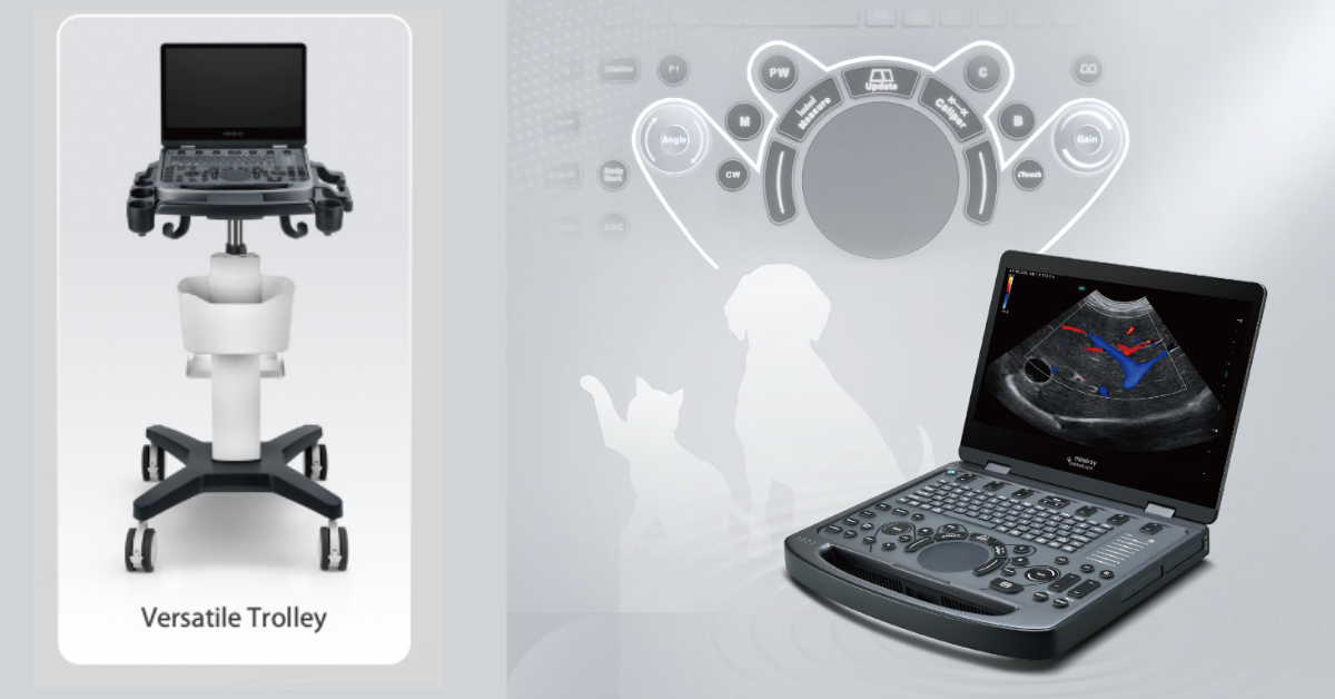
3. Portability & Durability
Lightweight and portable enough for in-clinic or off-site use, without a heavy cart.
Durable waterproof/dustproof control panel designed to tolerate hair, fluid, and accidental knocks — essential in high-volume vet settings.
Long battery life and easy probe switching keep daily workloads running smoothly.
GE LOGIQ e Vet
A laptop-style form factor makes it inherently portable, ideal for multi-room or field applications.
90-minute battery life between charges is standard on many configurations.
The E5’s design focuses on vet-specific ruggedness, while the LOGIQ e emphasizes proven portability and clinical durability.
4. Value & Warranty Considerations
Often, more aggressive pricing, including standard accessories like one transducer, cart, training, and a robust warranty.
With tools like SmartCalc and iReport included, practices find they get more built-in functionality per dollar.
GE LOGIQ e Vet
Carries a strong service and support ecosystem.
Pricing can be higher, especially when equipped with a full range of vetted veterinary probes and extended support plans.
5. Which One Fits Your Practice?
Here are a few quick scenarios:
General Practice:
The Vetus E5 often delivers excellent image quality, intuitive workflows, and substantial value. Its vet-centric software and reporting tools can save time in busy daily scanning.
Mixed Practice or Specialty Referral:
If you’re doing more cardiac or equine evals and want a platform with broad familiarity and flexibility, the LOGIQ e Vet might be a good choice.
Mobile / Field-Based Work:
Both units are portable, but Mindray’s rugged, purpose-built portability gives it a slight edge for rugged field use, while LOGIQ e’s laptop-style design and battery support make it ideal for point-to-point scanning.
The bottom line is that both systems have earned their place in modern veterinary practices. The E5 excels in intuitive vet workflows and built-in tools, while the LOGIQ e Vet brings a trusted, flexible platform with strong imaging fundamentals that spans diverse case loads.
The Mindray Vetus E5 stands out by delivering more practical performance for everyday veterinary work at a better overall value. Built specifically for veterinary workflows, it offers faster setup, one-click image optimization, cleaner abdominal and soft-tissue imaging, and built-in reporting that saves time on every scan.
Its rugged, vet-friendly design holds up in busy exam rooms, while advanced imaging tools come standard—without costly add-ons. For practices that want excellent image quality, efficiency, and ROI without paying a premium for a legacy platform, the Vetus E5 is the clear choice.

A Veterinary Ultrasound Buyer’s Guide

Choosing the Right Veterinary Ultrasound
For veterinary practices, investing in the right ultrasound system is more than just buying a machine — it’s about choosing a tool that fits your patients, workflow, and long-term goals.
Below is your guide to making smart, confident decisions when selecting the best ultrasound for your hospital.
1. Key Factors When Purchasing a Veterinary Ultrasound
Imaging Performance and Probes
The types of patients you serve — small animals, equine, or exotics — will determine your probe requirements. Look for technologies such as Doppler flow imaging, speckle reduction, and image clarity enhancements, and make sure probe switching is simple and efficient.
Workflow Integration and Veterinary-Specific Features
Ease of use is crucial, especially in a busy practice. Look for intuitive controls, veterinary presets, reporting tools, and connectivity to PACS or practice management software. If your practice includes mobile or fieldwork, portability should be a top priority.
Service, Warranty, and Support
The best technology is only as strong as the support behind it. Check for service plan options, local support, parts availability, and software upgrade pathways to ensure long-term reliability.
Investment and ROI
The initial investment is just the beginning. Include maintenance, training, probes, and consumables when evaluating overall value. Estimate how many scans you’ll perform weekly or monthly, and your revenue per scan, to calculate your break-even point and expected ROI.
2. Value-Focused Option for Small or General Animal Practices
For practices seeking high capability and a balanced investment, the Mindray Vetus E5 is an excellent choice.
Key Features
- Touch-sensitive gain adjustment for fingertip precision
- Intuitive control panel designed for veterinary use
- Advanced imaging: one-click optimization (iTouch), speckle-reduction (iClear), HR Flow, natural-touch elastography, and contrast imaging
- Lightweight and portable for in-clinic or mobile use
Why It Matters
This system offers strong value and a fast ROI. Its versatility across small and mixed animal applications — abdominal, soft tissue, cardiac, and reproductive — makes it a solid all-around investment for general practices.
3. Specialized Option for Equine and Large-Animal Practices
For equine or mixed-animal veterinarians, the Mindray Vetus EQ is a purpose-built system that delivers premium performance and portability.
Key Features
- Includes two transducers from options such as C5-1s convex, L13-3Ns linear, P4-2s phased array, or 6LE5Vs probes
- Extremely portable: weighs only 6.5 lbs and just 1.73″ thick
- 15.6″ LED monitor plus 12.3″ anti-glare touchscreen for barn or field use
- Preloaded equine presets: musculoskeletal, cardiac, abdomen, and reproduction
- Powered by ZST+ Zone Imaging technology for excellent image clarity and penetration
Why It Matters
Large animals require deeper imaging, a wider field of view, and durable equipment that performs well in challenging environments. The Vetus EQ is designed for these conditions — offering the portability, clarity, and ruggedness needed for on-farm or mobile work.
4. Key Questions to Ask Your Equipment Distributor
- What is included in the base package (probes, software, warranty)?
- What upgrade options are available (additional probes, software modules, reporting tools)?
- What service and support plans are offered (onsite vs. remote, response time, preventive maintenance)?
- How is training handled for your team?
- What are the consumable costs, warranty terms, and what happens if a probe fails?
- How compatible is the system with your current digital workflow (DICOM, PACS, practice management software)?
- What ROI timeline can you expect based on your case volume and patient mix?
5. Smart Decisions Made Easy
Ultrasound remains one of the most versatile and profitable diagnostic tools in veterinary medicine. Choosing the right system helps you improve patient care, expand diagnostic capabilities, and grow revenue.
Partner with a trusted distributor who provides not only the hardware, but also training, support, and integration guidance. That’s how your new ultrasound becomes more than just another machine — it becomes a vital tool for better diagnostics and sustainable practice growth.

8 Workflow Mistakes Undermining Your Diagnostic Equipment Investment

Diagnostic Imaging for Veterinary Practices
Explore our most requested systems, then review the workflow playbook below to maximize your ROI.
Avoid These 8 Imaging Workflow Errors in Vet Clinics
Investing in new diagnostic equipment, whether it’s a digital X-ray, ultrasound, or in-house lab analyzers, is one of the most impactful decisions a veterinary practice can make. But even the best technology can fall short if efficient workflows don’t support it.
It’s not uncommon for practices to purchase cutting-edge equipment only to find it underutilized, creating bottlenecks or staff frustration. The good news? These issues usually come down to workflow—not the equipment itself.
Here are eight common workflow mistakes that can quietly undermine your investment—and how to fix them.
-
Inadequate Staff Training
One brief demo at installation isn’t enough. Inconsistent or incomplete training leads to errors, slowdowns, and missed diagnostic opportunities.
The fix: Schedule comprehensive hands-on training for all relevant staff and create clear SOPs for each device. Assign a “champion user” to support ongoing training, maintenance, and onboarding.
-
Poor Equipment Placement and Room Layout
Cramped or inconvenient placement disrupts flow—staff travel farther, cross paths, and squeeze around tables, increasing time per case.
The fix: Map the workflow before installation. Place frequently used equipment near treatment/exam areas and ensure proper clearance for patients and staff.
-
Not Integrating With Your Practice Software
Without PIMS integration, teams waste time on manual data entry and risk transcription errors.
The fix: Work with your provider to ensure full integration. Enable direct image uploads and automatic results imports to save time and improve accuracy.
-
Overlooking Preventive Maintenance
Skipping maintenance or calibration causes inconsistent image quality and unexpected downtime.
The fix: Treat maintenance like patient appointments. Use a service plan for inspections, cleanings, and calibrations on schedule.
-
Not Adjusting Scheduling and Staff Roles
New technology changes patient flow. If schedules and roles don’t adapt, you’ll see idle time or backlogs.
The fix: Reassess after installation. Consider dedicated techs for diagnostics during peak times, or add “diagnostic blocks” to your calendar.
-
Ignoring Change Management
Team resistance can mean inconsistent use or reluctance to adopt new processes.
The fix: Involve the team early. Share the “why,” invite feedback, and celebrate quick wins to build buy-in.
-
Underutilizing the Equipment’s Full Capabilities
Advanced features—contrast tools, cloud storage, automated reporting—often go unused.
The fix: Schedule periodic check-ins with your distributor or manufacturer. Ask about updates, features, and best practices to unlock more value.
-
Failing to Measure Performance
If you don’t measure usage and outcomes, you can’t prove ROI or improve bottlenecks.
The fix: Track studies per month, time from imaging to diagnosis, and referral cost reductions. Use insights to refine workflows and highlight clinical/financial impact.
Supportive Workflow Makes a Difference
A successful diagnostic upgrade isn’t just about the technology—it’s about the workflow that supports it. With thoughtful planning, proper training, and regular review, your new equipment can enhance efficiency, patient care, and profitability for years to come.

What to Know About the Mindray Vetus E7 Ultrasound

Purchasing a new veterinary ultrasound machine is an exciting, yet complex, decision. A veterinarian may feel overwhelmed by choices. There are many systems available at all different price points, with different features—many of which are hard to compare side by side for vets who are new to performing ultrasounds.
In this article, we’ll look at some of the features of the Mindray Vetus E7 Ultrasound, a veterinary-specific model, to see if it might be a fit for your practice.
What Is the Mindray Vetus E7 Ultrasound?
The Vetus E7 is a laptop-type portable ultrasound unit made specifically for veterinary practitioners. It’s designed for use on both small and large animals, for abdominal studies, cardiology, musculoskeletal and small parts studies, and even some reproductive applications.
This 2022 model features a lot of new and advanced technology. Some of the key features that might appeal to a veterinarian are discussed below.
Features of the Vetus E7
While every practice has unique needs, these features may be beneficial to veterinarians in many clinical settings…
Small size and portability. The Vetus E7 is a laptop design, so it can be transported as needed. There is a 15.6-inch, high-resolution color LED monitor. The unit is 1.7 inches thick and weighs 3.0 kg (6.6 lbs) without the battery and 3.5 kg (7.7 lbs) with the battery. Battery life is about 1.5 hours on the laptop alone, or up to 8 hours with the included U-bank battery.
Durability and ease of cleaning. The unit is basically “sealed” in, making it more difficult for liquids and stray hairs to get inside the unit or under the buttons. This includes an anti-liquid touchpad that replaces the standard rollerball, and seamless keys/buttons. Materials are said to be durable and chemical resistant. It stands to reason that the seamless design might also be helpful for protecting the unit from dust and humidity when out in the field.
ZONE Sonography Technology+ (ZST+). This is the first laptop-based system to use the technology, which is supposed to provide excellent image clarity and quality. A simplified way to describe this technology would be to say that it uses software to process acoustic data in large “zones” at a fast speed. This large amount of acoustic data creates a detailed image and can help reduce tissue motion artifacts. Dynamic pixel focusing means good special resolution and the ability to focus at various depths without the user needing to manually adjust the focal point.
Dedicated veterinary presets and user-friendly workflow. Presets are available according to species (canine, feline, equine, bovine, ovine, and customizable), and further subdivided by body size (including dog ranges of <5 kg, 5-15 kg, and >15 kg). The iWorks feature offers smart scanning protocols that standardize the workflow and allow automatic addition of annotations, marks, and measurements. The company claims this can reduce exam time by 50% and reduce keystrokes by 80%, for faster and more efficient studies.
iScanhelper, a built-in learning tool that provides tips on how to scan, as well as anatomical illustrations, patient positioning and probe placement pictures, and ultrasound images for comparison with real-time scanning.
Additionally, the Vetus E7 system includes many standard features such as Doppler and compatibility with image storage software.
Conclusion
Is the Vetus E7 right for your veterinary practice? It depends on exactly what you’re looking for, what your practice plans to budget, and what you plan to use the machine for, i.e., what’s the return on investment based on the expected number and types of studies that would be performed at your practice.
Also, it’s important to ask your supplier questions to ensure you know exactly what you are purchasing. Check how many probes are included (and which types), whether there’s a warranty and what it covers, and anything else you might want to know. See if it’s possible to get a demo, too.
All that being said, the Mindray Vetus E7 does offer some exciting features for veterinarians. In addition to the practical considerations that make it easier to keep the machine clean, many vets would appreciate the features that may make it easier to learn and use this ultrasound unit, such as intuitive workflows and processing capabilities for high-quality images at multiple depths.

It’s common knowledge that ultrasound can require a lot of training and practice before a vet feels confident in the modality. So, anything that makes the process a little easier or more intuitive, and helps with obtaining high-quality images, can certainly be a plus.
Written by: Dr. Tammy Powell, DVM