
What is Fluoroscopy?
Fluoroscopy is an advanced imaging modality that is widely used in human medicine, although its potential in veterinary medicine is only just starting to be realised1. Like conventional radiography, fluoroscopy uses X-rays to produce an image, but in this technique, the image is produced as a video in real-time.
This moving image allows for a much greater range of diagnostic tests to be carried out and can also be used for a range of therapeutic interventions. This field is usually referred to as Interventional Radiography.
Fluoroscopic images are produced by an X-ray generator that produces either a continuous, strobed, or near-continuous, beam of X-rays.
The X-rays are captured by either an image intensifier or, in more modern digital systems, a flat panel detector. Image intensifiers need to be coupled via an optical distributor to a recording or viewing device such as a video camera or screen.
Flat panel detectors are similar to those used in conventional digital radiography (DDR systems) and are connected directly to a computer.
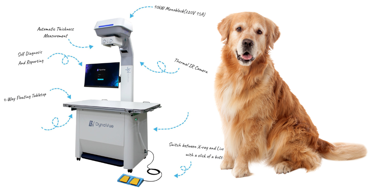
The two main types of fluoroscopy units used in veterinary clinics are portable C-arm units and R&F (radiography and fluoroscopy) rooms.
R&F units are made up of a stationary radiography table with an integrated fluoroscope. The x-ray generator has two separate tube heads, one is used for radiography and one for fluoroscopy.
Portable C-arms are more common in veterinary applications, being used for major surgeries in veterinary hospitals. Unlike an R&F room, a portable c-arm cannot take diagnostic X-rays, only fluoroscopy.
How is fluoroscopy performed?
Depending on the procedure being performed, the patient may be either anesthetized, sedated, or conscious.
In the case of conscious studies, animals are usually restrained within a box where they can easily sit up or lie down, as unlike conventional radiography there is no requirement for them to be perfectly still.
In practice, a change of position is often used to obtain different views and more detailed information. Contrast agents are commonly used to show function e.g. swallowing, or to highlight a particular anatomical area.
In interventional procedures, the patient would routinely be under general anesthetic, and a C-arm fluoroscope is used by the surgeon to visualize part or all of the procedure.
What are the advantages of fluoroscopy?
The big difference between fluoroscopy and traditional radiography is that fluoroscopic images are dynamic moving images, so can be used to assess the function as well as the structure of the body part being imaged.
This opens up a whole array of possibilities. Images produced by fluoroscopy tend to be less detailed than regular radiographs and, to counter this, contrast media are often used to delineate the required structures.
What conditions can fluoroscopy be used to help diagnose?
Many conditions are best diagnosed via fluoroscopy, where a static radiograph does not allow us to see function and ultrasound techniques don’t allow a large enough field of view or suffer interference from air or bone.
Studies that are commonly carried out may include;
Swallowing studies: Swallowing studies require the patient to ingest a volume of contrast material, such as barium, and the progress of this can be tracked through the throat and down to the stomach. This technique is considered the gold standard for investigation of the dysphagic patients.
Esophageal function: Fluoroscopy is useful in the diagnosis of functional and structural esophageal diseases such as megaesophagus and hiatal hernias.
Tracheal and bronchial collapse: A collapsing trachea may be visible on plain radiographs but is a dynamic condition and can easily be missed. Fluoroscopy is more effective at assessing the presence and degree of airway collapse.
Functional diaphragm disorders due to injury or disease of the phrenic nerve.
Localization of thoracic masses: Movement of the mass during normal respiration helps to differentiate peripheral pulmonary and thoracic wall masses.
Blood vessels can be assessed through angiography. Fluoroscopy allows radio-opaque dyes to be injected accurately into specific vessels, aiding the diagnosis of vascular ring anomalies and allowing procedures, such as mesenteric portography which is used to diagnose portosystemic shunts as well as assess the effectiveness of vessel attenuation at surgery.
Real-time evaluation of orthopedic implant placement intra-operatively.
Urinary dysfunction and intervention. Using contrast agents, fluoroscopy allows for easy diagnosis of urinary issues and the effectiveness of catheterizations or other interventions.
What conditions can be treated with the aid of fluoroscopy?
Treatment of conditions with the aid of fluoroscopy is termed Interventional Radiology and in the human field, this is the standard of care for many diseases.
The advantages of using IR as opposed to more traditional therapies include shorter anesthetics, lower perioperative morbidity and mortality, and reduced hospitalization times.
In some cases, conditions that were considered untreatable by other methods can be treated, for example, chemoembolization of unresectable tumors.
Veterinary medicine has some way to go before these techniques are adopted as widely as they are in human medicine.
There are certainly some difficulties and disadvantages in the use of Interventional Radiographic techniques, which include the degree of technical expertise required, specialist equipment needed, and the risk of undesirable radiation exposures to both patients and personnel.
These issues can be reduced greatly with the right training and equipment for the job.
IR techniques that are currently offered to veterinary patients include;
Treatment of cardiac and vascular diseases
Placement of devices within specific blood vessels for transarterial or transvenous embolization of vessels e.g. PDA occlusion, PSS shunt occlusion, or treatment of intractable hemorrhage
Vascular foreign body retrieval e.g. catheter fragments
Cardiac interventional procedures such as balloon valvuloplasty for congenital valve stenosis
Cardiac pacemaker implantation for treatment of arrhythmias
Pulmonary disease
Intraluminal tracheal stenting for treatment of tracheal collapse
Retrieval of tracheobronchial foreign bodies where surgery or endoscopic retrieval are not feasible
Cyanoacrylate embolization for treatment of recurrent chylothorax
Aspiration and biopsy of the thoracic wall and pulmonary masses
Management of neoplasms
Intra-arterial chemotherapy
Arterial embolization and chemoembolization to reduce tumor growth
Palliative stenting of neoplastic obstructions
Urinary disease
Urethral and ureteral stenting to overcome obstructions due to stones, strictures, and malignancies
Urethral catheter placement in cats with urethral rupture
What does the future hold for fluoroscopy
Veterinary fluoroscopy is a developing field of diagnostics.
It seems likely these techniques will become more widely available at both referral and first-opinion veterinary clinics. As expertise in this field progresses, more and more patients can benefit from this technology.
References
1. Weisse, C. W., Berent, A. C., Todd, K. L., & Solomon, J. A. (2008). Potential applications of interventional radiology in veterinary medicine. Journal of the American Veterinary Medical Association, 233(10), 1564-1574.
2. Gingold, E. (n.d.). Modern Fluoroscopy Imaging Systems. Image Wisely. https://www.imagewisely.org/Imaging-Modalities/Fluoroscopy/Modern-Imaging-Systems. Accessed 24/01/2024
3. Shaw, L., & Tudor, D. (2021). Fluoroscopy: Don’t Miss the Show. The Veterinary Nurse, 51–57.
4. Shalom NE, Gong GX, Auster M. Fluoroscopy: An essential diagnostic modality in the age of high-resolution cross-sectional imaging. World J Radiol. 2020 Oct 28;12(10):213-230.
5. Pollard, R. E., Marks, S. L., Cheney, D. M., & Bonadio, C. M. (2017). Diagnostic outcome of contrast videofluoroscopic swallowing studies in 216 dysphagic dogs. Veterinary Radiology & Ultrasound, 58(4), 373-380.
6. Fuentes, V. L., Johnson, L., & Dennis, S. (2010). BSAVA Manual of Canine and Feline Cardiorespiratory Medicine. BSAVA.
7. Pollard, R. E. (2012). Imaging evaluation of dogs and cats with dysphagia. International Scholarly Research Notices, 2012.
8. Bevan, J. M., & Taylor, R. A. (2004). Arthroscopic release of the medial femoropatellar ligament for canine medial patellar luxation. Journal of the American Animal Hospital Association, 40(4), 321-330.
9. Weisse, C. (2015). Veterinary interventional oncology: from concept to clinic. The Veterinary Journal, 205(2), 198-203.
10. Kim, M. Y., Kim, J. H., Kim, K. C., & Yoon, H. Y. (2022). The effectiveness of intraoperative mesenteric portography for preventing misdiagnosis of congenital absence of the portal vein in dog with extrahepatic portosystemic shunt: a case report. Acta Veterinaria Brno, 91(3), 267-272.
11. Meurs, K. M., Lehmkuhl, L. B., & Bonagura, J. D. (2005). Survival times in dogs with severe subvalvular aortic stenosis treated with balloon valvuloplasty or atenolol. Journal of the American Veterinary Medical Association, 227(3), 420-424.
12. Wess, G., Thomas, W. P., Berger, D. M., & Kittleson, M. D. (2006). Applications, complications, and outcomes of transvenous pacemaker implantation in 105 dogs (1997–2002). Journal of veterinary internal medicine, 20(4), 877-884.

