
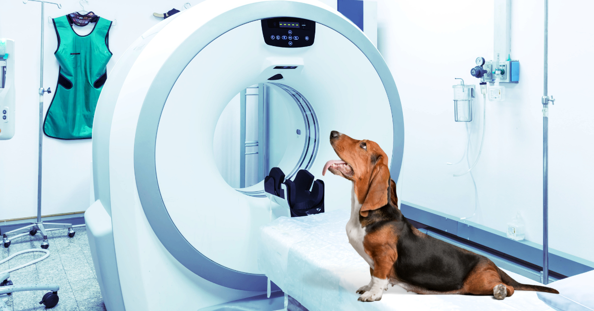
Cone beam computed tomography (CBCT) has been available in human dentistry for years, but it is now also entering veterinary medicine.
Some specialist vets who work with maxillofacial and dentistry cases have already started to use this technology. I
t provides detailed 3D images which can be helpful for oncology or trauma cases, as well as those suffering from dental disease. Here we are going to explore this imaging modality in more detail, including some scenarios in which you might choose to use it.
What is cone beam CT?
CBCT uses radiation, in the same way that normal computed tomography (CT) does. However, the shape of the x-ray beam is different. In CBCT the beam is in the shape of a cone, whereas normal CT has a fan-shaped beam.
The cone-shaped beam covers the area of interest, and the X-ray source only needs to pass once around the animal, being picked up by a sensor on the opposite side of the patient to create an image.
This means the average dose of radiation a patient receives is more similar to that of one thoracic radiograph than a conventional helical CT.
Due to the low amounts of radiation it emits, its lower purchase cost and the machine’s compact design, CBCT is becoming easier to access than normal CT. Its design makes it particularly ideal for imaging the maxillofacial area, which is a complicated area of anatomy to visualize.
Three reasons why a veterinary surgeon would want to use cone beam CT
Maxillofacial disease
As previously discussed, CBCT is commonly used for maxillofacial disease.
This imaging modality works well in small anatomical areas with high tissue density. This fits the bill for the maxillofacial area, which has several complicated structures including the teeth, upper and lower jaws, the temporomandibular joint, and the sinuses filled with turbinates. All of these structures can overlap and confuse diagnostic imaging, particularly in brachycephalic breeds.
CBCT can create a highly detailed, superimposition-free image that shows the area of interest in 3D. This gives a clear advantage over traditional, 2D dental x-rays.
A recent study into dental pathology in cats found that ‘CBCT provided more clinically relevant detailed information as compared to dental radiography’.
This was also the case in a 2018 paper on CBCT vs dental radiography in small to medium-sized brachycephalic dogs. These studies indicate that practitioners could be missing clinically relevant pathology when using dental X-rays alone.
As well as endodontic disease, other examples where CBCT may be superior to X-rays include –
Evaluating maxillofacial trauma
Assessment of temporomandibular joint disease, dislocation, luxation, or fracture
Viewing bone integrity associated with cystic lesions
Assessing the severity of oronasal communications and cleft palates
Evaluating the extent of oral tumors in hard tissue of the maxilla and mandible
Small and exotic animals
CBCTs can be used in examinations of the head and dentition of small and exotic species, as well as canine and feline patients.
A 2017 paper holds ‘CBCT to be superior to conventional CT for evaluation of normal dentition in rabbits’. Subtle changes in the periodontal ligament space can be detected more easily in rabbits, which can be an indication of emerging disease.
This could otherwise go unnoticed and be missed in conventional CT scans.
Whole body exams of many small, exotic animals may be possible with this technology too, further widening its appeal and usage.
Joint disease
CBCT can be used in extremities like the limbs to evaluate joint disease.
There are indications for its use in the musculoskeletal system including evaluation of fractures and dislocations in small bones and joints, and it can be used in conjunction with other imaging techniques to assess cartilage. It is easier to evaluate degenerative joint disease (like osteoarthritis) with CBCT than conventional radiography, particularly in areas where there is superposition of other bony structures.
This paper on CBCT in people with joint disease further demonstrates this point.
In veterinary medicine, a 2021 study in standing horses demonstrated that CBCT provided diagnostic imaging in a timely fashion. No doubt this modality will start to become more commonly used for joint disorders in a variety of species as veterinary practitioners start to utilize it further.
When would you not want to use cone beam CT?
The X-rays from the CBCT are not collimated.
They make a divergent, uncollimated cone meaning more scattered radiation is produced. This means there is a reduction in soft tissue contrast, and the resolution in areas like the thorax and abdomen is poorer, especially in large dogs.
Larger body parts cause greater scatter. Using CBCT in these areas will mean contrast is lower than if it is used in smaller areas, like the jaw or a limb.
This is different from normal CT scanners which have a collimated beam that runs as straight as possible, with little scattered radiation.
So normal CT should be chosen for soft tissue, thorax, and abdomen examinations (especially in larger cats and dogs) and CBCT would not be advised.
Equally, good radiation shielding – especially when a horizontal beam is likely – is necessary for imaging facilities utilizing this technique.
Summary
Cone beam CT shows great promise across multiple areas of veterinary medicine, but in particular for maxillofacial disease.
It provides highly detailed 3D images of areas that are traditionally hard to assess with conventional dental radiography. This helps veterinary surgeons to plan their treatment accordingly, which can only lead to better patient care and outcomes.
CBCT will probably be seen more and more frequently in hospitals, as its potential gains recognition by general practitioners.

