


Veterinary Endoscopy: Uses and Benefits for Patient Care

Minimally Invasive Procedures: The Benefits of Veterinary Endoscopy
Just like other advanced imaging modalities—such as veterinary CT systems, fluoroscopy, and more—endoscopy is becoming more common for use by general practitioners.
Is an endoscope a good value to add to a veterinary practice? How is it used, and how does it improve patient care?
In the next few articles, we’ll cover common uses and purchasing questions when it comes to veterinary endoscopy.
What Is an Endoscope?
An endoscope is a rigid or flexible tube that is used for imaging, diagnostic testing, and/or certain surgeries and procedures on veterinary patients.
The scope provides light and visibility (which may include high-resolution video on some models) to help the veterinarian better assess the patient’s anatomy and look for abnormalities. This veterinary equipment is typically hollow to allow the passage of tools and accessories—such as biopsy and retrieval forceps—to the area of interest.
Uses for Endoscopy in Veterinary Medicine
Standard uses may include non-invasively assessing anatomy, obtaining biopsy samples, and removing foreign objects.
A very common use of endoscopy in veterinary medicine is the evaluation of the GI tract, including the esophagus, stomach, and proximal duodenum. The scope would be inserted through the mouth and passed through the esophagus to visualize the inner portions of these organs without the need to perform surgery.
While evaluating the GI tract, a veterinarian might take note of abnormalities like inflammation, wounds or bleeding (examples: esophageal trauma or gastric ulcers), masses, strictures, foreign bodies, or other issues.
This visual exam can help with diagnosis, but biopsies are usually needed for a definitive diagnosis of many conditions, including inflammatory or neoplastic conditions. These biopsies can typically be obtained via the scope, so long as a full-thickness surgical biopsy isn’t required.
Tools or attachments can be used to retrieve and remove many types of foreign bodies. For a stricture, endoscopy can help facilitate balloon dilation therapy. It’s even possible to provide hemostasis with some scope accessories.
The colon, cecum, and parts of the ileum may also be evaluated via a colonoscopy. In addition to the GI tract, endoscopes may be used to evaluate other body systems such as the respiratory tract (examples: rhinoscopy or bronchoscopy) or urinary tract.
Similar to the upper GI tract, uses of the scope include visual assessment, biopsies, and minor procedures. For example, the tool can assist with traction and removal of nasopharyngeal polyps, or with biopsies of the bladder without the need to perform a cystostomy.
Additionally, laparoscopic-assisted surgery may be performed, especially for routine procedures like spays or even gastropexy. Scopes may also assist with feeding tube placements.
For exotic species like birds or reptiles, veterinary endoscopy can aid with gender determination, in addition to helping with diagnostic and minor surgical procedures.
Should a Veterinarian Refer to a Specialist for Endoscopy?
It’s probably never “wrong” to offer clients a referral—both for the best interest of the patient when indicated, as well as for liability purposes. If a general practitioner doesn’t have the right equipment or feels confident using the equipment, a referral is usually best.
However, it’s also true that endoscopy isn’t strictly limited to specialists like surgeons or internists anymore. It’s possible for general practitioners to gain experience using this modality and expand their offerings to their own patients.
That being said, a general practitioner shouldn’t simply order an endoscope and set up shop immediately. It’s important to research the equipment prior to purchase and then to gain lots of knowledge and hands-on experience on how to safely and effectively use it.
Consulting with a veterinary specialist who frequently uses endoscopy can be helpful. They can guide you on which types of scopes (there are many options out there) might be the best investment for your budget, for the widest range of uses on the types/sizes of patients your practice sees.
Then, pursue hands-on, guided instruction in the use of the new equipment. Some endoscopy systems include training, which is nice because it’s geared toward the specific model your practice purchased. There are also in-depth courses—many with guided, in-person instruction from a knowledgeable teacher—available at conferences, universities, or other continuing education venues.

With some due diligence and training, adding veterinary endoscopy can potentially be exciting and profitable to a practice that wants to provide a high level of care to its patients.
Written by: Dr. Tammy Powell, DVM

The Complete Small Animal Endoscope System
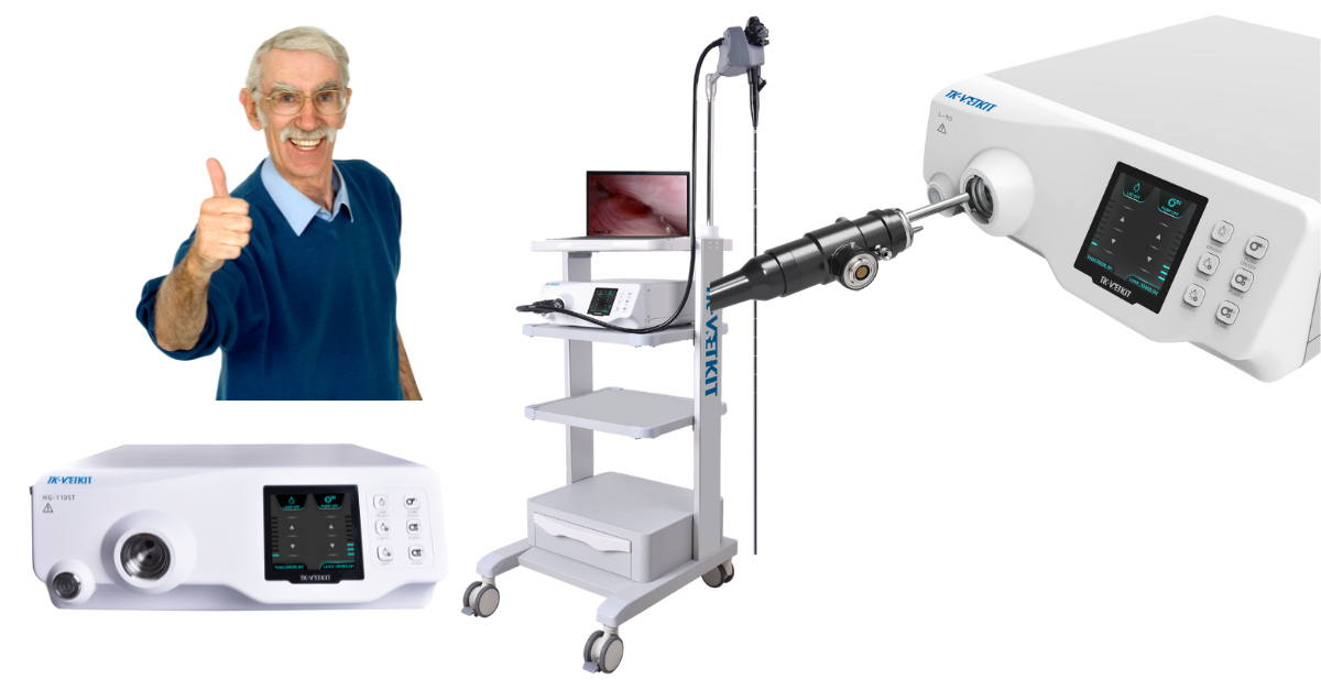
The Complete Small Animal Endoscope System$16,995 or $295 a month.
Are you looking for a reliable and affordable small animal endoscope system?
Look no further than our complete system, priced at only $16,995 or $295 a month.
With shipping included and a 3-year parts warranty, you can rest assured that your investment is protected.
Versatility: Important Uses Of The Endoscope In Small Animal Diagnostics
Our small animal endoscope system is an essential tool for the evaluation of animals with a variety of symptoms such as vomiting, diarrhea, and difficulty defecating.
It is also useful for obtaining biopsies of the stomach, intestines, and potentially esophagus. With the ability to remove foreign material and place feeding tubes, this system is an invaluable asset to any veterinary practice.
LED Light Source: Excellent Visualization And Sampling
Our system comes equipped with an LED light source generating a vivid, vibrant image. Accompanied by both a water and air pump, our scope provides excellent visualization and sampling of the gastrointestinal tract from the inside.
The scope can be easily plugged into a standard 110 outlet, making it easy to use in any practice.
Training Included: Endoscope Training Program
We understand that mastering the use of an endoscope can be challenging, which is why our Endoscope Training Program is designed to help you take your endoscopy skills to the next level. Our comprehensive program offers a step-by-step guide to building, setting up, and using an endoscope and its accessories with ease.
With live remote training using Team Viewer, you can learn from the comfort of your own practice.
Suction: Schuco Aspirators
Our small animal endoscope system is also equipped with a reliable and durable Schuco aspirator. The entire family of Schuco aspirators has evolved to meet the changes in standards and provide just the right aspirator for your suction requirements. With improved scratch and flake resistance, high vacuum range, and a vibration-free gauge, this aspirator is a must-have for any veterinary practice.
In conclusion, our Complete Small Animal Endoscope System is a versatile, reliable, and affordable tool for any veterinary practice. With its LED light source, Schuco aspirator, and comprehensive Endoscope Training Program, you can take your endoscopy skills to the next level.
Contact us today at 530-722-4977 to learn more.


Veterinary X-Ray Systems for Nonprofits and clinics

Many pet rescue organizations can greatly benefit from having radiographs available at their facility.
Here are some reasons to look into veterinary X-ray machines and how to acquire this equipment to offer diagnostic imaging to dogs, cats, or other pets at a nonprofit center.
Reasons to Invest in Veterinary Digital X-rays
Radiographs provide diagnostic value in many situations. Although the level of medical care provided by a nonprofit varies depending on their capacity, funding, staffing, and goals, x-ray images are one of the best tools that can be used on a wide variety of patients with a wide variety of medical conditions.
One example is taking radiographs of a pregnant dog or cat that arrives at the facility, to see how many puppies or kittens are expected and check that they are all delivered safely. Another common use is evaluating a pet for a potentially surgical medical problem, such as an intestinal obstruction. Radiographs can also help a veterinarian evaluate the extent of damage with traumatic injuries like fractured bones and help plan for treatment.
This is just scratching the surface. Radiographs are one of the most widely used diagnostic tools at veterinary clinics. Any rescue offering veterinary services (to facility pets awaiting adoption, or to client-owned pets) can certainly benefit from having a good X-ray system. Exotic pets, and even large animals like horses (typically with a portable X-ray machine), also frequently need imaging for a variety of medical conditions.
What About Digital Versus Film Radiographs?
Digital is largely favored nowadays. Specifically, veterinary DR systems (rather than CR x-rays) are a preferred technology.
DR radiographs offer several advantages, such as the speed of image acquisition (immediate), automatic settings, ease of image sharing, and reduced need for retakes thanks to quick image analysis and automatic settings that help adjust the image.
Additionally, there is no need to purchase further equipment to process images, unlike film and CR technology, which require a film processor and plate processor, respectively. Finally, as technology continues to advance, it’s easier to find replacement parts for newer tech like DR.
The Business Plan
With any new equipment purchase, it’s important to make sure the new machine and financial investment make sense for the organization.
Some private and government-owned nonprofits must provide a business plan or proposal to whomever they are held accountable, whether that is public/government regulations, board members, or other overseeing entities. Even if a formal report or proposal isn’t necessary, it still makes sense to create a plan to ensure the investment is financially feasible.
Here are a few factors to consider for a business and financial plan…
How will the equipment be used? Is it only for pets waiting to be adopted? Or will the organization offer services to client-owned pets in the community?
Which specific services will be offered? While a lot of X-ray studies are standard, the medical team may also have the option to offer contrast studies or other variations.
Who will perform the services? Vet techs can set up and take veterinary X-ray studies. And through experience, many technicians also gain some skill in reading images, or at least checking them for correct alignment and quality to make sure the image is usable. However, radiographic studies should be ordered by a licensed veterinarian. The vet is also the only one who can interpret the images, for making a diagnosis and treatment plan. Some nonprofits might already have qualified staff on their team. Others must consider hiring, either on a full-time or part-time basis or working with experts who are willing to volunteer their time.
Do any other equipment or supplies need to be purchased? A film processor or CR plate reader would be examples—although fortunately, neither is needed with DR technology. However, if the DR unit doesn’t come with a sensor, the organization must add that to its purchase list. Another consideration is sedation or general anesthesia. Chemical restraint is becoming favored in X-ray studies, both for patient comfort and staff safety. Finally, think about props such as positioning troughs.
Remember radiation safety. Don’t forget lead aprons, radiation badges, and other safety/compliance supplies. Setting up an X-ray suite can also require lead-lined walls and other radiation compliance factors in the local jurisdiction.
Will the nonprofit charge for veterinary x-ray studies—and if so, how much? If performing radiographs on adoptable pets, the cost should be factored into the operating costs of the facility. If offering services to client-owned pets, consider how much local pet owners can pay and how much the organization needs to charge per study to maintain financial health.
How to make the purchase of new equipment financially feasible? For many organizations, donations, grants, or a funding drive can really help with this. Think about any money the new equipment will bring in, as well as any associated new costs, such as equipment maintenance, interest/financing charges, staff time, and associated supplies or services.
How to educate pet owners on the value being provided? This is especially important if offering services to client-owned pets.
Are all local regulations and legal requirements for nonprofits in that state/jurisdiction being met?
Finding Veterinary X-ray Systems
A good strategy would be to invest in the best possible X-ray unit within the nonprofit’s budget. But where to look for one or find a good deal?
Used or refurbished X-ray systems may present an affordable option that is still in very good condition. Some will still have warranties and service plans in place, while others will not.
Some nonprofits benefit from donations of new or like-new veterinary equipment. This could come from other veterinarians in the area or even local human hospitals who are upgrading their equipment.
Some vendors or other organizations might offer new equipment as part of their own grant program, nonprofit arm, or effort to save on taxes. Or they might be willing to part with their demo or loaner machines for a very reasonable price.

Veterinary X-ray systems are one of the most universally used pieces of equipment that can help a lot of patients. With some planning and due diligence, it can be an excellent investment for any rescue or nonprofit that offers basic or advanced veterinary services to the animals in their care.
Written by: Dr. Tammy Powell, DVM

Veterinary Dental Equipment and X-ray for Nonprofits

Many pet rescue organizations, adoption centers, and other nonprofits use veterinary dental care to improve the health of pets being adopted or to provide a service to the local pet-owning community.
Here are some reasons to consider looking into veterinary dental equipment and offering dental services to dogs, cats, or other pets at a nonprofit center—as well as ideas for making it happen.
Reasons to Invest in Veterinary Dental Care
Along with obesity, dental disease is one of the most common preventable medical ailments in pets in the US. According to AAHA, most dogs and cats over the age of three have some degree of dental disease.
Dental disease can lead to periodontal disease, in which the structures that hold teeth in place are compromised. This can cause pain, infections, and tooth loss. Additionally, many pets suffer from common dental conditions like broken teeth, resorptive lesions (mainly in cats), and more.
Many of these conditions can be treated right there in the nonprofit or rescue organization. This could potentially offer benefits such as allowing pets to be more comfortable until they’re adopted or improving the mood and sociability of these pets by alleviating any dental pain they might be experiencing.
Additionally, many experienced or knowledgeable pet owners know that they would need to pay for dental treatment (along with associated anesthesia costs) for a pet that has dental or periodontal disease. So, in theory, a “clean dental slate” could possibly improve the adaptability of some pets. The pet’s better-smelling breath might help in that regard, too.
Some nonprofit organizations also offer low-cost veterinary services to the community, either to all pet owners or to those who demonstrate proof of a low income. Dental care could be a great way to improve the overall well-being of those pets.
The Business Plan
While it would be wonderful to offer every possible healthcare service to pets in need, whether those waiting to be adopted or those who already have homes in the community, any organization must do their due diligence and ensure the new equipment/service offerings make sense to them.
Some private and government-owned nonprofits must provide a business plan or proposal to whomever they are held accountable, whether that means public/government regulations, board members, or other overseeing entities. Even if a formal report or proposal isn’t necessary, it still makes sense to create a plan to ensure the investment is financially feasible.
Here are a few factors to consider for a business and financial plan…
How will the equipment be used? Is it only for pets waiting to be adopted? Or will the organization offer services to client-owned pets in the community?
Which specific services will be offered? With dental care, this might include dental cleanings, dental radiographs, and treatment of specific conditions such as extractions of diseased or injured teeth.
Who will perform the services? Veterinary dental cleanings, radiographs, and oral surgery require general anesthesia under the guidance of a licensed veterinarian. Radiographs and cleanings can generally be performed by trained vet techs, but under the direction and supervision of a veterinarian. Some nonprofits might already have staff who are qualified to perform these procedures. Others must consider hiring, either on a full-time or part-time basis, or working with qualified experts who are willing to volunteer their time.
Do any other equipment or supplies need to be purchased? As mentioned, general anesthesia is required. This is no problem for many organizations that already have surgical capabilities. But it might represent an additional investment for centers that are new to offering such services.
Will the nonprofit charge for veterinary dental services—and if so, how much? If performing dental services on adoptable pets, the cost should be factored into the operations costs of the facility. If offering to client-owned pets, consider how much local pet owners can pay and how much the organization needs to charge to maintain financial health.
Is the purchase of new equipment financially feasible? For many organizations, donations, grants, or a funding drive can really help with this. Think about any money the new equipment will bring in, as well as any associated new costs, such as equipment maintenance, interest/financing charges, staff time, and associated supplies.
How to educate pet owners on the value being provided? This is especially important if offering services to client-owned pets.
Are all local regulations and legal requirements for nonprofits in that state/jurisdiction being met?
Finding Veterinary Dental X-ray Machines and Equipment
A good strategy would be to invest in the best possible machine within the nonprofit’s budget. But where to look for one or find a good deal?
Used or refurbished dental X-ray systems or cleaning units may present an affordable option that is still in very good condition. Some will still have warranties and service plans in place, while others will not.
Some nonprofits benefit from donations of new or like-new veterinary equipment. This could come from other veterinarians in the area or even local human hospitals or dentists’ offices who are upgrading their equipment.
Some vendors or other organizations might offer new equipment as part of their own grant program, nonprofit arm, or effort to save on taxes. Or they might be willing to part with their demo or loaner machines for a very reasonable price.

With some planning and due diligence, many nonprofit pet organizations can find ways to offer veterinary dental services—yet another way to continue improving the health and quality of life of the animals they serve.
Written by: Dr. Tammy Powell, DVM

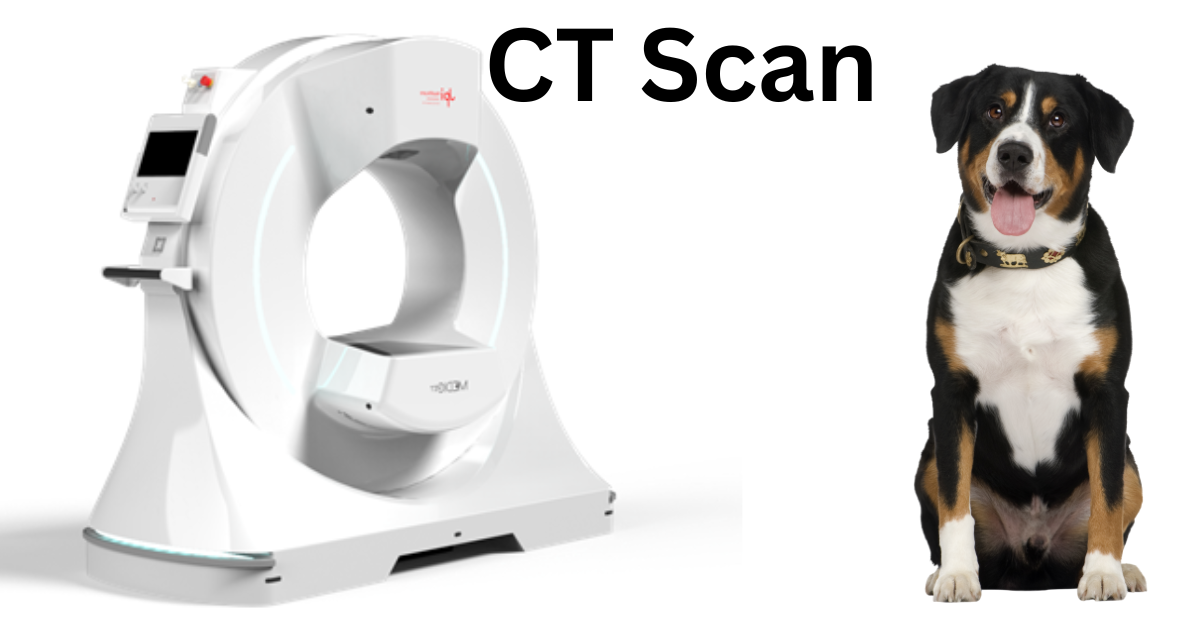
Veterinary CT Terminology and Technology, when buying

In recent years, veterinarians have had a lot to choose from in terms of CT system sizes, technology, combinations, and price points.
While it’s good to have options, it can also feel like “information overload” trying to choose the best machine for an individual practice. The following information may help with this important decision…
Conventional CT
Traditional, fan-beam-style CT scanners were the norm for a long time. And they still serve a valuable purpose. A fan-beam model is a good all-around option, especially for hospitals that see all different sizes of patients including large dogs.
Fan-beam CT machines work by taking image “slices” (cross-sectional images) that are picked up by an array of detectors. Then the patient is advanced further into the gantry (entryway) and another slice is taken—and so on until the area of interest has been fully imaged.
Slice Counts
Advances in fan-beam technology include systems that image multiple slices at once. Instead of a single slice, this could mean 4, 8, 16, 32, or even 64 slices at a time. These multidetector CT scanners have the advantage of being faster than a single-slice machine.
Although slice thickness factors in, as you can imagine, shooting a larger area (i.e., multiple slices) at once means the study takes less time. In some cases, this can reduce anesthesia or sedation requirements. But chemical restraint may still be needed for areas in which motion artifact is a bigger confounding factor, such as respiratory movements during lung studies.
As you can probably also imagine, the price of the machine goes up as the slice count increases.
Slice Thickness
There are pros and cons to both thinner and thicker image slices. Thinner slices allow for the acquisition of more details. Thicker slices allow for faster study times.
Helical or Spiral Scanners
Originally, the patient would be advanced into the machine, then stopped while the first image slice is obtained, then advanced a little further, and so on…
With helical models, the patient is continuously advanced without stopping. This results in the x-ray tube head moving in a spiral motion around the patient.
This can mean shorter study times. Helical models can also have great image quality, including 3D renderings.
Cone-beam Technology
Cone-beam CT is a newer, increasingly popular option. Instead of the images being captured on a narrow array of detectors like fan-beam technology, they are captured on a wider flat panel detector similar to those used for regular X-ray machines.
Veterinary cone-beam CT is a nice option because of the smaller footprint and lower price point. They’re also ideal for certain studies, like the skull, extremities, and some musculoskeletal views.
However, these small machines don’t accommodate larger patients or offer as much contrast and detail for soft tissue studies. And they may be more susceptible to motion artifact.
Portable CT Machines
Battery-powered, portable CT scanners are also available. They might be advantageous for busy hospitals with limited space, or for any practitioners who need to move their machine to the patient.
Many of these machines use standard electrical outlets for power or for battery charging, which is convenient compared to higher-powered machines. Some also have built-in shielding, which can decrease the requirement for lead-lined walls.
A practice should check the specifications for the specific machine they are purchasing.
Combo Machines
Some models offer combinations of CT, fluoroscopy, and even standard digital x-rays all in one. But are they worth it?
It depends. If there is a significant price advantage—and the image quality is great for ALL modalities—a combo could be a practical, space-saving option.
If one or both of these requirements aren’t met, a veterinary practice might find it’s better for them to focus on one modality at a time rather than purchasing a combination machine. It’s also important to consider whether there is a potential “bottleneck” in scheduling—for example, team members waiting to do a CT scan while someone else finishes radiographs.
Veterinary Radiologist Consultations
It’s exciting that CT technology is becoming more widely available and even used by some general practitioners. However, there is a large learning curve for any new imaging modality a practice adopts. Veterinary CT is no exception.
Specialists exist for a reason. Veterinary radiologists have significantly more knowledge and experience with advanced imaging modalities. A consultation with a radiologist can help with everything from pre-purchase planning (which model to purchase, requirements to set it up at a clinic, maintenance cost considerations, etc.) to doing teleradiology consultations on the images obtained from a CT scanner.
For teleradiology consultations, ask ahead of time if the radiologist has any specific requirements for the type of machine used, the settings, etc.
Investing In the Right Machine
As time goes on and more veterinary practices show an interest in CT scanners, there’s no doubt that new technology and options will be developed.

Decision makers at each practice must perform due diligence and talk to experts (veterinary radiologists, business/financial consultants, and state regulations for radiation safety) prior to purchasing and using a CT system, to ensure the machine they purchase is a good fit for them.
With that in mind, it’s an exciting time in veterinary medicine, as practitioners have yet another option for providing advanced patient care at their practice.
Written by: Dr. Tammy Powell, DVM

What Is the Cost of a Veterinary CT System?

Computed tomography (CT) seems to be gaining in popularity in veterinary medicine, since the diagnostic scans are beneficial in the workup and care of many patients. As the technology continues to advance, with more affordable options being marketed to veterinarians, CT scanners are even becoming available to non-referral or general practices.
Although veterinarians now have more CT models to choose from, is the price tag worth it? How much do CT systems actually cost, and can a veterinary practice make a profit by offering this service?
How Much Do Veterinary CT Scanners Cost?
As with any big-ticket equipment purchase, costs can vary greatly depending on the type of CT scanner, the model and manufacturer, deals from the vendor, whether it’s new or used or refurbished, and other factors. CT investments tend to cost more than a standard veterinary digital x-ray system. But the good news is, some newer models are certainly less expensive than previous ones.
According to Sound a new standard (fan-beam) CT machine can cost upwards of $500,000 or more, depending on the number of slices the machine produces. Manufacturer-refurbished models can run in the $100,000-200,000 range, or possibly significantly less if refurbished by a third party.
Portable models are listed from the high $100,000s to $500,000 or more—again, depending on the number of slices and whether the model is new or refurbished.
Cone-beam CT, on average, tends to be less of a financial investment than standard CT. That’s one of the reasons it has become popular, in addition to other factors such as taking up less space and producing better images for certain studies (particularly for skull/head studies and certain musculoskeletal conditions). According to Sound, a new cone-beam CT system can cost between $180,000-$250,000, whereas refurbished ones might start around $150,000.
While this smaller price tag is inviting, cone-beam does have its limitations. A veterinary practice must consider the pros and cons of each type of CT scanner and decide what they need for the patients they see and the types of studies they plan to perform.
What Additional Costs Should Be Expected?
Although the purchase price of a veterinary CT system is important, it’s certainly not the only number that factors into a purchase decision. Here are a few more to think about…
Shipping and installation. Check to see if these costs are included. However, keep in mind that some installation costs and logistics can’t be covered solely by the manufacturer or vendor.
Specifically, think about lead lining or shielding that the machine requires, as well as electrical requirements. For certain CT models, consultations with licensed physicists/radiation experts and electricians may be required, as well as remodeling of the CT suite, to accommodate the machine and meet local radiation safety regulations.
Ongoing maintenance and repairs. See if a warranty is included. Service and maintenance plans can also be a great option for many types of veterinary equipment.
However, service plans do tend to be on the pricier side for CT machines. Consider the cost of the plan, as well as the average cost of repairs, to see whether it makes more sense to pay for the plan or simply set aside some cash for unexpected repairs.
Consulting with a veterinary radiologist, or even the radiology department of a human hospital, may be helpful to determine which repairs are most common and how much they tend to cost.
Software. All digital imaging technology requires appropriate software to process, view, share, and store images. Since CT scanners take a lot of images per study, the file storage can be quite large. Make sure hardware and software can accommodate these files.
Explore which financing option makes the most sense for your practice. Consult a tax professional to determine which tax benefits you may receive from the purchase. And perhaps most importantly, contact a boarded veterinary radiologist for advice.
Think about the return on your investment. How much would your practice charge for a CT study? Are clients likely to say ‘yes’ to this price, including anesthesia costs? And how many studies could the practice realistically expect to perform in a week or a month? As much as a veterinary practice would love to provide every possible service to their patients, the business must make sure the purchase fits into their financial plan.

Veterinary CT systems represent an amazing and useful technology that can benefit many patients. Knowing the numbers, talking to experts, and having a plan in place can help a veterinary practice profit financially while also providing excellent patient care.
Written by: Dr. Tammy Powell, DVM

What to Know About Veterinary CT Unit Installation

When purchasing a CT scanner, veterinary practices should look at not only the purchase price but also the costs of installing and maintaining the unit.
Planning for the following factors can help a practice avoid surprises and smoothly purchase and integrate a veterinary CT machine…
Space Requirements
CT systems vary greatly in terms of size. Traditional, fan-beam CT scanners can be very large and require their own dedicated room. Some veterinary practices just might not have the space for these units, or they might need to do some extensive remodeling to accommodate them.
Other CT scanners, such as cone-beam or portable units, can be significantly smaller in size. This can make them attractive to veterinary practices that are limited on space.
Electrical Requirements
Some units can operate on standard electrical outlets. Others require a higher current—which might mean consultation with an electrician and rewiring are necessary.
As you might expect, the former is more common with newer, smaller CT units, while the latter is more common with large, traditional units that take a lot of power to operate. Portable units can even be battery-powered. But it’s crucial to check the specific electrical requirements for the unit you are purchasing.
Lead Shielding Requirements
Radiation safety is crucial with any x-ray modality. And CT is certainly no exception, especially since the amount of radiation produced with multiple slices can be large. In addition to personal protective equipment, shielding might include other requirements, such as lead-lined walls in the room in which the unit is housed.
As might be expected, a larger CT scanner generally has more lead shielding requirements than a smaller one. However, it again comes down to the requirements of the specific model of CT unit in which you are interested.
Also, while the specs of the individual machine are important, they’re not the only consideration. Every jurisdiction has its own radiation safety laws and regulations. Do your research and hire a radiation consultant prior to installing a new machine. They can instruct your practice on shielding requirements based on the radiation doses and expected usage, occupancy in the spaces outside the CT room, etc.
This often includes speaking to a veterinary radiologist, as well as a radiation physics consultant or licensed physicist who understands local legalities and how to keep your veterinary practice in compliance. Your local veterinary medical board may be able to point you in the right direction, too, in terms of regulations and finding licensed consultants to work with.
Doing this step prior to purchase can also help a veterinary practice understand what costs are involved in installing various machines and shielding, which might factor into the purchase decision.
Cooling Requirements and Additional Considerations
Some CT units, especially larger ones, can get quite warm during use. It’s important to ensure there is adequate cooling and ventilation in the room in which the machine will be used.
The flooring should be sturdy enough to support a heavy CT unit.
Also, in addition to the up-front costs (purchase and installation), it’s crucial to factor in long-term operating and maintenance costs. Some warranties and service plans are very pricy and designed for machines with heavy use. Other service plans (or even paying out of pocket for repairs) might be more practical for typical, light veterinary use.
Also, a veterinary practice team should always consider whether the CT system they are purchasing meets their expected usage requirements on the types of patients they see. For example, maybe a practice wants a smaller CT machine due to space limitations, but they also see a lot of large dogs that need abdominal scans, for which a larger CT unit would be ideal. It’s a matter of balancing these interests and finding the best solution.
Although the price tag plus additional costs can seem daunting, it helps to break down each consideration one at a time.
Then compare everything to the expected income from your new veterinary CT system to determine what are the most that your practice can or should invest.

This will greatly help with determining if CT is a good fit for your practice, and then selecting the right machine for your needs.
Written by: Dr. Tammy Powell, DVM

Vet CT Technology Comparison Traditional vs. Cone-Beam
As CT (computed tomography) technology advances, veterinarians have more options than ever to choose from for their clinic—including the increasingly popular cone-beam CT scanner.
So, is cone-beam technology superior to traditional CT units? It all depends on what a veterinary practice needs. Here are some factors that can help a veterinarian choose which veterinary CT system is right for them.
What’s the Difference Between Traditional and Cone-beam CT?
With traditional CT, images are created as a series of fan-shaped slices, which are picked up by a narrow array of detectors. Cone-beam CT units use a wider, cone-shaped beam with a flat panel detector or plate.
The simplicity of the plate means a cone-beam machine can be smaller and might have lower maintenance costs. However, cone-beam technology takes longer to acquire the images.
Both types of technology create a series of images that give a veterinarian a deeper look at a patient’s anatomy compared to standard radiographs. The total series of images essentially allows a 3D view of the area being studied, thanks to CT’s ability to eliminate the problem of superimposition. And newer cone-beam technology can create some impressive 3D renderings.
When Is Traditional CT Most Beneficial?
Here are a few situations in which a veterinarian might prefer a conventional veterinary CT unit…
Larger patients. Cone-beam, which were originally designed for studies of the head in humans, have a gantry or entry point that is relatively small. Thus, cone-beam CT might only be practical for cats, small dogs, exotics, or a larger patient’s head or extremities. Traditional CT, on the other hand, could potentially be used for full scans on larger patients.
Soft tissue differentiation. One big advantage of conventional CT is better soft tissue differentiation compared to traditional radiographs and even compared to cone-beam CT. Examples of uses could include visualizing individual muscle bodies and blood vessels, metastasis checks of the lungs, characterizing a soft tissue mass within an organ such as the liver, and detailed surgical planning.
Abdominal or thoracic studies. Due to a combination of soft tissue definition and accommodating larger anatomy, standard CT is usually the better choice for abdominal and thoracic studies.
Limiting motion artifact. Cone-beam units have slower revolutions, meaning motion artifact can be more pronounced. General anesthesia can help prevent this. However, when motion is a concern (such as when detailed thoracic studies are needed), traditional CT might provide an advantage. This is yet another reason why traditional CT is often preferred for thoracic studies.
When Is Cone-beam CT Advantageous?
Here are some situations when veterinary cone-beam CT might be a better choice…
Smaller footprint. Cone-beam tends to be much smaller than standard CT units. Some are even portable. In veterinary hospitals where space is at a premium, a cone-beam unit might be the only practical option. Additionally, some cone-beam models can be plugged into a standard wall outlet, which is very convenient.
Lower price point. On average, cone-beam CT units cost less to purchase than traditional CT units. It’s also important to look at ongoing maintenance costs regardless of which type of technology is being purchased. Cone-beam might come out ahead for saving on maintenance costs, too, especially since it’s often easier to find replacement parts or repair services for newer equipment models.
Skull and dental images. Cone-beam CT is ideal for studies of the head, since that’s the purpose for which it was originally developed. This could also include studies of the inner or middle ears, pharyngeal area, nasal passages, etc. Some have even suggested that it may replace dental radiographs in the future.
Small patients and small anatomy. Whereas traditional CT is ideal for thoracic and abdominal studies, cone-beam technology might take the lead with smaller anatomy, especially if it’s an area with inherent tissue contrast. Orthopedic issues of the limbs and paws are one possibility. Cone-beam CT can also be an option for small exotic patients who fit into the machine.
Every case is different, so a veterinarian should always use their own clinical judgment. Plus, new technology is being developed all the time, so there is even a third option—a “hybrid” CT model—available, giving veterinarians plenty of choices.

When in doubt, a vet can also consult with a radiologist for their recommendation. Some services even provide teleradiology consultations for CT scans.
Cone-beam is an exciting option that could be affordable and practical for a lot of veterinary clinics. However, each practice should consider all factors to decide which type of technology—and which individual model—is the best fit for their needs.
Written by: Dr. Tammy Powell, DVM

Enhancing Veterinary Care with CT Imaging Technology

While CT imaging was initially reserved for veterinary universities and referral hospitals, the technology is making its way into some general practices.
Like any technology, it becomes more compact, affordable, and practical to own over time, so a CT scanner might be a reasonable option for practices in some locations.
Here is an overview of CT in veterinary medicine, which will be followed by a deeper dive into the technology and its uses in the next few articles.
What Is CT?
CT stands for computed tomography, and it’s a specialty type of imaging modality. It uses x-ray technology, but instead of a single 2D image, the machine circles around the body to produce “slices” or cross-sectional images of the area being evaluated. Added up, these cross-sections allow the veterinarian to get more of a 3D look inside the body.
Depending on the technology being used, this could be performed by scanning a thin slice of the patient, then advancing them a little further into the machine (just a few millimeters at a time) and obtaining another slice, then repeating until the entire area under study has been imaged in this manner.
New technology features “multi-slice” imaging in which many cross-sectional images can be obtained simultaneously. As you might expect, newer technology can perform the task much faster.
Advantages of CT Scans
Some of the “pros” of CT technology, compared to other imaging modalities, include…
Overcoming superimposition. The cross-sectional nature of the images allows a veterinarian to see much more than they could with just a few traditional x-ray views. It reduces or eliminates the problem of superimposition. This is especially valuable for structures such as the skull, which is notoriously difficult to radiograph due to superimposition of the many structures inside the head, nose, etc. Another example might be a soft tissue mass in the abdomen that overlaps with structures such as the liver. CT imaging helps a vet see deep or superimposed structures more clearly.
Better tissue differentiation. Compared to conventional radiographs, CT allows better differentiation of tissues and more precise detection of x-ray attenuation, especially for neighboring soft tissue structures. Examples might include visualizing individual muscles, differentiating vasculature from surrounding tissues, or being able to see a mass in the brain or within an abdominal organ such as the liver or spleen. For these reasons, CT is often a great choice for evaluating musculoskeletal lesions and looking for/describing the extent of lung metastasis.
Contrast studies. These may be performed when more information or differentiation is needed. Although CT generally provides excellent detail, contrast studies may help further characterize a lesion.
Surgical planning. CT can help surgeons plan for a complicated mass removal—such as with nasal tumors, intrathoracic neoplasias, and more—by delineating and characterizing the full extent of the abnormal growth.
Disadvantages of Using CT
While CT is a great modality that is superior to conventional radiographs in many ways, every technology has its pros and cons. Here are some challenges to consider for using CT…
Anesthesia. Dogs and cats must usually be anesthetized for their CT scan. This allows proper positioning and prevents motion artefact. Even something as simple as normal respiration might create motion artefact. Ventilation may be controlled during the shot while the patient is anesthetized, to prevent motion as well as lung atelectasis that could be mistaken for a lesion.
Costs. Although more affordable than it once was, there’s no doubt that CT machines can be a significant financial investment—in terms of both the initial purchase and the upkeep. Given the cost of the technology, as well as the added costs of general anesthesia, some clients might be deterred by the price tag of a CT study. It may still be more affordable than MRI, though, in cases in which a CT study can be used in place of MRI (there are many).
Size constraints. This applies to both the patient and the hospital. Some CT models, especially smaller machines like cone beam CT, only fit patients of a certain size. Although they might still be used for the head or extremities of larger patients, this would eliminate some potential studies altogether on large dogs or other big patients. It’s also important to consider the footprint of the machine itself and where it will fit in the hospital.
Learning curve. CT images look a bit different than traditional x-ray images. Not to mention, most vets aren’t familiar with the evaluation of cross-sectional images. As with any new skill, it can take time and training to feel up to speed and confidently interpret CT studies. Plus, the staff must learn how to use the machine. Fortunately, there are many resources (texts, courses/training, etc.) to help. Also, teleradiology consultations are available for added support.

With all the potential uses of CT, it’s no surprise that it seems to be gaining in popularity and is available at some general veterinary practices. It will be exciting to see how the technology continues to evolve over time to advance veterinary medical knowledge and patient care.
Written by: Dr. Tammy Powell, DVM

Veterinary Equipment Considerations for Non-Profits

Non-profit veterinary practices can encompass a variety of different business models, each with its own unique equipment needs and purchasing considerations.
Here are some things to think about when planning a new non-profit or purchasing equipment for an existing non-profit organization.
Wish List
Some not-for-profit clinics help pet owners of limited financial means with basic wellness services like vaccinations, while others might include spay/neuter surgeries or even a wide range of care for illnesses. Other non-profits are rescue organizations helping dogs, cats, or other species, or even exotic animals overseas. And many other possibilities exist.
The first step is to envision the mission, goals, and practice style of the clinic. Which species will be helped? Which services will be offered—for example, are surgeries, diagnostic imaging, or dental care on the list?
If the clinic will be serving owned pets, are there eligibility requirements for the pet owners (such as proof of low income), or will everyone be able to access the facility’s care?
The plan might need to change based on a variety of factors, including local business laws and regulations and other practical considerations like finances. But having a clear vision for the practice will help new non-profit owners focus on their goals and top priorities when tough decisions must be made.
Budget, Funds, and Regulations
A clear goal or vision is crucial to starting a non-profit that helps pets or other animals. But unfortunately, the goal must fit into a realistic business plan to become a reality and be sustainable in the long term. Keeping the doors open is the best way to help most animals.
A business plan is a great place to start. This includes important considerations such as the budget. Equipment purchases, operating costs, staff compensation, insurance, rent and overhead expenses, marketing and fundraising efforts, tax considerations, and many other financial factors can make or break any business, including non-profits.
Compared to for-profit businesses, non-profits also have unique legal and financial requirements, which may have some variability between jurisdictions.
For example, there may be specific guidelines in terms of how the money is tracked and utilized and how (and how much) employees are compensated. And businesses might be required to hold regular member or shareholder meetings, with meeting notes submitted to a local regulatory body.
While there can be enormous tax benefits to having a non-profit business, there’s also the matter of figuring out how money will come into the business. Is there a small charge for services (low-cost versus free)? How much can be reliably raised in donations in that city or location? And are there grants or other helpful programs to apply for?
This can be quite complicated, with high stakes, so it’s smart to consult a business, legal, and financial professionals who have some familiarity with veterinary businesses.
Experts are there for a reason, and they can help create a plan for a financially feasible clinic.
Where to Buy or Source Equipment
With all of the above sorted out, a veterinarian will have a better idea of which equipment they can and should purchase for their new non-profit. At this stage—or, if searching for affordable equipment for an already-established non-profit—here are a few ideas for sourcing equipment…
Look for used veterinary equipment. Talk to local colleagues who are looking to upgrade their veterinary digital x-ray system, anesthesia monitoring equipment, or even smaller items like Tonopens or otoscopes. They might be willing to sell at a discount. Also, look for veterinary equipment selling sites such as usedvetequipment.com or explore sites like eBay.
Ask for equipment donations. It never hurts to ask! Perhaps other vets in the area, or even local human hospitals, are planning to get rid of old equipment when they upgrade. There could be tax benefits to them for donating, so it’s possible the exchange could be mutually beneficial.
Establish relationships with vendors. They might have demo or loaner models, or other gently used items they are willing to part with for a reasonable cost.
Hold a fundraiser. Explain to the local community which equipment you are looking to purchase and why, i.e., how it will help the pets you serve.
Look for grants. Some equipment providers or charitable organizations might be able to fund equipment for non-profit organizations.
Work with local veterinary clinics. Some of them might be willing to rent out their space and equipment to a non-profit or rescue organization. This could be a more economical alternative to purchasing equipment.
Besides purchase costs, consider other factors like reliability, the size of the equipment (for example, whether the new veterinary DR system will physically fit into the x-ray suite), portability if there is more than one location, and additional costs (shipping, installation, and warranties or costs of servicing/maintaining the equipment).

A little planning can go a long way toward finding equipment at a reasonable cost to help support a non-profit veterinary practice’s mission.
Written by: Dr. Tammy Powell, DVM

Veterinary Equipment Considerations for Mobile Vets

Starting a mobile practice is an exciting venture, but it comes with many important decisions to make—including which veterinary equipment to invest in.
Here are a few considerations for choosing equipment to help your mobile practice operate smoothly…
Patients and Services
The types (and sizes) of patients you see will be a big determining factor in terms of which equipment you need. This is also true of the types of services you provide. For example, will you be seeing horses and taking radiographs? Will you primarily be seeing small animals for wellness? Or will you have a hospice practice?
There are many options for what a mobile veterinary practice can look like. Envisioning it is the first step to determining what your practice requires in order to operate.
Regulatory and Safety Requirements
Look into any minimum requirements from your state veterinary board in terms of services to provide and equipment needed to provide those services.
Also consider safety regulations and best practices, especially for equipment like veterinary portable digital x-ray generators that will be used in the field rather than in a dedicated x-ray suite.
Brick-and-Mortar Availability
Is your mobile practice an add-on to your brick-and-mortar practice? If it’s a stand-alone business, is it possible to develop a relationship with a local practice for services such as radiographs?
Some practices might allow a mobile practitioner they trust to use their equipment for a fee. This isn’t always an option, and it’s certainly not a necessity for mobile practitioners who aren’t interested in this type of arrangement. However, the availability of equipment from a neighboring practice can be a win-win situation for both the mobile practitioner and the free-standing clinic—especially for newer mobile practices with a limited equipment budget and limited space. So, it might be worth looking into.
Mode of Transportation
Some mobile practices have a fully decked out practice van with all the bells and whistles. Others might simply carry their equipment in a standard vehicle for house calls or farm calls.
Either option (and many options in between these two examples) can work, depending on personal preference, local regulations, budget, and other factors. The point is, it’s important to think about the space you can utilize and what the transport conditions are like.
A large, climate-controlled mobile practice van might have enough space to safely store all types of equipment. A car might have limited space. And a truck might have plenty of room in the bed, but the equipment must be able to withstand temperature extremes.
Exposure to the Elements
For house call or farm call practices that must remove their equipment from the vehicle, exposure to the elements is an important consideration. A short walk from the car to a house for small animal practitioners might not be a huge deal. On the other hand, a long walk to a barn in snow, rain, or hot weather might be a true test of the equipment’s durability. Also, keep in mind dust, humidity, and bumps along the road.
Again, consider your unique situation to decide how durable your equipment needs to be, and how best to protect your investments.
Lightweight or Portable Equipment
If equipment must be carried or transported from the vehicle to the patient, consider how to do this most comfortably. Plan ahead to avoid exhaustion, back injuries, or other equipment-carrying issues. Equipment that is easy to carry is also less likely to be dropped.
Lightweight equipment is a plus, whenever it is still of excellent quality. Explore equipment that is designed to be portable.
Power Supplies
This might be more of a concern for farm calls or remote communities. But it is important to think about your equipment’s power supply and how you will keep it powered up throughout the workday.
A power cord is one option if you know the places you visit will have an electrical supply. Batteries are a convenient choice for more remote areas. Batteries are also a good option just for house calls to avoid being bothered with finding a plug or the possibility of someone tripping over or damaging a cord.
However, batteries have a limited lifespan, so you might need backups or recharging devices to take along in the vehicle. Batteries can also add weight to portable equipment.
Wi-Fi Connectivity
Nowadays, a lot of veterinary equipment connects to online software and practice management systems. If you’re away from reliable Wi-Fi connectivity, this might impact how your equipment functions and your ability to save images or other data. Consider having a wired connection, a reliable mobile network, or equipment that can be used without the internet as a backup.
Equipment is an important investment for any veterinary practice—and mobile practices are no exception.
Thinking about all of these factors can help mobile veterinary practice owner visualize their day-to-day operations and select the equipment that works best for them and makes their daily practice life easier.

Written by: Dr. Tammy Powell, DVM

What to Look for in a Veterinary DR System Warranty

Veterinary equipment purchases are important. They help a practice provide the desired level of care to patients. And while they generate revenue, they can also represent a significant financial investment—especially for larger purchases like veterinary digital x-ray systems.
Transparent pricing and billing
The purchase price of new equipment is a crucial consideration. But it’s also important not to overlook ongoing costs for repairs and maintenance—something that can really add up over the years if not appropriately planned for.
Thankfully, warranties and service plans can help protect your investment. But not all plans are created equal. Here are 10 factors to think about when it comes to a warranty or service plan for your new x-ray equipment…
Warranty length
The first thing to know is how long your new equipment will be covered. One to five years are common lengths for initial purchase warranties.
Warranty is renewable
Although a shiny new warranty is a great way to protect your x-ray unit, it’s important to think about what happens after the warranty expires. After all, many practices plan to use their DR system much longer than the first few years that are covered by the warranty. Some plans are renewable, while others are not. Although a renewable warranty can be great, it’s not necessarily a deal breaker if it’s not an option. The most important thing is to crunch the numbers and see what’s best for your practice’s budget.
Costs to renew a warranty
When it comes to crunching the numbers, the cost of renewing a warranty is a primary consideration. It can certainly be expensive to renew some warranties (assuming that option is even available), which can lead some practices to skip it and just pay for repairs and maintenance out of pocket. Knowing this information can help the practice decide what’s best for them.
What’s included?
Just because a warranty is in place, doesn’t mean that all repairs and maintenance will be covered! Ask the vendor or manufacturer specifically what is covered, rather than making any assumptions.
What’s NOT included?
It can be just as important to ask about common veterinary scenarios and see if they are covered. For example, spills (liquids such as urine or drool getting on the sensor), drops, and bite damage (especially for veterinary dental radiography) can happen in a veterinary hospital. Ask the vendor or manufacturer if these types of damages are covered.
Service and maintenance costs
Sometimes warranties simply cover damage and equipment malfunctions but don’t include routine maintenance. Either way, service and maintenance costs will certainly factor in once the warranty expires. Knowing what you are getting can help with future budgeting. Find out how frequently routine maintenance is recommended and how much it will cost. A separate service and maintenance plan might also be an option.
Timeline for repairs
When x-ray equipment breaks down, a practice might feel the frustration of having to refer patients, delay procedures, and miss out on income until their machine is up and running. Therefore, the expected time for repairs is important. See if loaner equipment is available to use during repairs, too.
Help with minor technical issues. Some issues can be resolved quickly with a simple phone call. A helpful tech support line, ideally available 24/7, can be a great thing to have.
Availability of replacement parts
Is the manufacturer going to continue making this model of equipment for some time? Or is the model becoming obsolete? It’s frustrating when a component of the machine or sensor needs to be replaced, but replacement parts are no longer being manufactured. It can also be very expensive. A lack of replacement parts means that new equipment might need to be purchased to replace something that’s broken down.
Advice from colleagues
Ask around for advice from other vets who have the same type of equipment and warranty or service plan. Veterinary forums are one option. You can also check with local colleagues or ask the vendor to put you in touch with others who have made the same purchase. Real-life experience is valuable for learning about any other questions you might not have thought to ask.
While it’s nice to have all the features listed above, that doesn’t mean a veterinary practice should rule out purchases that don’t have a “perfect” warranty. Instead, asking these questions can help a veterinarian gain a better understanding of the long-term costs of maintaining their equipment.

In addition to the actual purchase price, this information can help a practice minimize any unpleasant surprises and choose the veterinary DR system or other equipment that best meets their long-term needs and budget.
Written by: Dr. Tammy Powell, DVM

Pros and Cons of Film, CR, and DR Veterinary Systems

Different x-ray machines and modalities come at different price points. Technology accounts for much of the cost variations.
So, which technology is worth the cost, and which isn’t? If your practice is thinking about purchasing a veterinary x-ray unit, here are some considerations…
Pros of Film Veterinary X-ray Systems
Film radiographs have been a tried-and-true method for a long time. It’s what many veterinarians learned to interpret images on—and some practitioners find that by comparison, digital images are easy to “overinterpret” or find false positives for certain lesions.
Additionally, film x-ray systems and accessories often come with a cheaper purchase price. Like any technology, the price of older generations or models goes down as newer tech is developed.
Cons of Film Veterinary X-ray Systems
This tends to be the most time-consuming method of taking radiographs. Each film must be individually run through a developer. This developing process takes time in and of itself—and then that time can be compounded because a vet team must wait until a film is developed to know whether retakes are needed.
Plus, there is the added cost and maintenance of the developer, chemicals, and even the x-ray films that must be continuously purchased and eventually disposed of properly. There must be a separate dark room for developing images. And more moving parts mean more opportunities for something to break down.
A practice must also have enough room to store all their physical radiographs. There’s no backup copy if anything happens to the original, and sharing a film is much less convenient than sharing a digital file.
Pros of Veterinary CR Systems
CR (computed radiography) is a form of digital radiography, meaning it produces a digital image rather than a physical one. This makes storage and sharing of images simpler in many ways compared to film.
Images are produced after running the phosphor plate (which captures the image) through a plate reader. While this is typically faster than developing film, it still takes time. Some practices invest in more than one set of plates and readers to help improve efficiency.
Cons of Veterinary CR Systems
Although CR is typically faster than film, there is still time required to run the plate(s) through a plate reader. And for vets who are used to films, there can be a learning curve when adopting digital technology and learning to read digital images that show a high level of detail.
In terms of cost, this is often a middle option between older and newer technology (which could mean it’s more or less expensive, depending on what you’re comparing it to).
Pros of Veterinary DR Systems
DR (direct radiography) is the fastest method of taking a radiograph since there’s no developer or plate reader involved. Instead, the image is captured on a sensor and then directly produced on a screen just seconds after the shot is taken.
This saves time not only by eliminating the developing/reading step entirely but also by letting the team know right away if any retakes are needed. The newest technology is also more likely to have robust options for presets, to help the team set up a study with the right settings and exposure more quickly.
Digital veterinary software might allow the vet to zoom in or make measurements on the screen when interpreting images.
DR technology also tends to have the smallest footprint, since there’s no need for a separate developer (with its own dedicated dark room) or plate reader—just the generator, table, sensor, and hardware/software for the images. So, this can be a good option for smaller clinics with limited space.
Cons of Veterinary DR Systems
As with many types of veterinary equipment, newer technology tends to come with a higher price tag. However, just like other types of technology like digital cameras or personal computers, the purchase price starts to trend down as it becomes more widely available over the years. Digital x-rays are no exception, and prices have come down a lot since the technology first entered the market.
So, veterinarians might be able to find models within their practice’s budget, including used veterinary digital x-ray machines for sale. It’s also important to consider that a more efficient machine can help increase the number of studies per day or per week, which can help offset the purchase cost.
Which Type of Veterinary X-Ray Unit Is Right for Your Practice?
With all the pros and cons of each in mind, it really comes down to preference. And in addition to film, CR, or DR technology, also consider factors such as…
Expected x-ray usage or goals for income generated from the new machine.
Quality of images on all the different sizes of patients you see.
Financing options.
Warranty and service plans.
Availability of replacement parts over the next several years.
Software costs, updates, reliability, security, and compatibility with practice management software.
Veterinary DR systems certainly offer many advantages. And investing in the newest technology may help in terms of warranties and availability of replacement parts.

But every modality has its own pros and cons. By considering these, a veterinary practice will have a better understanding of what they’re getting for their money, which will help with choosing the right machine for their individual practice.
Written by: Dr. Tammy Powell, DVM

Veterinary Dental X-ray Features to Look For (That Have Nothing to Do with the X-rays…)

Diagnostic-quality images are the most important feature in a dental x-ray system. While buying something top-of-the-line isn’t always necessary, the images should be of sufficient quality for interpretation, which helps a vet create an appropriate treatment plan.
Assuming the machine produces great x-ray images, other factors can help a veterinary practice decide which system is the best fit for them. Here are a few factors to consider…
Film, CR, or DR
In addition to the generator that produces the x-ray beam, it’s important to think about where the image is picked up and how it’s processed. This article primarily focuses on veterinary digital dental x-ray, which can mean CR (computed radiography) or DR (direct radiography). Film is an option, too, of course. Here are some considerations for each modality…
Film is typically the most time-consuming since it takes time to develop each shot. It also consumes more materials than digital, including the films themselves and processing chemicals. This older technology can cost less initially, but supply costs over time must be factored in.
CR and DR both produce a digital image rather than a physical one. The biggest difference is the way the images are processed. DR sends the image directly to the viewing software just seconds after the digital sensor is exposed, so it is incredibly fast and convenient. The tradeoff is that DR is typically the most expensive modality. But this depends on the specific equipment being compared.
CR often costs a bit less, but it involves an extra step. Phosphor plates are used for the exposure, and they must be run through a processor to obtain the image and then wipe the plate clean for the next exposure. Although this is slower than DR, some veterinary practices improve efficiency by having multiple plate readers to allow more than one to run at a time.
Plate or Sensor Sizes
Many small animal veterinarians see patients ranging from 2-pound chihuahuas and small cats to large breed dogs of 150 pounds or more. Obviously, the teeth in these patients also vary widely in size.
For this reason, it’s desirable to have plates or digital sensors in more than one size—even several different sizes if possible.
There might even be additional applications, such as exotics radiographs, for certain sizes of sensors.
Local Regulations
Some jurisdictions or countries may have regulations on handheld units. It’s important to research first, prior to purchasing any new radiation-producing equipment. Also, the room or suite where the machine would be used should meet all safety regulations.
Setup and Installation
It’s important to consider where dental radiographs will be performed. Depending on the size and arrangement of the designated room, some installation options will fit well, while others will be limited by space constraints.
Common options include wall or ceiling-mounted, stand-mounted, or handheld veterinary dental x-ray units. For busy clinics with more than one prep or x-ray area, a portable handheld unit might be a good fit. When mounting a unit or considering an electrical supply, it’s best to have professional help to make sure everything is secured and safe.
User Friendliness
A user-friendly machine can improve efficiency. This means smoother practice flow, less frustrations and headaches, and potentially a higher number of x-ray studies performed per day or per week (and thus a better return on investment).
Anything that makes dental x-ray studies run more efficiently (minimum number of steps) and intuitively can help. Consider how patient and client information will be input. Look to see if the interface is intuitive and easy to understand. Presets can be very helpful, too.
Think about staff training. It’s no secret that dental radiographs (including that bisecting angle shot) can be a bit tricky at first for anyone who’s new to them. Some companies might offer a veterinary dental radiography training session or CE for team members after the purchase of a new machine.
Software Compatibility and Reliability
Digital veterinary software facilitates the viewing, storing, and sharing of radiographs—including dental ones. Good software makes these functions faster and easier, while software problems can waste time and create headaches.
In addition to basic functions, check how images are formatted, i.e., DICOM, jpeg, etc. Make sure the software is compatible with your practice management software. Ask about technical support, security/privacy/protection against hackers, and how the company handles any issues that arise.
Support
Durability is an important consideration. But even with durable equipment, sometimes accidents or glitches happen.
Protect your investment with warranties, a service plan, and/or 24/7 technical support. But remember that not all plans are created equal. See exactly what the plan covers. One common concern is bite damage (the sensors do go in the patient’s mouth, after all!).
Find out the expected timeline for repairs and whether loaner equipment is available in the meantime. And consider whether replacement parts are likely to be available for the foreseeable future.
Always start by making sure veterinary dental x-ray equipment meets its primary purpose: taking a good quality, diagnostic images.

After that, the purchase isn’t always an “apples-to-apples” comparison. Shop around and see if there’s a good system available with features that benefit your practice and make the process smoother and more efficient.
Written by: Dr. Tammy Powell, DVM

Recommendations From Your Fellow Veterinarians

Recommendations From Other Veterinarians Who Have Purchased
One of the most important things to consider when making a major purchase for your business is the recommendations of others who have already tried and tested the product.
That's why the endorsements from other veterinarians who have purchased our Digital X-ray system are so valuable.
Our Digital X-ray system has been tried and tested by numerous veterinarians, and the feedback has been overwhelmingly positive.
These professionals have seen firsthand the benefits and effectiveness of our system, and have been quick to recommend it to their colleagues.
So why are recommendations from other veterinarians important? Here are a few reasons:
They have first-hand experience with the product. Other veterinarians who have purchased and used our Digital X-ray system have firsthand experience with its capabilities and performance. They can provide valuable insights and perspectives that can help you make an informed decision.
They can speak to the reliability and durability of the product.
Our buyers can provide valuable insights into the long-term performance of our Digital X-ray system, helping you to make a confident and informed purchase.
They can share their own success stories.
In short, recommendations from other veterinarians who have purchased and used our Digital X-ray system are important because they provide valuable insights, perspective, and real-world examples of the product's effectiveness.
When it comes to making a major purchase for your business, it's always a good idea to seek out the recommendations of others who have firsthand experience with the product.
Are you interested in learning more about DirectVet from real customers who have experienced its benefits firsthand? Look no further!
Simply call 877-545-1202 and we will provide you with the names and contact information of satisfied customers who are willing to share their experiences with you.
Don't just take our word for it – see what other veterinarians have to say about our system.
We think you'll be glad you did.

Choosing the Best Veterinary Digital X-Ray System

Veterinary Digital X-Ray System Features to Look For (That Have Nothing to Do with the X-Rays…)
When choosing a digital x-ray machine for veterinary use, everyone can probably agree that diagnostic, good-quality images are of the utmost importance. That’s the whole point of using radiographs in the first place—to get accurate information about what’s going on with a patient.
However, assuming a machine produces great x-ray images on the sizes of patients you see, there are other factors that can affect practice flow, efficiency, and return on investment.
If you are choosing between two or more otherwise excellent machines, here are some additional factors to consider…
Generator and Table Size
Newer technology, including high-frequency generators, allow some x-ray units to be made smaller and more compact than previous models.
Many practices have limited space. So, if the table and system are still big enough to accommodate your largest patients, it might be advantageous to purchase an x-ray system with a smaller footprint.
Setup and Installation
Outside of minimum radiation safety standards for the x-ray suite or location, there are options when it comes to installing a new machine.
One consideration is the electric supply. Some newer systems provide the convenience of simply plugging into a standard outlet. Others might have specific electrical requirements, requiring consultation with an electrician or even rewiring the x-ray suite.
There’s also the matter of setting up the machine and ensuring it’s operating smoothly. Check if your purchase includes installation costs. Inquire about any shipping or transportation timelines and concerns.
User Friendliness
A user-friendly machine can improve efficiency. This means smoother practice flow, fewer frustrations, and headaches, and potentially a higher number of x-ray studies performed per day or per week (and thus a better return on investment).
User-friendliness can mean many different things. But basically, this is anything that makes a radiographic study run more easily and intuitively.
Often, it includes an easy way to input patient and client information. It could also mean intuitive interfaces for setting up a study based on species, views to be performed, and the patient’s measurements.
Presets are popular and can make setting up and performing a study even easier. For example, some systems will automatically adjust settings for the study at hand (for example, cat thorax or large dog abdomen).
Software Compatibility and Reliability
Digital veterinary software is important, since it’s how x-ray images are viewed, stored, and shared. It’s crucial for day-to-day operation (i.e., interpreting radiographs right after they are taken) and can be considered part of the patient’s medical record. Software problems create huge headaches and inconveniences.
The basic requirements are that software operates smoothly, efficiently, and securely. Any software can have glitches from time to time. But there should be confidence that the company prioritizes fixing any issues. Technical support should be available. Protection against hackers or other privacy invasions should be of the utmost importance, too.
Additionally, any software purchased along with your new x-ray system should be compatible with your practice management software. This will improve efficiency and eliminate many frustrations. Also consider how images can be formatted, i.e., DICOM, jpeg, etc.
Support
Veterinary x-ray equipment represents a significant financial investment. Protection of this investment can take many forms, such as warranties, service plans, and 24/7 technical support.
Ask about these things prior to purchase. In addition to service and repairs being available, remember to ask about the timeline for repairs and whether loaner equipment is available in the meantime to keep you up and running.
Finally, consider whether or not replacement parts are available—and likely to continue being available for the foreseeable future. An x-ray system investment is typically a long-term one, so it’s very frustrating (and expensive) to have no options for repair if a component breaks down.
Extra Bells and Whistles
Here are a few examples of “extras” a veterinary practice might look for on their new x-ray system…
Tools like measurements (possibly including preset, guided measurements like a vertebral heart score tool) within the software.
Horizontal beam capabilities, especially for practices that see a lot of exotic species.
Combination machines, such as one that also includes a modality like a fluoroscopy.
Everything you need for the new veterinary x-ray system is included in the purchase, such as one or more sensors, hardware, software, a workstation laptop, etc.
Access to training for the veterinary team, to help with learning the new system and improving efficiency.
Always start by making sure a veterinary DR system meets its primary purpose: taking good, diagnostic-quality images.

After that, the purchase isn’t always an “apples-to-apples” comparison. Shop around and see if there’s a good system available with features that benefit your practice and make the x-ray process smoother and more efficient.
Written by: Dr. Tammy Powell, DVM

Veterinary X-ray Cost: How Much To Charge for X-rays?

What to charge for digital X-rays
Pricing strategies can be challenging. But when appropriately managed, pricing can help the clinic meet its bottom line while also keeping clients happy.
Here are some things to consider when determining veterinary digital radiography costs…
Calculate Your Cost Per Radiograph or Study
This involves the sum total of many different factors, including…
The cost of your radiography equipment. Include the total monthly payment on the x-ray system if you financed it, as well as any service plan, software, or other ongoing payments.
If it’s easier, break down the monthly cost into weekly amounts or any increment that tells you how much income the equipment needs to bring in per day or per week.
Staffing costs. How many team members are needed per radiographic study? And how long are studies expected to take? This should give you a rough idea of the staff costs per study. Allow some margin of error for reshoots, as well as a bit of a learning curve in the beginning.
It would also be good to budget for team training. Any new piece of veterinary equipment (even a user-friendly one) requires time for people to build familiarity and maximize efficiency. While training requires an up-front time and cost investment, it will likely promote efficiency and maximize equipment usage and ROI in the long term.
Lost income opportunities. This is where things start to get a bit more complicated. It can help to work with a business professional, especially one familiar with veterinary medicine.
Opportunities lost basically means that during the time your team is performing radiographs, they are unable to do anything else, such as take in appointments. So, perhaps fewer appointments can be scheduled, which leads to a loss of income in one area (routine appointments) while gaining in another (x-ray studies).
Overhead costs. Every business needs to pay for its building, utilities, waste management, and other costs of daily operations. More than likely, this is already included as a percentage or markup in many of your products and services. Radiographic studies should also take this into consideration, as it’s a necessary cost of doing business.
For radiographs specifically, this might also factor in patient positioning aids, protective lead aprons and gear for team members, and safety/regulatory compliance such as radiation badges.
How Much Income Will Radiographic Equipment Produce?
Here are a few considerations that may help you come to an expected dollar amount…
How many radiographs does your practice expect to perform per day/week/month? Also, could this number be increased? For example, maybe more studies will be performed after teaching team members how to convey the value of radiographs to clients, or thanks to a new veterinary DR system that improves efficiency and allows for more studies in less time.
What do you expect that clients will pay for a radiographic study? This is based on knowing your area (including factors like cost of living) and your clientele.
Remember, it’s important to factor in a client’s perception of what a service is worth. This is called value-based pricing. For example, some vets note that clients are willing to pay more for emergency services (emergency x-rays) versus routine or preventive care (senior wellness screenings), because the perceived value is higher to them.
If your practice discovers that costs are high compared to what clients will likely pay, it might be necessary to lower costs as much as possible. For example, maybe buying used veterinary equipment would be the best option, so long as it is in good condition.
Learn the going rate in your area. Never discuss prices directly with other clinics, as this can be considered price fixing and run against antitrust laws. However, it’s usually okay to have a team member call and “secret shop” certain prices. If you’re not sure what’s permitted or not, check with a legal professional.
Also, keep in mind whether your practice is focused on high-end service or on offering the best prices relative to other practices in your area. It’s not always an “apples to apples” comparison, even for local competitors.
Consult helpful resources. Many vets refer to AAHA’s Veterinary Fee Reference as a general guideline for pricing services. Keep in mind recommendations must be adjusted to factors at your clinic or your local area. Also, investigate continuing education resources for practice management, which often include pricing strategies. The Veterinary Information Network (VIN) offers practice management courses, and many conferences might also have this specialty available.
Consider Combining Costs When Relevant
Here are some examples for consideration…
Lower prices for each set of additional images. A lot of the up-front cost involves setting up the study. But once the patient is measured and ready to go, additional views generally go faster than the first ones. So, a five-view study might include a set price for the first two views, then a lower cost per view for the next three. Or a GI contrast study—which might involve at least 10 shots—could include decreased costs for such a large number of views, or even be priced as a package deal.
Combine radiographs with other services—such as sedation or surgical procedures—when relevant. A sedated or anesthetized pet is often easier and faster to position for their x-rays.
Bundle other services when appropriate, such as senior wellness checks that include bloodwork and imaging to screen healthy pets.
Adjust Prices as Needed
Pricing strategies can be an art as much as they are a defined calculation. So, it’s important to be adaptable over time. Circumstances might change in your community. And routine price increases are expected in most industries due to the real costs of inflation.
Consult a Business or Financial Professional
This can all get quite complicated. And most veterinarians and team members much prefer working with animals to managing a business.
Professionals exist for a reason, so be sure to use them to your advantage as needed. This could include business consultants, financial professionals, or others relevant to your situation who are familiar with veterinary practices.

Doing some research and using available resources can help a veterinary practice develop the most effective pricing strategy—and balance client expectations with a good ROI on their new veterinary equipment.
Written by: Dr. Tammy Powell, DVM

Top 10 Tips for Choosing a Veterinary Dental X-ray Machine
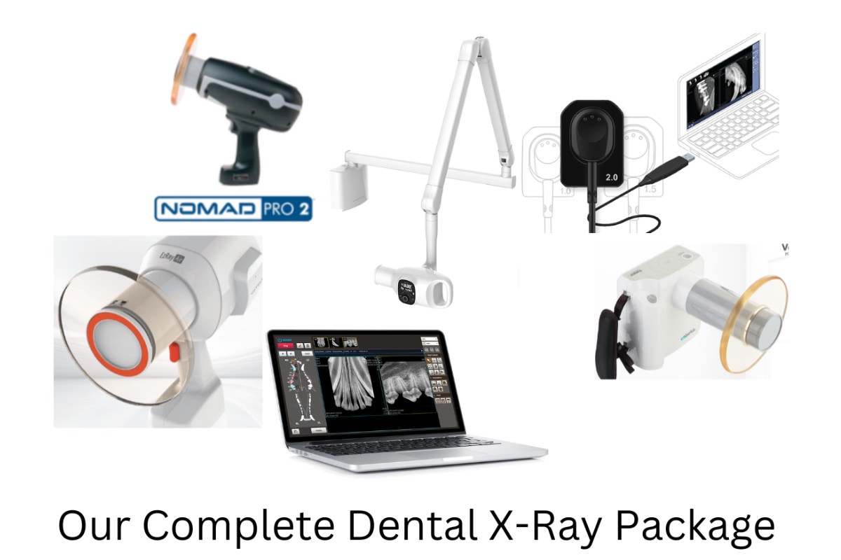
Dental radiographs have become an essential part of veterinary dentistry, often being upheld as the standard of care. That’s because dental x-rays are the only way to diagnose the full extent of dental or periodontal disease.
They help uncover unexpected lesions, assist with planning surgical extractions, and serve as part of the medical record.
Here are 10 factors that can help a busy veterinary practice decide how much to invest and which veterinary dental x-ray equipment is right for them…
Cashflow/return on investment plan. It’s not unusual for veterinary practices to report a significant increase in dental income after investing in dental radiography equipment. That’s because of an increased ability to detect pathology that needs to be treated.
However, each practice has a unique clientele, practice style, patient load and degree of busyness, and financial situation.
Developing a business and financial plan for dentals can help. Think about expected usage of the new equipment, how much to charge in your local area, and other factors. This will help a practice determine how much to invest in their dental radiography equipment.
Image quality and repeatability. Dental x-ray images need to clearly show fine details. A veterinarian must be able to distinguish small changes around the periodontal ligament and root apex, or other fine details, to determine the degree of pathology that is present.
Ask colleagues for advice. See which machines they have, whether they are happy with their purchase, and what they do or do not like about their x-ray system. When planning for a purchase, ask vendors or manufacturers if a test period with a trial machine is available and try it on different size patients to ensure it produces diagnostic images.
Technology: Film, CR, or DR. Although film is becoming less common in all x-ray modalities, there are some practitioners who prefer manually developing dental films and can do so efficiently. But for many veterinarians, digital is the way to go—it’s just a matter of choosing between CR (phosphor plates that use a plate reader to produce a digital image) or DR (a sensor that directly produces a digital image).
Cost is one consideration, as CR technology tends to be less expensive—although it does require the purchase of one or more plate readers. However, DR can be more efficient since there’s no need to run the sensor through any type of developer or plate reader.
Components and sizes. In addition to a generator, the practice will need plates (for CR) or sensors (for DR). Depending on the manufacturer, another important difference between these two technologies comes down to sensor/plate size. CR typically has more of a range of sizes available, which can be helpful for practices that see large dogs, small cats and dogs, and everything in between.
Before purchasing, check which size sensors or plates are available and ensure this will meet your needs.
Mounted versus handheld/portable. Veterinary dental x-ray generators can be mounted to carts or a wall. There are also handheld veterinary dental x-ray generators available. A mobile practice or one with limited space might appreciate a handheld unit, which has a smaller footprint and less wiring/installation concerns. It also allows for flexible angles, potentially without having to move the patient around as much to get all necessary shots.
On the other hand, a handheld unit is at a higher risk of drop damage. And safety must be considered, as veterinary radiology in general is moving toward hands-free restraint and having operators out of the room (if possible) when the image is generated. Some countries or jurisdictions might have restrictions on portable units, too, so be sure to double check before purchasing.
User-friendliness. There’s a learning curve for anyone new to dental x-rays—both for the team members taking the shots, and for the veterinarian interpreting the images. So, no matter which system you purchase, it’s important to invest in some type of training.
That being said, some systems are certainly more intuitive to use than others. Some teams would prefer to have a veterinary digital dental x-ray system with species-specific presets that make it easy to capture all shots at the correct settings.
It also helps to have a generator that is easy to move and set in the right position and angle, whether that’s an ergonomic portable machine or a mounted generator on an arm that stays where it is put.
The durability of ALL components. Common “accidents” in the veterinary setting include drop damage, water damage, and bite damage to the plate or sensor. Look for systems designed to stand up to daily veterinary use.
Warranty, service, and maintenance. Speaking of damage, sometimes accidents happen even when all reasonable precautions are taken. And you don’t want to lose out on your investment if that occurs.
A warranty is very nice to have, although it should cover common veterinary practice-related damage, including bite damage. Ask about warranty costs, coverage, and renewal options.
Also, look for ongoing costs related to service and maintenance, whether loaner equipment is available during extended repairs to equipment, and whether tech support is available.
Veterinary dental x-ray software compatibility. Software is needed for saving images to practice management software or electronic medical records, as well as for viewing and sharing the images. Make sure the dental x-ray equipment you plan to use is compatible with your practice’s software.
Financing and money considerations. Is purchasing the equipment outright an option? Or does financing make more sense in terms of cash flow? Remember to check on tax benefits as well.

Research and discuss everything with decision-makers at the practice. Ask for suggestions from team members who will be using the dental x-ray equipment. Consult business, tax, or financial professionals as needed.
Although any veterinary equipment purchase is a significant investment, it can also be a way to bring new income to the practice—not to mention boost patient care and improve workflow and efficiency.
Written by: Dr. Tammy Powell, DVM

Tips for Selecting the Ideal Veterinary X-Ray Solution

A veterinary DR system (digital direct radiography) is a common wish list item for veterinary practices. But how to know when it’s worth the cost? How much to invest? And what’s the best veterinary digital radiography system?
The answer will vary from practice to practice. In addition to looking at just the purchase price, here are 10 important considerations that can help a veterinary practice choose a new x-ray system that’s perfect for their needs…
Cashflow/return on investment plan
Knowing how much income a veterinary digital x-ray machine will bring in can guide the decision on how much to budget for the purchase.
Factors to consider include how busy the practice is (and expected to be in upcoming years), how much clients are willing to spend on their pets, and more. A veterinary business or financial consultant can help create a financial plan for new income and expenses and determine how much it makes sense to spend.
It’s also helpful to research different financing options
Where to purchase. Many veterinarians start by speaking to vendors they know well. This might bring extra perks, such as discounts for bundling services (say, purchasing an x-ray machine and lab services from the same provider). Just be sure to check the contract for minimum ongoing purchase requirements (such as the number of tests performed per month or per year) and see if this makes sense for your practice or not.
You might find deals from other sellers, too. Look for reviews and reputability, along with looking at the x-ray system itself. There are also many used veterinary digital x-ray machines for sale on sites such as eBay or usedvetequipment.com.
Imaging capabilities
Of course, you’ll want to make sure the machine has all the technical specifications your clinic needs, for all the patients you see. A feline-only hospital would have very different needs from a large animal mobile practice.
For small animal general practices, it’s advisable to test the machine on a cat’s limbs and a large dog’s abdomen and thorax, to make sure images stay high-quality and diagnostic at these different size ranges.
Ease of use
Although there’s a learning curve for any new veterinary equipment, some machines are much easier than others to learn.
Look for intuitive interfaces that will be easy for the team to start using right away. Presets for different types of species and x-ray studies are also very helpful, reducing errors and saving time.
Shipping and installation costs. Ask about potentially “hidden” costs such as shipping and installation, as well as electrical requirements (some require rewiring at the practice).
Support and replacement parts availability. Where is the company located? Is there a 24/7 tech support line? Also, find out about the availability of replacement parts, especially for older models.
Ongoing costs. Inquire about a warranty, including how long it lasts, what it covers, and if it can be renewed (and how much that costs).
Loaner machine
Also, look for information about service and maintenance requirements. See if a loaner machine is available if repairs will take a long time.
Digital veterinary software compatibility. It’s common to integrate DR images directly into practice management, image viewing, and sharing, or electronic medical record software. To avoid headaches and extra work, make sure the x-ray system you are purchasing is compatible with your practice’s software.
To avoid future expenses as much as possible, ask if software upgrades are included and if tech support or a guarantee is provided in case of any malfunctions.
Finally, ask about the privacy/security of the software
Any extra requirements or options. For example, maybe your clinic needs a portable system or horizontal beam capabilities. Or, maybe the practice would benefit from a digital x-ray unit that also has fluoroscopy capabilities.
Tax benefits. Sometimes, tax benefits alone can help a practice’s bottom line and justify an equipment purchase. Always consult a tax professional on this matter to file correctly, avoid pitfalls, and gain the maximum benefit.
It’s important to discuss everything with decision-makers at the practice and to consult business professionals as needed. It can also be helpful to include team members who would be using the x-ray system: they might have great ideas on what would make the practice flow go better and thus maximize the ROI on the new equipment.

Although a large equipment purchase is a significant investment, it can also be a way to bring new income to the practice—not to mention improve patient care and save staff time.
Written by: Dr. Tammy Powell, DVM

Small Animal Veterinary Ultrasound Probes: What to Look For
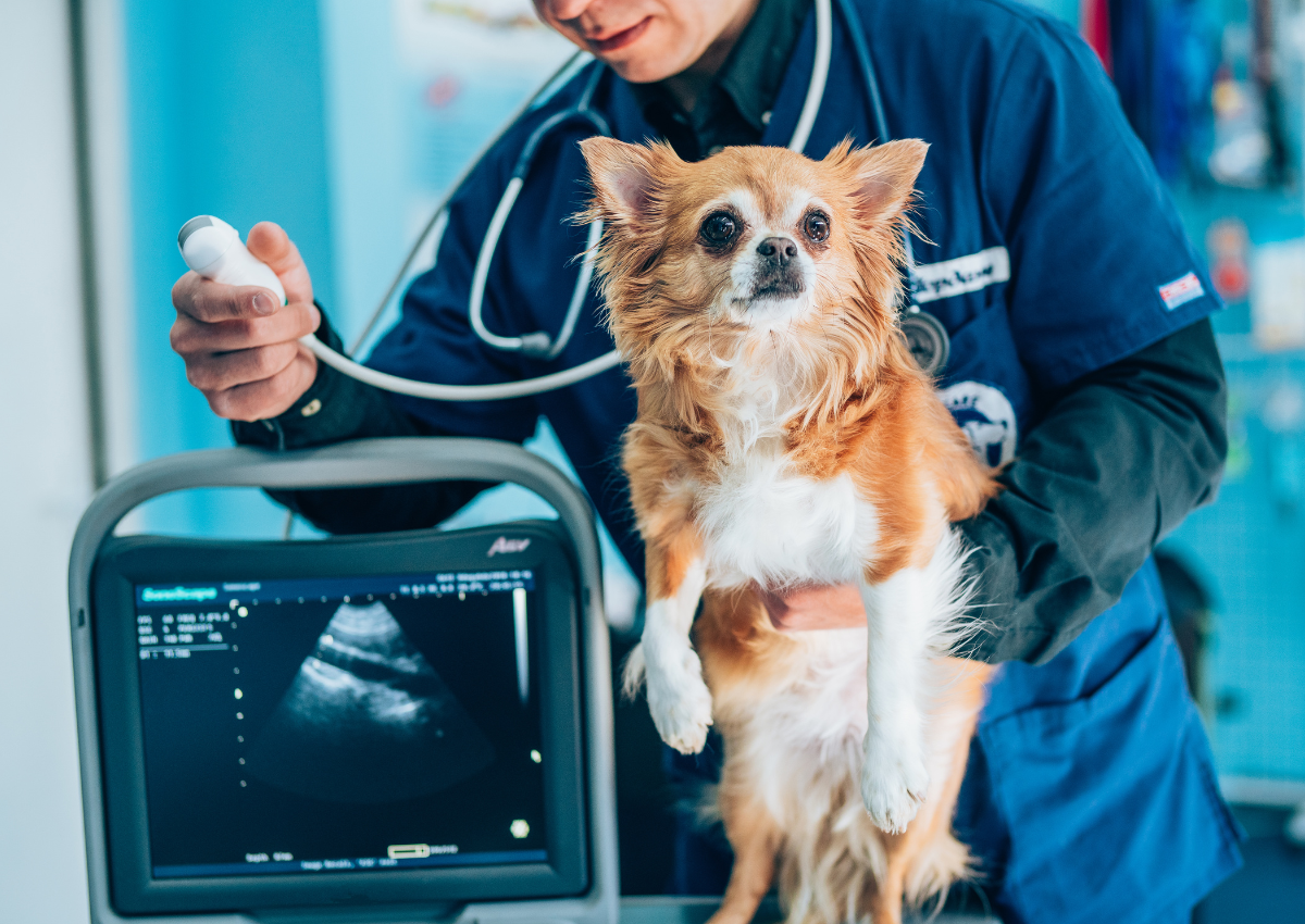
Ultrasound probes, or transducers, are a necessary part of any veterinary ultrasound system. Without a good quality probe, veterinarians would have difficulty producing high-resolution images with their ultrasound machine.
But since probes can also be a big financial investment, it’s important to select one that’s a good fit for your practice.
How Do Veterinary Ultrasound Probes Work?
These devices have piezoelectric crystals built into the probe’s working surface, which convert electrical energy into sound waves. These sound waves interact with tissues at different depths within an animal’s body, reflecting back to be picked up by those same crystals. The information is transmitted back to the ultrasound machine to produce an image for interpretation.
However, not all probes are created equally. Different probes work better than others for different patients or types of studies.
Goals for Your Veterinary Ultrasound System
What will the doctors, specialists, technicians, or other team members be using the ultrasound for?
If specialty ultrasounds, like echocardiograms and detailed abdominal studies, are performed frequently, it probably makes sense to invest in high-quality probes. On the other hand, if the ultrasound is used primarily for cystocentesis, the fanciest probe on the market might not be a great investment.
Some practices might want to invest in handheld or portable veterinary ultrasound units that connect to a smartphone or tablet. This is probably best in scenarios like triage with a focused check for fluids (A-FAST or T-FAST).
Of course, another important consideration is the type of patients your practice sees. A feline-only practice might have different needs than a practice that sees large dogs.
Goals may change over time and with additional experience. It’s not necessary to buy all the probes upfront, before practitioners are ready to use them. But it might make sense to select an ultrasound machine with additional functionalities that offer room to grow, so more probes can be added in the future when they are needed.
Veterinary Ultrasound Probe Types and What They’re Used For
Here are some important factors to consider when choosing a transducer…
Frequency and depth. Frequency is measured in MHz. Higher frequency probes offer better image resolution.
Depth refers to how deeply the sound waves penetrate into tissue.
While each of these factors are important in their own right, they are often grouped together because they are inversely related: a higher frequency probe has shallower penetration, and vice versa.
Thankfully, newer technology means many probes offer an adjustable range of frequencies and depths, so a single probe can be used on a variety of patients. There are even some that auto-adjust when viewing different depths.
Size. Probe size generally refers to the “footprint,” or the area of the probe in contact with a patient. For cats and small dogs, a smaller probe is more easily placed under the ribs, which offers an advantage for seeing certain organs. Footprint is also relevant for cardiac studies since the probe must be placed between ribs.
Shape. This refers to the shape of the footprint of the probe, and properties that tend to be associated with that category. The most commonly used probes in small animal medicine typically fall into one of these categories…
Linear. A linear probe has a footprint in a straight line. This creates a square or rectangular image with less distortion at the edges. Typically, these probes are higher frequency and low depth. Ideal for surface structures (like muscles, joints, and ligaments), some practitioners prefer linear probes for abdominal studies in cats and small dogs. A smaller, hockey stick-shaped version is also available, which may be a good fit for exotics or small structures like the eyes.
Convex and microconvex. Convex probes have a curved footprint. They vary in frequency and can be used for a variety of studies. They create a more triangular image, with some distortion at the edges.
Microconvex probes, which have a smaller footprint, are probably the most commonly used, versatile probe in small animal medicine. They’re often a good fit for those who want to invest in a good “all around” probe for cats and most dogs, for abdominal and basic cardiac exams.
However, a standard convex probe might be needed to see deep structures in very large dogs.
Phased array. This is considered a more “advanced” probe. It’s a common choice for echocardiography. It facilitates easier passage of the beam between ribs, while still generating high-quality images. Phased array probes also offer a higher frame count, for documenting small changes with the heart’s movements. They typically have several functions, including Doppler.
Additional Considerations
As with any equipment investment, it’s important to research the equipment itself and consider factors like financing, tax advantages, warranties, and service plans.
Talk to veterinary ultrasound suppliers. Ask to try out the machine you’re considering. Many offer a sample or demo machine that you can keep in the clinic for a short period of time, or at least can offer a demo in your office. Speak to colleagues or check veterinary forums for recommendations, too.
And of course, remember to protect your investment. Dropping a probe can cause permanent damage. Have a system to prevent this, including proper storage, as well as a tie or leash to hold the probe to the operator’s arm in case of accidental drops, so it won’t hit the floor.

All things considered, an ultrasound probe can be a great investment in patient care and generating income for the practice. But since they can be pricey, it’s important to choose wisely and find a probe that’s a good fit for your practice’s needs.
Written by: Dr. Tammy Powell, DVM