


How to Overcome Saying Goodbye to Their Canine Patients

When the veterinary community is mentioned, we often envision a team of compassionate and dedicated individuals who work tirelessly to care for our beloved furry friends.
While that is true, many people may not realize the emotional toll of the job, especially the difficult task of saying goodbye to patients we often grow very close to.
No matter how experienced we are, euthanizing a dog is never easy.
It is a decision that weighs heavily on the hearts of those involved.
Choosing to say goodbye means they must come to terms with the fact that they’re ending a life that is loved and cherished.
The bond between a dog and its parent(s) is truly special, and facilitating the end of that bond is heartbreaking.
The emotional burden of euthanizing dogs is a leading cause of compassion fatigue and burnout among veterinary teams.
Compassion fatigue is a type of emotional exhaustion that comes from caring for others suffering. When faced with the difficult task of putting down a dog, it can take a toll on even the most resilient of people.
The constant exposure to heartache and grief can leave us feeling drained and overwhelmed, making it difficult to continue providing the level of care that we pride ourselves on.
Veterinary teams can face several challenges when euthanizing dogs.
They may face:
Moral stress: This can result from a veterinarian’s recommendation differing from a pet parent’s. For example, an owner may not want to euthanize their dog, but the Veterinarian believes the pet is suffering. Alternatively, an owner may request euthanasia when the Veterinarian feels there’s a viable treatment option.
Ethical challenges: Whether euthanasia is considered an ethical challenge may depend on the reasons for the request.
Emotional weight: euthanasia is emotionally taxing, and it can be hard to focus on the medical or moral reasons for the goodbye.
What’s talked about least is what comes after euthanasia…
…More often than not, we have appointments to follow. We must gather, switch gears, and move on to our next patient.
This transition can be incredibly challenging as we put aside our emotions and focus on providing the best care for the next family.
This ability to compartmentalize our emotions and remain present for each patient requires special resilience and strength.
To help with these challenges, some recommend:
Take time to prepare the owner before the procedure. Be sure to set expectations and share what is within and outside the veterinary team’s control.
Setting clear expectations from the start can help prevent unwanted surprises and improve the experience.
Schedule some “flex” time after euthanasia appointments. Give yourself and your team breathing room before seeing your next patient.
Start a tradition. Some hospitals decorate a small rock for each patient who passes in their care and leave it near the entrance or a nearby tree. This is a meaningful practice for pet parents and veterinary teams to ensure their beloved pet (or patient) is not forgotten.
Despite the emotional hardships that come with the job, we continue to show up daily, providing compassionate care for our patients.
Your dedication to your community and your unwavering commitment to the well-being of your patients is truly inspiring.
Veterinary staff are scarcely recognized or appreciated for the sacrifices they make daily to offer our support and compassion.
The heavy toll of euthanasia does not go unrecognized.
Despite the emotional challenges we face, we remain committed to the health, wellness, and needs of the patients who draw us in every day.
Take this moment to know you’re appreciated. Your tireless work to care for your patients and our community is acknowledged and valued.


Providing Mobile Veterinary Euthanasia Services

Helping families with their pets
As veterinary professionals, we are faced with the difficult task of helping pet owners decide to say goodbye to their beloved companions.
Euthanasia is a compassionate and ethical part of our profession, but it can be a profoundly emotional experience for both the pet owner and the veterinary team.
In recent years, mobile euthanasia options have become increasingly popular as a way to provide a more comfortable and familiar setting for pets and pet parents during this difficult time.
These services offer a personalized and compassionate approach to end-of-life care, allowing pets to pass peacefully at home surrounded by their loved ones.
Offering mobile euthanasia services provides comfort and convenience to pets and pet owners.
In-home comfort: It’s a gift to enable pets and their parents to be surrounded by friends, family, and an environment familiar to them. Knowing their pet is comfortable is critical to pet parents during their last moments. Allowing them to snuggle up in bed with their favorite blanket and the comforting scents of home around them makes for a more calming experience.
Low Stress: Often, pets become anxious and uneasy when visiting a veterinary hospital, and being able to say goodbye in the comfort of their home can help alleviate some of the anxiety and fear accompanying the process.
Flexibility: Mobile services allow pet owners to choose the time and location that best suits their needs, making the experience more personalized and meaningful.
By offering this service as an option, you can meet the needs of pet owners who may need help traveling to a clinic or prefer the privacy and comfort of their homes.
Clients are grateful for your extra care and consideration during such a difficult time.
From a business perspective…
Offering mobile euthanasia services can be a valuable addition to a veterinary practice.
Personalizing the experience: With the well-known struggle of compassion fatigue in our community, having the opportunity to provide a personalized and comfortable experience to your patients and clients can bring a sense of ease to you, too.
Trust & relationship building: Building and maintaining good relationships with your clients is paramount to a successful business.
You’ve heard the classic Maya Angelou quote:
“People will forget what you said…but people will never forget how you made them feel.”
Providing them with a service where they can grieve in the comfort and psychological safety of their homes speaks volumes about your business morals.
Incorporating mobile euthanasia into your practice's services embodies compassion and empathy, as it significantly reduces the pet's stress by allowing them to pass peacefully at home.
This service alleviates the pet's suffering and provides immense comfort to pet owners, sparing them the distress of transporting their beloved companion during a difficult time.
However, balancing this compassionate approach with practical business considerations is essential.
Offering mobile euthanasia involves additional costs, such as travel expenses, time management, and ensuring the availability of necessary equipment and medications on the go.
Carefully evaluating these factors ensures that the service remains sustainable. This allows you to continue providing empathetic care to pets and their owners while maintaining the financial health of your practice.
When structuring your pricing, make sure you consider:
Commute time and distance (gas, mileage, etc.)
Time taken away from other patients and hospital duties
Time necessary to gather and pack/unpack equipment, meds, and other necessities into the vehicle
Additional staff needed (Veterinary assistant or Nurse)
…Now, if you’re feeling a sense of guilt for adding an upcharge to these services, remember that you’re offering a service to your community that many so desperately want to be able to utilize.
By embracing this compassionate approach to care, we can comfort and support those in need during one of life's most challenging moments.

Finding Work-Life Balance as a Mobile Veterinarian

“Struggling to balance work and life as a mobile veterinarian? You’re not alone. Our demanding, unpredictable days often leave us overcommitted and burnt out. Discover practical tips to prioritize, delegate, and say “no,” fostering a healthier work-life harmony while providing exceptional care.”
Finding work-life balance as a mobile veterinarian can feel nearly impossible.
Despite the loving patients, supportive community, and medical mysteries that keep us returning for more – we often find ourselves overexerted and burnt out.
We’re no strangers to long, demanding, unpredictable workdays and patient needs.
We’re also no strangers to neglecting or compromising pieces of our personal lives for the job.
Chronically overcommitting is our forte. And a patient in need is our kryptonite.
As veterinary professionals, we’re naturally type-A, empathetic folks who aspire to make life a little better for our patients and their owners/parents.
Although we don’t like to admit it, the imbalance between work and home life is something we’ve normalized but not something we must continue to settle for.
So, how do you break the cycle?
Let’s not pretend it’s easy to do – it’s certainly not. But it is doable.
It helps to start small. We don’t have to eat the whole watermelon at once.
Making drastic changes can inadvertently have the opposite effect, causing added stress and overwhelm.
One of the most impactful and, inadvertently, most challenging things we can do is to say “no.”
Raise your hand if you struggle using this word …
If that’s you, you’re not alone
You may have heard the phrase “No is a full sentence”. It’s time we start using it as one.
Regrettably, veterinary professionals want to help in any way we can.
We hate to disappoint or feel like we’ve let someone down.
By saying “yes” to one thing, you may not realize that you’re saying “no” to another in that same breath.
For example, by saying “yes” to that last-minute, non-emergent appointment, you’re saying “no” to eating dinner with your family.
Saying “no” starts to become a little easier when you have a good grasp on your priorities and can delegate appropriate tasks to others.
If you could use a little boost in these areas, Try starting here:
Prioritizing: Give this a try - find 15 minutes to sit down and intentionally think about your routine priorities.
Start by taking a sheet of paper and drawing a line down the center.
Label the left side “Professional” and the right side “Personal.”
Take 5 minutes to brainstorm your professional priorities (patient appointments, inventory, etc.).
Use the next 5 minutes to repeat the same exercise for the personal side.
Take a look at your list. With a highlighter, highlight the ones that bring you joy.
If by the end of this exercise, you're alarmed by the lack of joy you experience daily. It’s a good sign that it’s time to make some tweaks.
Delegate: Ask yourself: what tasks can I have someone else take over? These can be work-related tasks or personal ones. Do you have a team member looking for opportunities that you can empower to take on something new? Believe it or not, most of us find it easy to overfill our plates but struggle to take something off.
Let’s look at some ways to disconnect after a long day.
Use the alarm on your phone
You can wrap up your day by using the alarm feature on your phone.
Set an alarm for 30 minutes before you’re scheduled to finish and another for 30 minutes after.
When the first alarm goes off, start planning your exit.
If you’ve not started making your way home by the time the second alarm goes off… it’s time to go.
There will undoubtedly be days when you cannot abide by this rule, but for most days, try to make this a new personal rule (apply the 80/20 rule if you can)!
On the car ride home…
Whether just around the block or an hour-long trek back, it can be the perfect space to start disconnecting.
Consider this…
Listen to a podcast: find a podcast that helps you to switch your brain off from work mode. Do you love a good mystery, some comedy, or personal development? You can get a recommendation from a friend or search for topics you're interested in.
A quick hello: Use this time to connect with friends and loved ones. It doesn’t have to be lengthy, but you’d be surprised what a friendly voice can do to boost your spirits. Try to avoid work-related topics.
In your free time, if you haven’t joined one of the many online communities of veterinarians through social media sites, consider joining. (This can also be a small but helpful way to break up your day between appointments.)
Sometimes, just having a brief conversation (or even reading conversations) with those who wear similar shoes can be validating and relieving.
Remember…Your community needs you.
Not only for the world-class veterinary care you provide but also for all the value you bring to the world outside of working hours.
Our selfless ambition to improve the lives of pets and their parents is often an unintentional bend toward perfectionism, which can negatively affect our mentality.
If you, or someone you know in the veterinary community, are struggling with ongoing overwhelm, burnout, anxiety, or depression, rest assured you have a shoulder to lean on. Here are just a few of the many resources committed to our well-being:
· NOMV (Not One More Vet): https://www.nomv.org/

Boost Morale in Your Practice: Three Effective Ways To Help

““Veterinary professionals face compassion fatigue, burnout, and poor culture. This blog offers practical tips for boosting team morale and well-being, reducing turnover, and enhancing your practice’s reputation. Learn how to start with good news, connect work to purpose, and gauge team happiness effectively.””
3 ways to Improve Culture and Reduce Compassion Fatigue in Your Practice
It’s undeniable that compassion fatigue and burnout are two of the highest-ranking issues working in the veterinary field.
Poor culture doesn’t fall far behind.
We’re constantly faced with the emotional toll of caring for sick and injured patients and distressed or grieving pet parents.
While it is essential to have empathy and compassion for the patients we care for, it’s equally important to take care of ourselves and support our teams.
Improving practice culture and team wellbeing is not easy and certainly doesn’t happen overnight.
Taking a step back to recognize the need for positive change in your hospital can help to:
Decrease staff turnover & increase satisfaction
Enhance your practice’s reputation
Improve staff wellbeing & encourage work/life balance
There are lots of resources floating around about “how to improve team morale” and “what to do to prevent burnout.”
Many share advice like “set boundaries,” “focus on team wellbeing,” and “practice self-care.”
All of this is valid advice, but there’s a gap between what we’re being told and how to apply it.
So, let’s start to get a little more specific…
Here are three ways to improve culture and reduce compassion fatigue in your practice – AND – ways you can apply them.
1. Start with Good News!
Team culture significantly contributes to how you view and experience your work.
You can start to sway a more positive team culture by baking good news into the day.
Practices that start their day/shift with good news experience:
Higher rates of engagement
A better sense of team building, connection & community
Increased emotional resilience
What type of good news are we talking about…
Choose your adventure here!
Good news could be related to work or professional development. (I placed my first urinary catheter yesterday!)
It could be something personal. (My partner and I just closed on our new house!)
It could be something that happened over the weekend or something they’re looking forward to. (I just booked a cruise for the end of the year!)
Or, good news can be sharing something new you’ve learned. (Yesterday, I realized that a group of geese is called a “gaggle”! Who knew?!)
What might sharing good news look like?
Good news can be sprinkled throughout your day wherever it makes the most sense to your practice. It doesn’t have to be lengthy or detailed—short and sweet will do the trick!
Here are a few ideas to get you started:
Rounds: If your hospital does rounds at the start of each shift, ask each person to start by sharing good news before diving into patient updates.
Team huddles: if your practice starts each day off with a team huddle, start the meeting by sharing good news! (Have a large staff? Consider asking for three volunteers each day to share to keep it brief)
Whiteboard: don’t have daily gatherings with your team? Consider setting up a whiteboard in a common area (the break room, treatment area, etc.) and ask the team to write their good news at the start of their shift.
The Washington Post stated that 7/10 Americans suffer from negative news fatigue in their article “The Good News Effect. “
The article concerns what we often consume through news and social media.
While we can’t control what our team consumes through their many downloaded platforms, we can be intentional about bringing good news into their day.
Encouraging the brain to think about something positive, even during times that feel draining or overwhelming, can have a long-term effect on wellbeing.
2. Link work + Purpose
It’s a sad, common misconception that you need to be saving the world to feel like what you do is meaningful.
All too often, we hear phrases like “I’m just a receptionist,” “I’m just a Vet Tech,” or “I just work in general practice.”
We either lack pride or feel judged by our roles as if we have little value.
As veterinary leaders, we can help our teams (and often ourselves) flip the script.
Every role in the hospital holds value, and every person is important. It’s our job to empower our teams to take pride in their purpose.
How can we do that?
You can be creative with this one! Here are a few ideas to get you started:
Create a “why” or “purpose” statement: reserve a little time at your next team meeting to give the team 5-10 minutes of silence to think of and jot down why they come to work every day. What’s fulfilling to them? What inspires them? What impact do they feel they make to the hospital or veterinary community?
Create Vision Boards: this could be fun for a team meeting or outing activity and can be done the old-fashioned way (paper, magazines, scrapbooking material) or digitally, using a free digital creative space or even a Pinterest board. Have the team build their “vision” behind why they love their jobs and how they hope to see it grow.
Tip: These activities can be done individually and as a team.
For example, you may have each team member create individual vision boards and then create a team vision board as a group. A personal vision board might include their goal to learn how to perform an ultrasound and some photos of their pets since they’re their inspiration.
The team vision board might include photos of favorite patients, getting that new intensive care unit for the treatment area, and getting AHAA Certified.
3. Scraps in the bucket
Do you have a pulse on your team’s culture and overall happiness at work?
It can be challenging to gauge, especially in hospitals with a large staff or multiple departments.
Can’t I send out a survey?
Sure, surveys are undoubtedly helpful.
But what if you could get a pulse on how many good days the team experiences vs the not-so-great ones in a fun and interactive way?
Here’s an idea you can try:
Most hospitals have abundant scrap paper (even in the era of trying to go “paperless”). Why not put it to use, even after it’s been used?
Put two buckets in a common area (near the lockers, breakroom, or the hospital’s exit in the back).
Label bucket 1: I had a good day!
Label bucket 2: Tomorrow will be better (or “I’ve not had a great day”, or simply “bad day”).
Have the team crumple a piece of scrap paper and toss it into one of the buckets before they leave.
This can be a fun way to determine whether your team has more good days than bad or vice versa overall.
Don’t have a lot of scrap paper laying around?
First of all, bravo! Feel free to use an alternative. Some practices have used things like ping pong balls.
One practice got creative, buying two rubber dart boards and extra darts (the ones that won’t destroy the walls, of course) and placing them in the breakroom.
One was labeled “I had a great day” and the other “I had a bad day”.
All these activities can help foster a positive work environment, nurture team building, and bring a little fun into the mix.
Reducing compassion fatigue and improving culture isn’t a “one size fits all” solution; it requires an intentional approach and a gentle reminder that we’re all in this together.

Mental Health: Combat Compassion Fatigue & Stress

““As a veterinary community, we prioritize patient care, but the job’s demands strain mental health. Recognize and address issues like compassion fatigue. Find support through NOMV, AVMA, and AAHA to ensure well-being. Remember, asking for help and prioritizing your health is okay.””
Caring for Pets, Coping with Stress: The Mental Health Challenges of Veterinary Professionals
As a veterinary community, we dedicate our lives to caring for our patients and prioritizing their well-being.
We have a deep passion for helping our patients and their parents, but often, the demands of the job can take a toll on our mental health.
From handling complex cases to managing the emotional burden of poor diagnosis and euthanasia, it's easy to feel overwhelmed and burnt out.
Not to mention the stress of owning, managing, and maintaining a successful practice.
The veterinary profession, where so many rely on your care, makes it even more crucial for veterinarians to have their own support system.
It's a tragic reality that suicide is a serious concern within the field, with 1 in 6 experienced suicidal ideation since leaving veterinary school.
It feels cliché to sy, “It's important to prioritize your mental health,” but just as you prioritize the health of your patients, your well-being is paramount to a fulfilling work life.
Compassion fatigue is a genuine issue that so many of our peers face…
…And unfortunately, it’s grown a sense of normalcy within our community.
Working in a high-stakes field like veterinary medicine can feel like it comes at a price, but recognizing signs and symptoms early within ourselves and our teams can help keep that cost low.
One of the most essential things leadership teams can do is acknowledge the existence of mental health and wellbeing issues in the profession and provide support and a sense of teamwork and community for their teams.
A shift happens when compassion fatigue and burnout start to kick in.
You may experience symptoms like:
Decreased energy
Overwhelming feelings of burnout/disengagement
Generalized anxiety or depression
Difficulty concentrating and sleeping
Rumination about recent cases with unfavorable outcomes
Thoughts of harming yourself to escape problems
These same symptoms are ones to watch out for in our peers, too. They can easily brush aside and chalk up to having a “bad week, month, or year.”
This is why it’s so important to recognize the warning signs and have some strategies in your back pocket to help curb the mental burden these symptoms cause.
To start, you may have to ask yourself or your peers some challenging questions.
What might be the root of these symptoms?
What’s within my control to change? What’s outside of my control?
Am I putting more pressure/stress on myself than is necessary?
As you start to answer some of these questions, here are a few things that may help that are within the realm of your control:
Avoid perfectionism– always seeking perfect solutions exacerbates stress and anxiety.
Avoid comparing yourself to others– it’s said that comparison is the thief of joy. Comparison leads to envy and a lack of self-esteem. Focus on recognizing and continuing to strengthen your talents and skills.
Set boundaries– Serving your clients is essential, but there’s a fine line between being helpful and allowing work to overstep into your personal life. Achieving a healthy work-life balance will promote your desire for longevity to continue the work that brings you joy.
Prioritize yourself– Make time for activities that bring you joy and relaxation, whether exercising, spending time with loved ones, or engaging in a hobby. Stop making excuses. Taking breaks and setting boundaries with work can help prevent burnout and replenish your mental and emotional reserves.
Find connections: whether it's talking to fellow colleagues, seeking therapy, or joining a support group, having a safe space to share your feelings and experiences can make a world of difference.
In addition to seeking support and practicing self-care, there are resources available specifically for veterinarians dealing with mental health challenges.
Organizations like Not One More Vet, the American Veterinary Medical Association, and AHAA offer resources, support, and access to mental health professionals who understand veterinarians' unique challenges.
NOMV emphasizes raising mental health awareness in the veterinary profession and research to reduce suicide rates of veterinary professionals. Through an online network, NOMV provides support and healing efforts to show you that you are not alone.
As part of their work, NOMV also works with veterinarians to build a support network of therapists and other mental health resources.
Similarly, American Veterinary Medical Association (AVMA) provides well-being tools and resources such as:
Wellbeing Assessment for Veterinarians A self-care planning guide
Steps to create a healthy working environment AVMA Journals Collection on Wellbeing
Promoting a healthy work culture of support and care increases productivity and feelings of self-worth. AVMA also informs veterinarians about how to receive help and encourages the use of preventative measures regarding mental and physical health.
You can also find a wealth of helpful well-being articles on the AAHA website geared toward mental health and well-being for the veterinary community (check out: AAHA Guide to Veterinary Practice Team Wellbeing).
Remember, it's okay not to be okay, and asking for help is a sign of strength, not weakness.
So, take a moment today to check in with yourself and your team and prioritize mental health.
We all deserve to feel supported, cared for, and valued.
Take this as a gentle reminder that you’re not alone in this journey, and resources and coping strategies are available to help you navigate the challenges of the job with compassion and resilience.

Overcoming compassion fatigue for Veterinarians

““Veterinarians often face compassion fatigue, stemming from the emotional toll of their work. Recognizing signs like burnout and emotional exhaustion is crucial. By setting boundaries and prioritizing self-care, vets can manage this fatigue. Learn practical strategies for maintaining your well-being and delivering exceptional care in our latest blog.””
Compassion fatigue is a genuine and often overlooked issue many veterinarians face in our work.
The demanding nature of the job, the emotional toll of caring for sick and injured patients, and the pressure to provide the best possible care can all contribute to feelings of overwhelm and exhaustion.
Thankfully, there are strategies that veterinarians can use to overcome compassion fatigue and continue to provide the high level of care that their patients deserve.
First and foremost, it’s essential to recognize the signs of compassion fatigue (also known as empathetic distress) in ourselves and those we work closely with. These signs may include:
Feelings of burnout
Emotional exhaustion
Cynicism
Decreased sense of personal accomplishment
Once we recognize these signs, we can take steps to address and manage the things we’re most struggling with.
Setting clear boundaries is an act of self-compassion and respect.
Setting Boundaries is one of the best (often most challenging) strategies for overcoming compassion fatigue.
Learning to say no when feeling overwhelmed, giving yourself permission to take breaks, and prioritizing self-care are often frowned upon in our field.
Veterinary teams are filled with caring, empathetic individuals who are notorious for overcommitting and overburdening themselves, making it difficult to set boundaries with colleagues and pet parents.
Setting some self-boundaries is a great place to start! Ask yourself:
What limitations can I set around being contacted during personal time?
What can I delegate to a colleague or team member to reduce feeling overwhelmed?
How can I empower my team to handle challenging situations so I’m pulled in less frequently?
What can I do to prioritize myself and the things that bring me joy?
Having even so much as a general answer to some of these questions can help you start to set some boundaries to reduce feeling overstressed.
How can you set boundaries with pet parents?
Setting a boundary with your clients can feel uncomfortable at first. To get started, you’ll need to identify the types of behavior you’ll tolerate and those you won’t.
For example, you might be willing to tolerate a pet parent sharing their frustration over an extended wait in the lobby.
Still, you’re unwilling to tolerate yelling, foul language, or verbal abuse toward you or your staff.
Next, you’ll need to kindly yet firmly communicate the boundary you’ve set. This part can feel a little uncomfortable if it’s not part of your daily practice.
For example, “Thank you for sharing your feedback, Mr. Smith. While I understand your frustration, my team and I will not tolerate abrasive behavior. I’d like to ask that you reduce your tone, or I’ll have to move on to my next appointment.”
Setting boundaries with your clients demonstrates compassion for your team, encourages respect from pet parents, and protects your mental health.
Tip: This can be a great team-building activity at a staff meeting.
By allowing the team to collectively establish acceptable and unacceptable behavior (both internally and from clients), you empower each of them to have a voice. You can equip them with strategies to use when facing a difficult situation.
Consider adding boundary-setting and reinforcement strategies to your next team huddle so your team feels prepared to enforce the boundaries you’ve agreed upon.
Self-care is a close second for the best strategies to overcome compassion fatigue.
Self-care means taking the time to prioritize your own physical, emotional, and mental well-being. Making daily choices to engage in activities that bring joy and relaxation, like:
Exercise (going to the gym, rollerblading, dance, martial arts…)
Hobbies (reading, coaching, golfing, DIY projects)
Spending time with loved ones
All too often, we feel dedicating time to ourselves feels selfish, or “we’ll get to it tomorrow,” but over time, it harms our interactions with our staff, patients, and clients.
Studies have shown that regular self-care enhances Veterinarians' ability to continue caring for patients compassionately, safely, and more accurately (decreasing mistakes and enabling critical thinking).
Remember, it is not selfish to prioritize your well-being…
…it’s necessary to continue to show up as the best version of yourself for your patients and clients. If we don’t take a moment to recharge ourselves, we won’t have enough juice left to help those who need us most.
Wellbeing Resources Geared Toward the Veterinary Community
The American Veterinary Medical Association (AVMA) hosts a number of CE-accredited trainings and webinars and offers a free wellbeing assessment along with some helpful self-care tools
Not One More Vet (NOMV) is an organization that works tirelessly to support the well-being of the veterinary community
Suicide and Crisis prevention Hotline offers free, confidential support for those in distress
Don’t allow yourself or your team to suffer alone. Don’t wait. Reach out. Speak up.

Burnout vs. Compassion Fatigue in Veterinary Medicine

““Discover the differences between burnout and compassion fatigue in veterinary medicine. Understand how prolonged stress and emotional tolls from treating sick animals can impact well-being. Learn effective self-care strategies and leadership roles in maintaining a healthy work environment.””
Working in the field of veterinary medicine is incredibly rewarding.
We provide comfort and treatment to patients with illness, disease, and injury.
We’re privileged to nurse sick patients back to health and experience the joy of being reunited with their families.
…But our line of work also has its fair share of challenges.
One of the biggest obstacles that veterinary professionals face is the risk of burnout and compassion fatigue.
Both can significantly impact the mental and emotional well-being of those working in the industry, but it is important to understand the differences between them.
Let’s start with burnout.
Burnout is a state of emotional, physical, and mental exhaustion caused by excessive and prolonged stress.
In the veterinary space, feelings of burnout can occur when we are constantly faced with demanding workloads, long hours, and emotional distress from dealing with sick or injured animals. It’s chronic and happens gradually over time.
Burnout can lead to feelings of:
Frustration
Exhaustion
Cynicism
A sense of detachment/alienation from the job
Burnout can also manifest as physical symptoms…
You might see or experience symptoms such as fatigue, headaches, insomnia, and a lack of self-care.
On the other hand…
…compassion fatigue is a specific type of burnout
Compassion fatigue (also known as empathy fatigue) occurs when individuals are regularly exposed to the suffering/trauma of others.
In our field, compassion fatigue can develop as a result of repeatedly witnessing the pain and suffering of patients, as well as the emotional toll of dealing with grieving pet parents.
Compounded stress results from an ongoing wish to relieve suffering from patients and pet parents (often followed by feelings of failure), which can lead to feelings of hopelessness, guilt, and a decreased ability to empathize with others.
The very nature of our work exposes us to the (often) sad reality of patient decline and owner distress or loss.
Burnout and compassion fatigue are serious issues that can profoundly impact the well-being of veterinary professionals.
Veterinary teams must recognize the signs and symptoms of these conditions and take steps to prevent and address them.
One way to combat burnout and compassion fatigue is through self-care practices.
Self-care means preserving time for personal well-being, such as:
Regular exercise
Healthy eating
Adequate rest
Actively participating in hobbies outside of work
Spending time with friends and family
If you’re struggling with burnout and/or compassion fatigue, seek support from colleagues, friends, and mental health professionals immediately.
Building a solid support network can help us cope with the challenges of the job and prevent feelings of isolation and overwhelm.
Additionally, veterinary organizations and leadership teams play a crucial role in addressing burnout and compassion fatigue by offering resources such as:
Counseling services
Stress management workshops
Flexible work schedules
As leaders, we need to prioritize our own well-being to help maintain a supportive and healthy work environment for our teams and empower them to continue providing the best care for our patients.
By recognizing the signs of burnout and compassion fatigue and taking proactive steps to address these issues, we enable our teams to continue making a positive impact in the lives of our patients and pet parents.
Well-being Resources Geared Toward the Veterinary Community
American Veterinary Medical Association (AVMA) hosts a number of CE-accredited trainings and webinars and offers a free wellbeing assessment along with some helpful self-care tools
Not One More Vet (NOMV) is an organization that works tirelessly to support the well-being of the veterinary community
Suicide and Crisis prevention Hotline offers free, confidential support for those in distress
Don’t allow yourself or your team to suffer alone. Don’t wait. Reach out. Speak up.

3 Signs of Compassion Fatigue in Veterinary Medicine

““Discover three signs of compassion fatigue in veterinary medicine. Learn to recognize physical, emotional, and behavioral symptoms like exhaustion, anxiety, and withdrawal. Awareness and early intervention are critical. Explore resources from AVMA and NOMV to support well-being.””
As Veterinary caregivers, we’re deeply motivated to serve our patients and deliver the best possible care.
We’re no strangers to the phrase “we don’t deserve pets.” Their unwavering love and loyalty are gifts we’re so fortunate to experience.
The American Veterinary Medical Association (AVMA) defines the human-animal bond as “a mutually beneficial and dynamic relationship between people and the animals that are influenced by behaviors that are essential to the health and well-being of both.”
This bond brings joy, warmth, and laughter to the workday, and being surrounded by like-minded, compassionate coworkers makes the work environment that much brighter.
But, with the stress of being understaffed, working long hours, and repeated exposure to trauma and grief…
…it’s easy for a bright environment to feel dark.
All too often, veterinary teams struggle with feeling overworked.
Pair that with bearing witness to abuse, neglect, and euthanasia… It’s easy to see why compassion fatigue is so common among our peers.
Compassion Fatigue is a result of extreme exhaustion unique to compassionate caregivers regularly exposed to loss or trauma.
There’s a growing awareness that caregivers of all kinds should make sure they make time to care for themselves.
Why?
It’s the same reason you’re told on a flight to put on your own oxygen mask first before helping others.
At the risk of sounding cliche, the phrase “you can’t pour from an empty cup” couldn’t be more accurate.
Like our feline patients, veterinary teams often hide signs of being overwhelmed. They often appear stoic, professional, and productive, which can make recognizing signs of compassion fatigue difficult.
The more we know about compassion fatigue, the better we can identify the symptoms…
…and step in to support our peers when they need us most.
Let’s take a moment to unpack three common signs of compassion fatigue to be on the lookout for:
#1: Physical signs
Compassion fatigue might physically present itself.
Let’s take Dr. Isaacs, for example.
Since her associate is on maternity leave, she’s trying to maintain 3x of her normal caseload, and this week, there’s been an unusual amount of euthanasia on the schedule.
You notice she’s been wearing the same scrubs for the past three days and hasn’t taken her usual lunch or snack breaks.
You overhear a client comment on her tired appearance during their pet’s physical exam. Dr. Isaacs casually shares that she’s not been sleeping well and has had intermittent headaches.
Little red flags are going up. She’s showing common physical signs that she’s experiencing compassion fatigue including:
Exhaustion & headaches
Changes in sleep patterns or appetite
Lack of self-care
Stomachaches & digestive changes
#2: Emotional signs
It’s not uncommon for compassion fatigue to appear more emotionally.
Take Ashley, for example.
Ashley has been a Veterinary ER nurse for 6 years and is well experienced. She has a great reputation for acting quickly, calmly, and rationally during patient crises.
Lately, you’ve noticed she’s been hypervigilant about her patients and hasn’t been as social with the team. When patient outcomes aren’t successful, she becomes easily overwhelmed, emotional, and negative.
Sudden mood swings or overreacting aren’t uncommon symptoms.
You might also witness:
Increased anxiety or sadness
Feelings of guilt or helplessness
Difficulty concentrating
Hypersensitivity or insensitivity to emotional experiences
#3 Behavioral changes
For this one, let’s talk about Daniel.
Daniel has only been a receptionist for a few months but has learned the ropes quickly.
Due to a recent heat wave, the hospital has seen an influx of patients suffering from heat-related complications, many of whom have had unfortunate and unexpected goodbyes.
Although Daniel has been very friendly and easy to work with up to this point, you notice his patience threshold decreasing. He often disappears to the breakroom for extended periods and has been very quiet during his shifts.
Since his time in practice has been so limited, it might surprise you to learn that he’s struggling with compassion fatigue.
He’s demonstrating common signs like:
Isolation or withdrawal
Irritability
Lower tolerance to frustration
Compassion fatigue doesn’t have boundaries
It doesn’t discriminate against your time or experience in practice or the role you serve at the hospital.
If you’re experiencing some of the symptoms listed above, know you’re not alone. Do your best to be open and transparent with your team about your feelings and what you might need to recover.
As a leadership team, we want to foster a culture that supports the mental and emotional well-being of our employees.
At the very least, we want to ensure that our team is aware of and has access to resources that could significantly impact their well-being.
Wellbeing Resources Geared Toward the Veterinary Community
The American Veterinary Medical Association (AVMA) hosts a number of CE-accredited trainings and webinars and offers a free wellbeing assessment along with some helpful self-care tools
Not One More Vet (NOMV) is an organization that works tirelessly to support the well-being of the veterinary community
Suicide and Crisis prevention Hotline offers free, confidential support for those in distress
Don’t allow yourself or your team to suffer alone. Don’t wait. Reach out. Speak up.

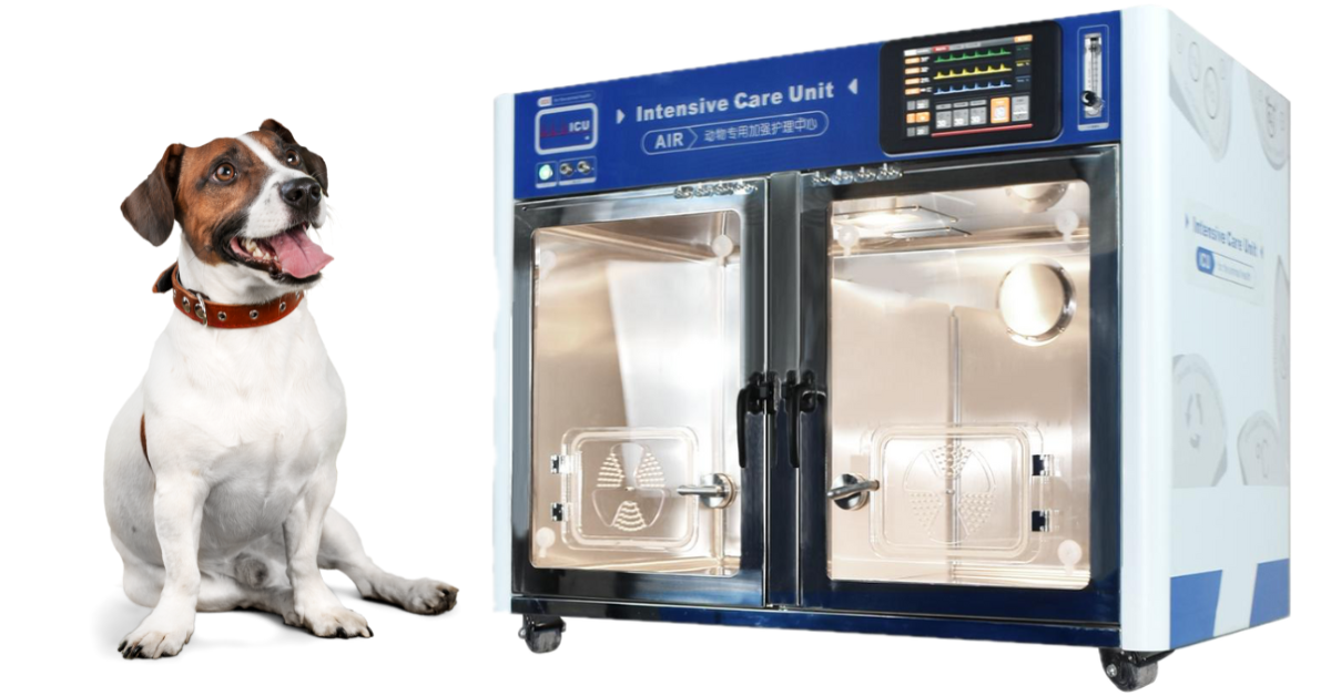
Elevating Veterinary Patient Care Protocols with ICU Cages
Picture this… Let’s call it scenario 1:
Rocco, a canine patient you’ve come to know and love, comes in for a routine dental.
After completing a pre-anesthetic exam and reviewing his recent lab work, Rocco was led to the treatment area for anesthetic induction and his dental procedure.
His dental visit was thankfully uneventful. He had a basic cleaning and one minor extraction. He’s ready to move on to recovery!
As Rocco wakes up from anesthesia, he’s thermogenic (his body temperature is below average) and tachycardic (his heart rate is severely elevated).
As veterinary nurses attempt to intervene, Rocco’s anxiety increases, and he begins thrashing and vocalizing. Through the kennel door, one of the nurses notices the color of his tongue slowly change from bright pink to a light pale purple, and they attempt to administer oxygen.
Rocco’s responding fearfully and won’t allow the team to come close. The veterinarian, who’s already started seeing afternoon appointments, is pulled from a room to assist.
Let’s pause here…
A few red flags are going up:
1. The patient is overly stressed and because of his fearful response, intervention, and treatment are delayed
2. The Veterinarian is pulled away from appointments, causing delays for other patients needing care
3. Clients are getting understandably upset due to extended wait times
The snowball effect continues…
So, let’s switch gears and take a look at scenario 2:
Rocco, who you know and love, comes in for his routine dental as scheduled.
The pre-anesthetic exam and labs look good – so he’s off to his procedure.
The procedure goes as planned. A simple cleaning and a single extraction. Easy peasy, lemon squeezy.
Rocco has been moved to the recovery area, where a temperature-controlled ICU cage awaits him. The warm environment helps slowly bring his body temperature back to normal.
The soft-close plastic doors reduce the unfamiliar noises of the hospital, helping keep Rocco’s anxiety at bay. With clear visibility, his veterinary team can keep close tabs on his progress.
After his swallow reflex returns and he’s safely extubated, the treatment technician uses the external control panel to turn on some supplemental oxygen for Rocco as he recovers.
After a short time, Rocco’s vitals are in a healthy range; he’s bright, alert, and ready to head home!
Although your hospital may not offer emergency or ICU services…
ICU cages still bring incredible value to patient care and staff monitoring in general practice.
They increase hospital workflow and efficiency by helping to reduce complications, minimizing unnecessary staff attention, and minimizing disruptions to the daily schedule.
In our last scenario, having the ICU cages as part of their recovery protocol helped:
· Improve patient recovery
· Reduce appointment interruptions
· Ease observation and monitoring
As time passes, more veterinary practices are investing in ICU cages to enhance their patient care protocols and elevate their standard of care.
Now more than ever, our furry friends are considered family members more than they are “house pets.”
The love and companionship they bring to our lives are unmatched, and as their veterinary care team, we have the exciting opportunity to provide a comfortable and safe experience.
Learn More about ICU cages - https://newvetequipment.com/intensive-care-unit

Maximizing Veterinary Patient Recovery with ICU Cages

Processes and Procedures: Achieving Optimal Outcomes for ICU Patients
In our ICU units, the primary objective is clear: to ensure our patients survive, recover, and return to their owners in the best possible health.
Given the critical nature of their conditions, this goal is often challenging, but by adopting strategic approaches and effective management techniques, we can significantly optimize patient outcomes.
Collaboration among veterinarians, veterinary technicians, and pet owners is essential to this process.
Establishing Objectives
The initial step is to define the desired outcomes and set clear expectations for all parties involved.
This may include goals such as reducing patient mortality rates, accelerating mobility post-surgery, or enhancing data recording efficiency.
These professional aims will differ from the clients' priorities, which typically focus on the pet's quality of life.
Early in the case, it's crucial to engage with pet owners to understand their primary concerns and objectives.
Whether their goal is to get their dog back to running long distances, encourage their cat to eat again, or provide palliative care for a pet in its final days, understanding these aims is the first step toward achieving them.
Staffing Appropriately in Veterinary ICUs
Effective management of an ICU hinges on efficiency, both among staff and within care procedures. Ensuring that an ICU is appropriately staffed is crucial.
Each team member should understand their role and perform their duties to the best of their abilities. Additionally, staff should be ready to assist colleagues when needed and prioritize their own physical and mental well-being.
Research in human medicine has demonstrated that nurses significantly impact patient outcomes and survival rates in hospitals. We can draw a parallel in veterinary medicine, recognizing that the expertise and efficiency of veterinary technicians are vital to the success of any shift.
A 2020 study reinforced this, examining errors and patient outcomes in three U.S. veterinary hospitals. It found that variations in veterinary technician staffing levels and experience directly influenced the occurrence of errors.
This underscores the importance of appropriate staffing in maintaining high standards of care and improving patient outcomes in veterinary ICUs.
Communicating Effectively in a Veterinary ICU
Effective communication is crucial in any team environment, but it is especially vital in a busy, fast-paced, and stressful ICU setting.
ICU patients often have complex, multi-factorial needs, making thorough and precise communication essential.
This involves not only verbal communication, such as running 'rounds' at the start of each shift, but also ensuring that paperwork—including hospital charts, medication charts, and owner consent forms—is meticulously maintained.
Small details, like checking off when a medication has been administered or highlighting a known allergy, can significantly impact patient care.
Ensuring legible handwriting and clearly labeling any symbols used can enhance accuracy. Assigning one or two people to oversee each case can help minimize discrepancies caused by communication errors.
Rounds are beneficial for keeping the entire team updated on the critical aspects of each case. However, detailed handovers should ideally occur directly between team members responsible for the patient to avoid potential miscommunication.
Learning from mistakes
Veterinary medicine is a volatile business and despite everyone’s best efforts, things can and will go wrong.
In order for the system to continue to function positively, it is vital that mistakes are not ignored or shrugged off – this does absolutely not mean that a blame culture should be encouraged, but means that any mistakes, or even near-misses, should be documented and learned from in order to ensure they don’t happen again³.
It is worth noting that these shouldn’t be limited to drastic events such as the death of a patient, but could be as simple as a set of blood results going missing.
An anonymous system is one common way to achieve this, by which the information is fed back to senior staff who then can relay it and any solutions to the team, with or without a staff meeting.
If members of staff have been involved in a difficult case or one with a negative outcome, they should also be encouraged to attend ‘debrief’ meetings in order to process the situation, and to help support their mental health.
This is much more commonly done in human medicine but should also be promoted within the veterinary field.
Having checklists and SOPs in place for situations such as drawing up drugs or dispensing medications, which center around one person ‘doing’ and another person ‘checking’ can help to reduce errors in dosing.
It is vital that training is also kept up to date and any new members of staff are trained in these protocols as soon as possible.
Auditing
Carrying out clinical audits can help bring to light any potential issues and areas in which patient care or outcomes could be compromised.
An example of a clinical audit could be evaluating ICU patient temperatures after they have undergone an operation, to ensure they are being managed appropriately and are consistently within expected limits. I
f this audit was to reveal that maybe temperatures in the post-operative period are below the recommended range, it should prompt changes to be made to improve the situation, such as implementing the use of active or passive warming devices.
However, probably the most important part of this process is to re-audit after a certain period of time, to ensure improvements have taken place.
One model that summarizes this is the PDSA model – Plan, Do, Study, Act⁴.
Plan to carry out a change
Do carry out a change
Study the results of carrying out the change
Act to make any further adjustments
Post-event audits can tie in nicely with evaluating mistakes or near-misses in the practice and provide a structure to learn from. But again, there should never be any blame placed on any individual or group of individuals.
Looking at the bigger picture
Veterinary medicine is constantly evolving, and we are discovering new and exciting methods to aid in our diagnostics and treatment of our veterinary patients.
However, sometimes, we need to take a step back and look at how getting the basics right can form a stable foundation on which to build a patient’s recovery.
Although surgery and drug therapy will make up a large proportion of the treatment an animal may receive, they are not the only things we have in our armoury to improve outcomes.
Nutrition: The Foundation of Your Pet's Recovery
Proper nutrition is essential for the recovery of your pet, whether a cat or dog. Without appropriate and sometimes specialized nutritional therapy, other medical or surgical treatments may not reach their full potential.
The Benefits of Complementary Therapies
In addition to nutrition, complementary therapies like physiotherapy, hydrotherapy, laser therapy, and acupuncture have shown significant benefits for your pet's recovery. These therapies are effective not only for mobility issues but also for a range of other conditions.
Adopting a Multi-Modal Approach
Utilizing a multi-modal approach in treatment increases the chances of achieving our primary goal: ensuring that the pet survives, recovers, and returns to you in the best possible health.
Maximizing ICU Potential
The ICU is a complex environment but also one that offers immense opportunities. By leveraging the full capabilities of veterinary staff and medicine, we can transform dire situations into positive outcomes for your beloved cat or dog.
References
1. Curtin LL. An integrated analysis of nurse staffing and related variables: effects on patient outcomes. Online J Issues Nurs. 2003;8(3):5. PMID: 14656199.
2. Hayes GM, Bersenas AM, Mathews K, Lane WG, LaLonde-Paul DF, Steele A, Avellaneda A. A multicenter observational study investigating care errors, staffing levels, and workload in small animal intensive care units. J Vet Emerg Crit Care (San Antonio). 2020 Sep;30(5):517-524. doi: 10.1111/vec.12991. Epub 2020 Sep 12. PMID: 32918379.
3. Quality improvement for patient safety and a better practice culture; Pam Mosedale and Mark Turner; The Veterinary Nurse 2022 13:4, 156-161
4. https://todaysveterinarypractice.com/practice-management/measuring-patient-outcomes/
3 Reasons Why ICU Cages are Critical to Veterinary Recovery

Unsurprisingly, ICU (intensive care unit) cages have gained popularity in veterinary hospitals and are slowly being considered a standard part of inpatient care.
The rate of pet ownership continues to experience steady growth, resulting in a rising demand for veterinary care and services.
Veterinary ICUs, specialty & emergency hospitals, and general practices have expanded hospital treatment areas to accommodate ICU cages and support the incoming demand.
Market research indicates that ICU cage popularity will continue to grow due to their crucial role in providing intensive care to ill, injured, or post-operative patients.
With a controlled environment for monitoring and supportive care, ICU cages are the ideal stress-free environment for patients to recover comfortably.
Let’s take a look at three main reasons ICU cages are so critical to veterinary recovery.
#1 Convenient Patient Monitoring and Supportive Care for Inpatients
There is no “one-size-fits-all” approach to monitoring and supportive care, and it’s safe to say you can never have too much.
Age, weight, disease process, pain profile, and other factors can vary the level of supportive care our patients need.
To be cautious, monitoring should continue even after the patient is considered " normal.”
ICU Cages offer:
Soft, close, clear plastic doors for easy visual observation
3-1 lighting systems with adjustable brightness for exams, observation, and therapy
Silent air systems for quiet oxygen therapy
Built-in IV support and Nebulizer
#2 Comfortable Anesthetic Recovery
Since post-anesthetic recovery isn’t always straightforward, diligent monitoring can reduce the risk of complications.
Anesthesia (and surgical procedures) can profoundly impact a patient’s thermoregulatory system. Small changes in a patient’s body temperature can encourage recovery or harm cellular and tissue function.
Hypothermia following an anesthetic procedure is one of the most common complications found in canines and felines.
ICU cages use an active warming approach in a temperature-controlled environment, which reduces the risk of hypothermia and helps to increase recovery times.
#3: Providing the Best Standard of Care
Now more than ever, pet parents are willing to seek advanced diagnostic and treatment options for the best possible care.
With a growing awareness of advanced veterinary medicine, please ensure your practice has the best supportive equipment for your incoming patients.
Your patients and clients deserve the peace of mind of knowing your practice offers housing geared toward safety and comfort.
This will allow your team to provide hassle-free observation and monitoring for each patient.
Learn More - What Does An ICU Cage Unit Do For A Critical Pet Patient? https://newvetequipment.com/blog/what-does-an-icu-cage-unit-do-for-a-critical-pet-patient

Equipping a 21st century veterinary hospital with ICU

““Equip your veterinary clinic’s ICU with the latest in critical care technology. Our guide covers essential ICU equipment, from cutting-edge ICU cages and syringe pumps to multiparameter units and defibrillators. Ensure optimal care for your sickest patients with our top recommendations.””
ICU for the veterinary clinic
The intensive care unit (ICU), or critical care unit, is where some of our sickest patients end up spending much of their time.
With that in mind, it is important to ensure that your practice is equipped with appropriate equipment to help provide the best care for patients.
Whether you're starting from scratch or revamping an existing ICU, it's important to take advantage of everything on your shopping list. We have put together some 'must-haves' for bringing your ICU into the 21st century.
ICU cages
Cutting-edge care should also consider patient comfort, where ICU cages come in.
ICU cages provide a safe and sterile environment. These units can be carefully controlled to provide the right temperature and oxygen concentration for a whole array of patients and conditions, including:
Newborns
Animals with infectious diseases
Postoperative patients
Critical care cases
Cardiopulmonary diseases
Elderly patient care
A good unit also features easily adjustable lighting for patient monitoring and therapy.
Thanks to features like soft-close, clear plastic doors, these cages should allow patients to be safely monitored while reducing disruption. Built-in IV support, nebulization facilities, and silent air systems all help elevate the care of these patients further.
Syringe pumps (syringe infusion drivers)
In addition to drip pumps (used for careful intravenous fluid administration), syringe pumps should also be featured in your ICU.
These can help ensure that constant-rate infusion medications are safely delivered, and they can also be used for intermittent dosing regimens. This all helps ensure accurate medication delivery.
A Crash Cart
A well-organized crash cart is essential for any ICU.
It should contain emergency medications such as epinephrine, naloxone, atropine, and dexamethasone, as well as key pieces of equipment such as a defibrillator and multiparameter unit (or individual monitors like a pulse oximeter and electrocardiograph).
At a minimum, it should also be stocked with intravenous catheters, giving sets, intravenous fluids, and tape. Your crash cart must be checked daily to ascertain whether anything needs replenishing.
Defibrillator
While rigorous staff training in cardiopulmonary resuscitation (CPR) is essential for any well-run ICU, a defibrillator could help significantly in critical cases.
Defibrillators work by delivering an electric shock to the heart to “break” the arrhythmogenic cycle in cases of VF (ventricular fibrillation) or VTac (ventricular tachycardia).
Defibrillators have been shown to increase the probability of ROSC (return of spontaneous circulation) in crashed patients.
They could make all the difference when managing the resuscitation of a patient with a life-threatening cardiac arrhythmia.
Multiparameter units
Multiparameter units can provide a wide range of information about your patient.
These units can observe vital signs like blood pressure, oxygen saturation, and electrocardiograms (ECGs). This information is essential for animals under anesthesia, during the postoperative period who are critically ill.
Ultrasound machine
Ultrasound machines can provide a lot of information about the emergency patient. A point-of-care ultrasound (POCUS) can be done at the patient's cage or as part of their initial emergency triage.
AFAST and TFAST (abdominal and thoracic-focused assessment with sonography for trauma, triage, and tracking) will benefit various cases.
This includes animals that present collapsed, with respiratory distress or with abdominal pain, or those that have experienced trauma.
Due to the valuable information that can be gleaned from these examinations, an ultrasound machine should be considered an essential kit in your ICU.
Mechanical Ventilators
Mechanical ventilation may be needed in highly sick cases.
These machines are vital to helping severely hypoxemic patients (PaO2 <60mmHg) despite receiving oxygen therapy, hypercapnic (PaCO2 >60mmHg), or at risk of impending respiratory failure.
Various machines exist, from essential ‘anesthesia’ ventilators to complicated human care unit ventilators. Your ICU would benefit from one of these machines to care for animals in extreme respiratory distress, which could include cases such as –
Aspiration pneumonia
Severe heartworm cases
Congestive heart failure
Intoxications
Trauma cases
Intracranial disease
Suction machine
A suction machine is well worth having on standby in your ICU to help clear airways in cases of respiratory distress and to manage patients with temporary tracheostomy tubes. It also helps to improve the field of vision during a severe hemorrhage.
Glucometers
Don’t overlook the basic glucometer!
Not only are these essential for helping to monitor your diabetics, but they provide crucial information for many of your other patients, too. Glucose monitoring in neonates is essential, as well as helping you to manage your toxicity patients (e.g. xylitol poisoning), animals with suspected insulinomas and those with malnutrition.
Good lighting
Good lighting is essential when examining and stabilizing critically ill patients.
LED exam lights provide longevity, low heat emission, and high performance. The right light will be easy to clean and adjustable and should be designed to reduce shadows on the examination area.
Whether you go ceiling-mounted, wall-mounted, or floor-standing, the right light will make all the difference for your ICU.
Final thoughts
Hopefully, our helpful guide has helped you focus on your veterinary hospital's needs.
If you require help with some of the items on your shopping list, don’t hesitate to contact us!
We can provide an array of equipment, including intensive care unit cages, veterinary monitoring equipment, LED lighting, ultrasound machines, and more.

Caring for Critically Ill Pets in the ICU: Cage Guidelines

Understanding Critical Care
What comes to mind when you hear “critical care unit”? Trauma? Life-threatening illness? Life-support?
Just as we have emergency and critical care services for people, the same applies to our pet patients.
…And just as we’d expect exceptional care for our loved ones, we take the same approach for our veterinary patients.
Veterinary ICUs (intensive care units, also known as critical care units) are caring and compassionate environments specifically for patients suffering from injury, illness, or disease.
They offer lifesaving care services to high-risk patients who often experience better outcomes thanks to advanced care equipment and team training.
ICU Standard of Care
We can all agree that patients deserve the highest quality of care, but how can we be sure we’re delivering? By having a caring staff? Prompt medical intervention?
Standard of care means having a set of guidelines that define the appropriate treatment for a particular health problem.
What enhances a hospital's standard of care is having caring and knowledgeable staff who are dedicated to patient care and increasing their odds of recovery.
It can also mean having a diverse suite of diagnostic and care equipment ready for treatment intervention, and comfortable monitoring.
What if Our Practice Doesn’t Have a Dedicated ICU Department?
Not every practice is equipped to handle critical cases as well as a dedicated veterinary ICU, but every hospital has the opportunity to provide the best standard of care for their patients.
Standard of care starts with 2 steps.
Step 1: A knowledgeable veterinary medical team
Step 2: Having the right equipment at our fingertips for proper care
Let’s start with ICU cages for example…
ICU cages have gained popularity in hospitals with services ranging from emergency & critical care to specialty and general medicine.
Which makes sense when you think about it…
Think about the number of patients you’ve seen recently that would benefit from anesthesia recovery in a quiet and well oxygenated space.
Or, what about the c-section puppies who need a temperature controlled, safe space to nest while their momma recovers.
Whether you have a patient in need of cardiopulmonary disease management, or just want a calm space for an anxious patient to decompress after diagnostics, an ICU cage offers features for a safe, comfortable stay.
Not all ICU cages are created equally, here’s some things to look for…
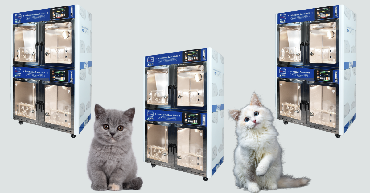
Space flexibility
Your hospital likely treats patients of varying shapes and sizes. Finding an ICU unit with a removable divider lets you treat two patients side by side, or provide individualized care.
o Finding open real estate for new equipment isn’t easy. Investing in an durable Veterinary ICU unit with an expandable design allows you to add a second story to provide quality care to more patients.
Streamlined control panel
Ensure you can easily customize and monitor temperature and oxygen levels for tailored patient care.
Patient treatment & monitoring
Look for a veterinary ICU unit with a built-in medical nebulizer & IV support.
Avoid ICU units that don’t offer silent air systems and soft-close plastic doors.
Some ICU units offer 3-in-1 lighting systems with adjustable brightness and timing functions. White light can be used for clear examinations, warm light for monitoring and observation, and blue light for therapy.
Disease control can’t be overstressed. Look for units that use animal-friendly PHI (photo-hydro-ionization) technology for real-time disinfection and deodorizing.
Although specific equipment may not fall under a typical definition of a “standard” of care, your hospital’s standard is what you make of it.
Investing in your team and the tools available to them will undoubtedly improve your standards and can increase the hospital's income.
Learn More - What Does An ICU Cage Unit Do For A Critical Pet Patient? - https://newvetequipment.com/blog/what-does-an-icu-cage-unit-do-for-a-critical-pet-patient

What is an Overexposed X-Ray and What do they Look Like?

With almost all types of veterinary diagnostic imagery, the effort is in the prep work!
That means getting the correct measurements, exposure settings, and positioning off the bat leads to the best results.
In a previous blog, we discussed why we get underexposed X-rays and how to identify them.
However, it’s easy to overcompensate when adjusting exposure settings and get the opposite effect: overexposed X-rays.
With proper knowledge, practice, and technique, getting a well-balanced exposure rate is simple!
What is an Overexposed X-Ray?
Overexposed X-rays are a result of X-ray settings being too high (using an increased kVp and mAs). This causes too much energy build up in the primary beam.
With excessive energy build-up, the patient absorbs little energy, and an overdose of radiation is absorbed into the detector. A detector starts white and darkens as it’s met with radiation.
As a result, the image will be overexposed (or too dark) to be considered diagnostic. The X-rays are essentially “burning” the plate, causing significant internal structures and tissues to appear less visible.
As you can imagine, this inhibits a clear interpretation by the Veterinarian or Veterinary Specialist and delays a proper diagnosis and treatment.
How Will I Know the X-Ray is Overexposed?
It’s not hard to identify veterinary X-rays that have been overexposed.
Here are a few characteristics that will stand out:
· Image has a darkened appearance
· Poor visibility of internal structures
· Blur or distortion
· Scatter Radiation (reduces image contrast and clarity as a result of the x-ray changing direction –“scattering” - when it meets the patient)
To be appropriately interpreted by a Veterinarian or Veterinary Specialist, you’ll likely need a retake to visualize lesions, growths, or other abnormalities clearly.
Why is My X-ray overexposed?
Capturing an overexposed image is frustrating. Now that we’re pretty clear on what they look like, let’s talk about what may have caused it:
· Incorrect kVp or mAs settings (if settings appear correct, consider re-measuring to confirm measurements were accurate)
· Forgetting to move the x-ray tube head to the correct position (if it’s too close to the cassette, it’s easy to cause overexposure)
· Possible technical error
If I Need a Retake, How Can I Avoid Another Overexposed X-Ray?
Here are a few easy-to-follow tips to avoid overexposed X-rays with your next image:
1. Correctly measure the anatomical part of the patient you’ll be radiographing using a set of calipers (remember, it’s best to measure in the position they’ll be in for the image)
2. Confirm X-ray settings using your veterinary X-ray technique chart
3. Ensure the x-ray tube head is in the correct position (raised if the plate is on the tabletop or lowered if it’s in the bucky tray)
4. Don’t forget to collimate (helps to reduce scatter radiation for a better image quality)
For best results, use computed radiography software tools to reduce artifacts, distortion, and scatter radiation (not to mention hassle-free transfers of DICOM or JPEG images for diagnostic review).
The Quick Take Away
If we think of X-ray exposure like a toaster, when the toaster produces too much energy, we’re left with toast that’s burnt to a crisp.
Just as we’d need to decrease the time on our toaster for a better breakfast, the x-ray settings will need to be reduced on the machine for a balanced diagnostic image.
We want to aim for images that have:
· Sharpness (in focus, clear, and distinct)
· Visual characteristics of density and contrast (adequate representation of tissue density)
· Clear visualization of internal structures
· No distortion or artifacts (no patient motion, technical errors, unnecessary objects, etc.)
Avoid the hassle of retakes by putting effort into the prep work—accurate measuring, exposure settings, and positioning—and getting back to what truly matters to you and your patients.

Why We Get Underexposed X-rays and How to Identify One
Why are Clear Veterinary Diagnostic Images So Important?
Proper diagnostic imagery is the bedrock of developing lifesaving treatment protocols for our patients.
Beyond diagnosis, x-rays enable Veterinary professionals to monitor the effectiveness of ongoing treatment, making adjustments based on the patient’s response (which can affect the patient’s long-term prognosis).
A poor-quality image can snowball into a delayed diagnosis, postpone treatment plans, and defer patient care.
For this reason, it’s important to identify the characteristics of a diagnostically valuable x-ray.
Various factors contribute to an image’s ability to be considered diagnostically worthy. To get started, look at one of the most common causes of retakes: underexposed X-rays.
What Makes Up a Quality Veterinary Diagnostic Image?
Before we dive into what characteristics to look for in an underexposed image, let’s first talk about qualities that make up an image with a balanced exposure rate.
An image with adequate quality will display:
Sharpness (the image is in focus, clear, and distinct)
Visual characteristics of density and contrast (adequate representation of tissue density)
Clear visualization of internal structures
No distortion or artifacts (no patient motion, technical errors, unnecessary objects, etc.)
Proper knowledge and use of a veterinary X-ray technique chart enables consistent exposure rates and image quality.
What is an Underexposed Veterinary X-Ray?
Underexposed X-rays result from X-ray settings being set too low.
When x-ray settings are too low, insufficient energy builds up in the primary beam. With too little energy meeting the X-ray detector (the tool that captures the image, also known as a cassette), the image appears radiopaque (meaning white to light gray).
A few other characteristics you’ll notice in underexposed X-rays are:
Lack of density and contrast (making the image appear “cloudy)
Poor visibility of internal structures
Blur or distortion
Scatter Radiation (reduces image contrast and clarity as a result of the x-ray changing direction –“scattering” - when it meets the patient)
The result of these characteristics will likely require a retake (increasing radiation exposure to both the patient and those capturing the image).
It’s no secret that X-ray retakes are less than ideal. Not only do they increase exposure risks, but they also decrease productivity and hospital efficiency and delay diagnostic review and treatment planning.
When an X-ray image lacks proper exposure, it is considered underexposed and less diagnostically valuable (or not at all).
How to Avoid an Underexposed Veterinary X-Ray
Here are a few easy-to-follow tips to avoid underexposed X-rays with your next patient:
1. Correctly measure the anatomical part of the patient you’ll be radiographing using a set of calipers (for best results, measure in the position they’ll be in for the image. For example, lateral recumbency)
2. Using the measurement, refer to your veterinary x-ray technique chart for correct kVp and mAs settings
3. Ensure the x-ray tube head is in the correct position (raised if the cassette is on the tabletop or lowered if it’s in the bucky tray)
4. Use the collimator on the veterinary X-ray generator to reduce unnecessary scatter radiation
Use image-enhancing algorithms and software tools offered by computed radiography to reduce artifacts, distortion, and scatter radiation for best results.
Risks of Poor Quality Veterinary Diagnostic Images
Just as a good diagnostic image can result in a successful treatment plan, a poor-quality X-ray can create barriers and delays in a confident diagnosis.
Inadequate exposure during an X-ray can lead to:
Misdiagnosis
Inconclusive diagnosis
Failure to resolve discrepancies in interpretation
Something as simple as an underexposed image can lead to potentially life-threatening consequences and compromised patient care.
The Quick Take Away
Think of X-ray exposure like a toaster—if the toaster doesn’t produce enough energy, when it’s finished, you’re left with nothing but a warm piece of white bread.
Just as we’d need to increase the time on our toaster, the machine's X-ray settings will need to be increased for a balanced diagnostic image.
Enhancing Safety in Your Veterinary Radiology Room
Dosimeter badges and a proper X-ray or CT room shield plan are the best ways to protect your veterinary team from radiation exposure.
Want to learn more? For more information on radiation safety at your practice, check out the links below:

How to use a Veterinary X-Ray Technique chart

Mastering Veterinary X-Ray Techniques
Quality diagnostics are the foundation of a confident patient diagnosis. It’s no surprise that veterinary radiography is one of the most commonly used diagnostic tools when confirming (or ruling out) a patient diagnosis.
Although the terms “x-ray” and “diagnostic image” are often used interchangeably, to rule an image as “diagnostic”, it should display quality contrast and dynamic range and avoid any artifacts.
To achieve these characteristics, let’s start with the foundation of what produces a quality diagnostic image: a technique chart.
The Purpose of a Technique Chart for Veterinary Radiography
A digital radiography technique chart is a standard guide to producing the optimum exposure for a diagnostic image.
Put another way, it’s a table of predetermined exposure settings that, when used accurately, ensure consistent image quality and patient exposure.
A reliable, user-friendly chart is important for obtaining clear diagnostic imagery with consistent radiographic density and contrast.
Why do Technique Charts for Veterinary Digital Imagery Matter?
Patient care and wellbeing is always top priority and proper treatment starts with a confident diagnosis.
Veterinarians and Veterinary Specialists rely on quality diagnostic images to rule out suspected diagnoses and confirm findings to develop an accurate treatment plan.
With many veterinary practices upgrading to quality veterinary X-ray software, there’s a little more forgiveness in under/overexposed images (with veterinary X-ray technology advancements, we can manipulate image contrast); however, not all exposure imperfections can be corrected.
Having a precise image from the start is crucial to a proper evaluation.
What Factors Make Up a Technique Chart?
Let’s break it down…
Most technique charts are segmented anatomically by thorax (chest), abdomen, spine, and extremities (arms, legs, tail) since each area varies in density. The measurement of the anatomical body part will determine the exposure settings.
What exactly are exposure settings?
Exposure settings include:
· kVp (kilovoltage peak), which controls the penetrating power of the x-ray beam (or how energetic the x-rays are)
· mAs (milliampere-seconds), which controls the number of x-rays.
Typically, the higher the mAs, the better the image quality; however, it’s important to be judicious with these settings because they can also contribute to the patient's radiation dose.
How to Use a Technique Chart for Veterinary Digital X-Rays
It all starts with accurate measurements. Begin by using a set of calipers to measure the anatomical part of the patient you’ll be radiographing.
Tip: for best results, measure the patient in the position they’ll be in during the radiograph (ex: right lateral recumbency) and measure from the point of x-ray entrance to exit.
Once you’ve got your measurement (in cm), consult the technique chart to determine the kVp and mAs to set the X-ray generator.
Depending on your veterinary hospital’s X-ray system setup, you may also need to determine whether the X-ray cassette needs to be placed on the tabletop or in the bucky tray (also known as the “film tray”).
You can place your technique chart aside and manipulate your tube head and lighting in preparation for your image.
Tip: Remember, the tube head may need to be raised if the cassette is on the table top or lowered if it’s in the Bucky tray.
Remember to place a left/right marker to indicate patient positioning (for example, if the patient is in right lateral recumbency, an “R” label should be included on the image).
Once you’ve confirmed your settings are correct, the cassette and tube head are in the correct position, the label is placed, and the patient is in position, you’re ready to capture your diagnostic image!
Making Adjustments to Exposure Factors
Sometimes adjustments are necessary for a clear visual of injuries or problem areas. Here are a few tips to consider before your retake:
If the initial radiograph is too light, try increasing the kVP by 15% (mAs remain the same)
If the initial radiograph is too dark, try decreasing the mAs by 15% (mAs remain the same)
Want to learn more?
For more tips on achieving quality diagnostic veterinary images, check out these articles below:
Warnings!
Don’t participate in x-ray diagnostic imaging if you’re pregnant to avoid harmful radiation to your little one.
Avoid using old, run-down, or defective protective gear. If cracks, tears, or fraying appear, replace your veterinary radiology protective supplies.
Additional Resources:

Contrast Enhances Veterinary Cone Beam CT for Pet Care
What is cone beam computed tomography?
Cone Beam Computed Tomography (CBCT) is an imaging technique that is widely utilized in human medicine.
Its unique characteristics make it especially suited to the fields of orthopedics, image-guided radiation therapy, interventional radiology, maxillofacial surgery, and dentistry.
Still, its benefits are only just starting to be realized within the veterinary world. CBCT is capable of producing detailed 3D images that are proving particularly suited to a range of veterinary applications such as maxillofacial surgery (especially trauma), dentistry, imaging of exotic species such as rabbits, and for orthopedics, in particular, imaging of joints and extremities.
CBCT is a specific form of Computed Tomography (CT). As with regular CT imaging, CBCT utilizes a moving beam of X-rays that are detected by a digital X-ray detector.
A series of images are obtained from various angles around the patient which can then be used to generate a 3D image using complex algorithms via specialist computer software.
The main difference between CBCT and regular CT, as its name suggests, is that CBCT utilizes a divergent beam of X-rays which form a cone shape. This allows the X-ray beam to cover a larger area in a single pass.
Regular CT machines in contrast take “slices” through the patient using a flat fan-shaped beam of X-rays that require several passes over the area of interest to acquire sufficient information.
What are its advantages and disadvantages?
The specific characteristics of the CBCT system mean that it has the advantage of being faster than a standard CT scan and exposes the patient to a lower radiation dose due to only a single pass being required to produce an image.
In addition, CBCT machines tend to be smaller and less expensive to purchase, making them more accessible. The images produced by CBCT can also show substantially superior spatial resolution compared with regular CT.
This means that images are clearer and more detailed, allowing better visualization of small structures and abnormalities, particularly within bony tissues.
There are however some disadvantages to the technique. Due to the shape of the beam and the lack of collimation, CBCT scans tend to produce more scatter radiation.
The consequence of this is inferior contrast resolution in soft tissues - meaning that subtle lesions of soft tissues, resulting in only slight differences in contrast, are more likely to be missed.
The scatter also increases the radiation exposure in the surrounding areas and potentially to operating staff. This scatter is amplified when CBCT is used for larger body parts such as the thorax or abdomen.
For these reasons, CBCT tends to be used for smaller body areas such as the head and bones, particularly the extremities. It is beneficial for imaging exotics and their dentistry.
Why is it important to use contrast in CBCT?
The use of contrast is common in many imaging modalities to further enhance and widen their application.
CT typically uses non-ionic contrast media (e.g. iohexol), commonly injected intravenously but can also be injected directly into a body cavity under investigation, such as a joint space.
Contrast is used to highlight specific areas of interest, especially those that may be usually hard to see. In CBCT, iodinated contrast media that are injected intravenously will highlight vessels and soft tissue structures supplied by these.
This has the potential to increase the diagnostic utility of CBCT massively.
The addition of contrast studies in addition to plain CBCT scans is crucial as it will both provide additional information and overcome some of the limitations of the technique.
In orthopedic examinations, the addition of contrast by direct injection into the joint provides high-resolution images of the articular surfaces aiding the assessment of lesions found in degenerative joint disease for example.
In dentistry and maxillofacial surgery, intravenous contrast techniques that highlight blood vessels allow the surgeon to assess vascular structures better and in particular any abnormalities of the vasculature that could be potentially affected by any planned procedures.
Contrast also improves the ability of CBCT to detect and assess soft tissue lesions, for example, neoplasms presenting at an early stage.
Cone beam CT is a relatively new diagnostic modality in the veterinary world.
However, its unique properties and affordability mean that it is likely to be more widely used in the future.
Contrast techniques can greatly increase the range and accuracy of diagnostic procedures possible with these systems and these techniques will likely continue to develop, offering new and exciting opportunities.
References:
[1] Kumar M, Shanavas M, Sidappa A, Kiran M. Cone beam computed tomography - know its secrets. J Int Oral Health. 2015 Feb;7(2):64-8. PMID: 25859112; PMCID: PMC4377156.
[2] Venkatesh E, Elluru SV. Cone beam computed tomography: basics and applications in dentistry. J Istanb Univ Fac Dent. 2017 Dec 2;51(3 Suppl 1):S102-S121. doi: 10.17096/jiufd.00289. PMID: 29354314; PMCID: PMC5750833.
[3] Kim, M., Kim, B., Choi, H., Choi, Y., Oh, S. H., Kang, J., Lee, S., Kang, J., Kim, G. T., Choi, Y., & Hwang, E. (2015). Intravenous contrast media application using cone-beam computed tomography in a rabbit model. Imaging Science in Dentistry, 45(1), 31. https://doi.org/10.5624/isd.2015.45.1.31
[4] Posadzy, M., Desimpel, J., & Vanhoenacker, F. (2018). Cone beam CT of the musculoskeletal system: clinical applications. Insights Into Imaging, 9(1), 35–45. https://doi.org/10.1007/s13244-017-0582-1
[5] Riggs, G. G., Cissell, D. D., Arzi, B., Hatcher, D. C., Kass, P. H., Zhen, A., & Verstraete, F. J. (2017). Clinical Application of Cone Beam Computed Tomography of the Rabbit Head: Part 2-Dental Disease. Frontiers in veterinary science, 4, 5. https://doi.org/10.3389/fvets.2017.00005
[6] Bregger, M. D. K., Koch, C., Zimmermann, R., Sangiorgio, D., & Schweizer-Gorgas, D. (2019). Cone-beam computed tomography of the head in standing equids. BMC Veterinary Research, 15(1), 289. https://doi.org/10.1186/s12917-019-2045-z
[7] Myller, K. A., Turunen, M. J., Honkanen, J. T., Väänänen, S. P., Iivarinen, J. T., Salo, J., Jurvelin, J. S., & Töyräs, J. (2017). In Vivo Contrast-Enhanced Cone Beam CT Provides Quantitative Information on Articular Cartilage and Subchondral Bone. Annals of biomedical engineering, 45(3), 811–818. https://doi.org/10.1007/s10439-016-1730-3
[8] Hamard M, Sans Merce M, Gorican K, Poletti PA, Neroladaki A, Boudabbous S. The Role of Cone-Beam Computed Tomography CT Extremity Arthrography in the Preoperative Assessment of Osteoarthritis. Tomography. 2023 Nov 29;9(6):2134-2147. doi: 10.3390/tomography9060167. PMID: 38133071; PMCID: PMC10747585.

Large Animal Endoscopy: The Future of Veterinary Medicine

Unlocking Veterinary Insights: The Importance of Large Animal Endoscopy
Endoscopy has been widely employed in equine veterinary hospitals for many decades, particularly in the assessment of the upper respiratory tract of competition horses.
Its use is not restricted to this area, however. As the available range of endoscopes, tools, and accessories expands, so does the availability and application of endoscopy in large animal general practice.
Alongside its use in horses, the large animal endoscope may be a valuable tool for veterinarians working with production animals (particularly those of high economic value), camelids, farm pets, and zoological species.
Reasons to Perform Large Animal Endoscopy
As with small animals, large animal endoscopy may be undertaken for both diagnostic and therapeutic purposes. Cases benefiting from endoscopy may include those with the following:
Structural abnormalities such as strictures may be identified and possibly treated endoscopically
Conditions associated with exercise intolerance or poor athletic performance
Traumatic injuries, for example, bolusing injuries of the pharynx or esophagus in production animals, rectal tears in the horse post-examination
Ingestion or inhalation of foreign material which may be identified and retrieved endoscopically in most large animal species
Neoplastic disease visualization, biopsy, and, in some cases such as ethmoid hematomas, treatment
Parasitic disease identification and sampling
Infectious diseases such as guttural pouch mycosis or sinusitis in horses
Inflammatory disease for example Equine Asthma (EA) where bronchoalveolar lavage samples may be valuable
Gastric ulceration especially in horses
Large animal endoscopy may be used in the following organ systems:
Upper respiratory tract - including nasal passages, paranasal sinuses, guttural pouches, pharynx and larynx
Lower respiratory tract - including trachea and bronchi
Oral/dental assessment in all species
Upper gastrointestinal tract - including esophagus, stomach, and proximal small intestine
Lower gastrointestinal tract including the rectum and colon
Urinary tract
Reproductive tract
Large animal endoscopes
Large animal endoscopy most often employs a flexible endoscope (either a fiberscope or video endoscope) of diameter and length appropriate for the patient and organ system under investigation. Dynamic endoscopy is also now available, using a portable, wireless, flexible endoscope to assess an animal during exercise.
Large animal endoscopes require a light source, an air pump for insufflation, irrigation, and suction, as well as a monitor to receive the image in the case of video endoscopes. Fiberscopes and rigid endoscopes may be connected via a camera system to a display monitor.
Instrumentation for large animal endoscopy
Instruments used in large animal endoscopy may include the following, some of which are available either as single-use or reusable items:
Biopsy forceps – with various tips to suit the tissue to be biopsied. In this case, single-use forceps may be advantageous in gaining optimum biopsy quality as the cutting edge will always be sharp.
Grasping forceps – including rat-toothed, alligator jaws, or multi-pronged forceps
Retrievers, snares, or baskets – for the retrieval of foreign objects
Polypectomy snares – with cutting wire for the removal of polyps and other small growths
Cytology brushes – for collecting cell samples, particularly from the lower airway or gastrointestinal tract
Catheters with injection needles - for aspiration biopsy or flushing and aspirating fluid e.g. bronchoalveolar lavage
Guttural pouch probe – to help navigate the endoscope into the guttural pouch
Equine laryngeal forceps – particularly useful during trans-endoscopic laser surgery
Coagulation electrode – where hemostasis is required.
Other applications
Rigid endoscopes (or telescopes) are also widely used in large animal veterinary clinics. Applications may include
Dental assessment
Arthroscopy – for diagnosis and treatment (e.g. meniscal injury)
Thoracoscopy – for diagnosis and treatment (e.g. pleural lavage in pleuropneumonia cases)
Laparoscopy – for diagnostics (e.g. organ visualization and biopsy), elective surgery (e.g. cryptorchid castration or ovariectomy in horses) or therapeutic surgery (e.g. abomasopexy in cattle, neoplasia in all species, inguinal ring closure in equine hernia patients or nephrogenic space ablation after entrapment colic).
Artificial insemination and embryo transfer work
Theloresectoscopy – for diagnostics and minimally invasive teat surgery in cattle
Rigid endoscopes are passed through a trocar or cannula into the required space. Operating instruments for use during laparoscopy and thoracoscopy may include scissors, grasping forceps, biopsy forceps, injection cannulas, a palpation probe, needle holders, and knot tyers. Specialist instruments are available for embryo transfer in horses and cattle and for toggling procedures in cattle.
Instruments for use during arthroscopy may include probes to assess articular cartilage and subchondral bone; curettes for debridement; osteotomes and elevators for separating bone fragments; rongeurs; grasping forceps; arthroscopy punch for resection of cartilage, synovium or soft tissue masses; chisels; scissors; and drainage cannulas.
Conclusion
Large animal veterinary practitioners are presented with some specific challenges when diagnosing and treating our patients.
They are often, by definition, large in size which can limit the use of some diagnostic techniques such as abdominal radiography.
Frequently our patients would be at high risk under general anesthesia both in terms of risk to the patient and potential risk to the handler(s) too. In our livestock patients, in particular, financial limitations may preclude the use of advanced imaging modalities.
Large animal endoscopy represents an accessible, practical, and economically viable solution in many of these cases, both in terms of diagnostics and therapeutics.
Additional resources
The Veterinary Endoscopy Society - https://veterinaryendoscopysociety.org/
Equine endoscopy - https://adobevetcenter.com/equine-endoscopy/
Equine gastroscopy https://www.veterinary-practice.com/article/equine-gastroscopy-a-complete-perspective
Gastrointestinal biopsy in the horse https://journals.sagepub.com/doi/full/10.1177/10406387221085584
Transendoscopic URT laser surgery in the horse https://www.sciencedirect.com/science/article/pii/S0749073917302870
Livestock endoscopy - https://adobevetcenter.com/livestock-endoscopy/
Endoscopy in cattle https://pubmed.ncbi.nlm.nih.gov/22134601/#:~:text=Parts%20of%20the%20upper%20respiratory,prognosis%20of%20different%20pathological%20conditions.
Theloscopy in cattle https://www.sciencedirect.com/science/article/abs/pii/S0749072004000854?via%3Dihub
Dosimeter Badges in your clinic: Key FAQs Answered
What are veterinary dosimeter badges?
Veterinary dosimeter badges are small devices worn by veterinary professionals to monitor their exposure to radiation in workplaces where diagnostic imaging procedures are performed on animals.
Why are veterinary dosimeter badges necessary?
Veterinary dosimeter badges are crucial for ensuring the safety of personnel who work with radiation-emitting equipment, helping to monitor exposure levels, and preventing potential health risks.
How much does a dosimeter badge cost?
The cost is $88.00 per person per year. The price includes quarterly replaced dosimeter badges and quarterly lab reports.
Do I need to return the badge whenever I get a new quarterly badge?
Yes, you must return all the dosimeter badges every quarter following our return process.
What happens if I don’t return a badge or if I don’t return it on time?
Returns must be on time to maintain the monitoring process which may lead to increased costs and regulatory issues. To avoid this, there is a $50 per badge, per quarter late fee. This ensures compliance, data accuracy, and the effective management of dosimetry programs, prioritizing worker safety.
How do veterinary dosimeter badges work?
These badges contain sensitive materials that change when exposed to radiation, allowing for measuring radiation exposure over time.
Who needs to wear veterinary dosimeter badges?
Anyone working with radiation-emitting equipment in veterinary settings, including veterinarians, technicians, and radiology staff, should wear dosimeter badges.
How often should veterinary dosimeter badges be worn?
Dosimeter badges should be worn whenever there is a possibility of radiation exposure, following workplace protocols and regulations.
How are veterinary dosimeter badges read and analyzed?
Badges are collected quarterly and sent to our specialists for analysis, with reports provided to the veterinary facility.
Are veterinary dosimeter badges reusable?
No, dosimeter badges are single-use devices and should be replaced after analysis.
Are veterinary dosimeter badges transferable?
No, each dosimeter badge is person-specific and nontransferable.
Are there regulations or guidelines for veterinary dosimeter badges?
Yes, our services are governed by The National Voluntary Laboratory Accreditation Program (NVLAP) which provides guidelines and regulations for the use of dosimeter badges to ensure safety.
How can I obtain veterinary dosimeter badges for my practice?
You can obtain dosimeter badges from https://form.jotform.com/221224959076157
Who should I contact for more information about veterinary dosimeter badges?
For further assistance or questions about veterinary dosimeter badges, please get in touch with our customer service team support@newvetequipment.com or call at 530-722-4977

Avoid X-Ray Over-Exposure: Expert Tips for Veterinarians

Sharpen Your Skills: Mastering X-Ray Technique to Avoid Overexposure
X-rays are workhorses in the veterinary world, helping diagnose countless conditions in our furry (and feathered, and scaled) friends. But for those perfect diagnostic images, proper exposure is crucial. Over-exposed X-rays, while easy to spot, can lead to a whole host of problems. Don't worry, though, this blog is here to help!
Why Does Exposure Matter?
A good X-ray is like a perfectly balanced recipe: all the elements need to be just right. Over-exposure, like adding too much salt, makes the image too dark and difficult to interpret. This can lead to missed diagnoses and even unnecessary repeat X-rays, exposing your patients and staff to additional radiation.
Culprits of the Over-Exposed Image
Several factors can contribute to over-exposed X-rays in your clinic:
Distance: A slight change in the distance between the X-ray source and the animal can significantly impact exposure. Ensure proper positioning and consult your exposure chart for the correct distance based on the animal's size and the area being imaged.
Technical Tweaks: Incorrect kV and mA settings can also play a role. Higher kV settings lead to more powerful X-rays that can penetrate deeper, potentially overexposing the film. Adjusting these settings based on the animal and the desired image is crucial.
Missing Grids: Grids help filter out scattered radiation, improving image quality and reducing exposure. Ensure you're using the appropriate grid and factoring it into your exposure calculations.
Tips for X-Ray Success
Here's how you can ensure your X-rays are picture-perfect:
Exposure Charts: These handy tools provide recommended kV and mA settings for various animal sizes and body areas. Consult them religiously!
Exposure Indicators: Most digital systems come equipped with these, helping you identify overexposed images. Learn how to interpret the readings and adjust settings accordingly.
Regular Maintenance: Keep your X-ray machines well-maintained for optimal performance and accurate exposure.
Remember:
Over-exposure isn't just about image quality; it's about patient and staff safety. By following these tips and staying vigilant, you can ensure your X-rays are both diagnostic and safe.
Bonus Tip: Consider exploring X-ray machines with features like automatic exposure adjustments based on animal size, like the DynaVue Duo. This can minimize errors, optimize imaging, and ultimately, enhance the care you provide to your animal patients.