


Choosing the Right C-Arm: Comparing Today’s Top Veterinary Models
Choosing a C-arm is one of the most significant equipment decisions a veterinary practice can make.
Between mobile and mini systems, varying dose technologies, and wildly different footprints, it can feel overwhelming to determine what actually matters for your clinical needs.
Whether your practice focuses on orthopedics, ER, general surgery, or mobile specialty work, the right C-arm will strike the perfect balance between image quality, ease of use, safety, and cost.
This guide breaks down the key differences and highlights what to look for.
Mobile vs. Mini C-Arms: Which One Fits Your Practice?
Mobile C-Arms
These full-size systems are best suited for:
High-volume orthopedic hospitals
Specialty centers performing complex soft tissue or vascular interventions
Clinics with spacious ORs and high caseloads
Advantages and Considerations:
Full-size mobile C-arms offer a larger field of view and greater versatility for advanced interventional work, often with features such as DSA. However, they come with clear trade-offs—a larger footprint, higher cost, and greater space and training requirements to integrate smoothly into a practice.
Mini C-Arms
Initially designed for human extremity imaging, veterinary-focused mini C-arms have evolved into highly effective tools for small-animal orthopedics and foreign-body procedures.
A strong example is the Orthoscan Tau 1515, which features a compact, all-in-one footprint that fits seamlessly into ORs that were not originally planned to support fluoroscopy.
Advantages and Considerations:
Mini C-arms are highly maneuverable and cost-effective, making them ideal for general practices, ortho-focused GPs, and mobile surgeons who need a small footprint and easy mobility. Their compact size does come with limitations, including a smaller field of view, limited full-body imaging, and the potential absence of advanced features such as DSA—Flat-Panel Detectors vs. Image Intensifiers.
Flat-Panel Detectors (FPDs)
FPDs are now considered the gold standard due to:
Higher resolution
Lower noise
Better consistency over time
Reduced distortion
Many modern C-arms use CMOS flat-panel technology, which delivers sharp, bright images with enhanced detective quantum efficiency to reduce noise and improve image quality at lower doses
Image Intensifiers
Image intensifiers are an older technology. While functional and often cheaper (especially refurbished), they:
Degrade over time
Produce more distortion at the edges
Offer lower contrast and resolution
For practices focused on orthopedic precision, flat-panel detectors are usually worth the upgrade.
Image Resolution and Dose: What Truly Matters
Resolution
Look for systems that offer:
High pixel density
High signal-to-noise ratio
Clear visualization of fine bone detail
Dose Reduction
Radiation safety is a critical consideration for veterinary teams. Modern systems use a combination of hardware and software to reduce dose.
Digital Subtraction Angiography
DSA or Digital Subtraction Angiography is most relevant for:
Vascular studies
Shunt evaluations
Pacemaker lead assessment
While not every C-arm includes DSA, practices planning to expand into advanced soft-tissue or cardiac procedures may want to prioritize it.
Footprint, Maneuverability, and OR Workflow
Clinics with limited space, or those that move between multiple Ors, should focus heavily on footprint and ease of movement.
If your OR space is tight or you anticipate moving between rooms, a mini C-arm may significantly improve workflow without sacrificing clarity.
What Does “Value” Look Like for Your Type of Practice?
For general practices, value often comes from cost-effective imaging, a small footprint, and easy day-to-day use. At the same time, orthopedic-focused GPs tend to prioritize high-resolution images, low dose, and workflow efficiency. This makes a flat-panel mini C-arm an excellent fit, especially for clinics performing frequent TPLOs where a newer detector pays off quickly.
Emergency and critical care hospitals benefit most from speed, reliability, and the ability to guide foreign body retrieval or urgent orthopedic stabilization, and may choose either a mobile or mini system depending on their caseload.
Specialty hospitals usually require full capabilities, including a large field of view, advanced software, and often DSA, making a full-size mobile fluoroscopy unit the most appropriate choice.
Meanwhile, mobile surgeons see the highest ROI from compact, portable, durable systems—typically mini C-arms designed with veterinary-first software and rugged construction for use across multiple clinics.
What Actually Matters?
When choosing a C-arm, the most important considerations are:
Does it deliver the image quality you need for your caseload?
Can your team use it quickly and confidently?
Does the footprint fit your clinic’s workflow?
Will the dose profile keep your team safe?
Is the price aligned with the value and procedures you plan to offer?
In the end, choosing the right C-arm is about finding the balance that fits your practice: image quality that supports the procedures you perform, technology your team can use confidently, and a system that integrates smoothly into your physical space and workflow.
Whether you’re upgrading your orthopedic capabilities, expanding your ER tools, or simply looking to improve surgical precision, the right C-arm can elevate the standard of care you deliver every day.


Who Needs a C-Arm? A Breakdown of Veterinary Practices That Benefit Most
How C-Arms Improve Outcomes in Modern Veterinary Care
As more veterinary hospitals expand their surgical and interventional capabilities, the question isn’t just “What does a C-arm do?”, it’s “Do we actually need one?”
C-arms provide real-time fluoroscopic imaging that can elevate surgical accuracy, open the door to new procedures, and streamline workflows. But like any major equipment purchase, they’re most valuable for practices that can truly utilize them.
If you’re trying to determine whether a C-arm fits your caseload and growth plans, this guide breaks down which hospitals benefit most.
1. Orthopedic-Focused General Practices
If your hospital performs a high volume of orthopedic procedures, a C-arm can quickly become indispensable.
Orthopedic benefit:
Real-time visualization during fracture repairs
Precision alignment for TPLOs and other corrective osteotomies
Ability to confirm implant placement instantly
Fewer retakes, fewer anesthesia adjustments, and fewer post-op surprises
In orthopedics, a few millimeters can make the difference between a smooth recovery and a complication. Fluoroscopy reduces guesswork and increases surgical confidence.
Common Cases Include:
TPLO and TTA procedures
Long bone fracture repair
Luxating patella corrections requiring implant placement
Angular limb deformity corrections
Joint evaluations
For surgical GPs looking to elevate their ortho offerings or reduce their reliance on referral hospitals, a C-arm can be a turning point for clinical capability.
2. Specialty Hospitals
Specialty practices tend to see the broadest range of cases in which fluoroscopy improves outcomes. Their higher caseload also spreads the cost across more procedures, making ROI easier to calculate.
Specialty benefits:
High surgical volume
Advanced ortho, soft tissue, and neurological procedures
Interventional radiology availability
On-site specialists who rely on real-time imaging
Common Cases Include:
Complex fracture repairs
Vascular and cardiac procedures
Tracheal stent placement
Pacemaker implantation
Subtotal colectomy foreign body checks
Urethral and ureteral stenting
For these hospitals, a C-arm isn’t just helpful, it’s often a clinical requirement.
3. Emergency & Critical Care Hospitals
ER/ICU environments regularly encounter cases where fluoroscopy changes the course of treatment within minutes.
ER / ICU benefit:
Ability to identify and remove GI foreign bodies rapidly
Real-time visualization for unstable orthopedic injuries
Support for urgent cardiac or vascular interventions
Faster decision-making when minutes matter
Common Cases Include:
GI foreign body retrieval (particularly linear objects)
Urethral obstruction diagnostics
Fracture stabilization
Pericardiocentesis guidance
Contrast-based urinary or vascular studies
In ER settings, a C-arm reduces delays, minimizes patient movement, and improves efficiency during high-acuity cases.
4. Mobile Surgeons and Traveling Specialists
Mobile surgical teams often work with varying setups and caseloads, so easy positioning and real-time imaging can drastically improve workflow consistency across clinics.
Mobile/Travel benefit:
Standardized imaging, no matter where they operate
More predictable surgical efficiency
Ability to offer specialized procedures to general practices
Mobile surgeons offering TPLOs, fracture repair, or interventional procedures often rely on a C-arm to maintain high standards across diverse environments.
Understanding DSA: When Digital Subtraction Angiography Matters
Some C-arm systems include DSA (Digital Subtraction Angiography)—a powerful imaging tool that reveals blood flow with exceptional clarity.
What DSA Does (in simple terms)?
DSA subtracts pre-contrast images from post-contrast images to highlight only the motion of contrast material. In practice, it produces obvious vascular images in which bones and soft tissue are “subtracted out.”
When Veterinary Practices Use It:
Diagnostic vascular studies
Portosystemic shunt (PSS) identification
Cardiac pacemaker lead visualization
Fluoroscopy-guided biopsies where vasculature is a concern
Why DSA Matters:
Reveals tiny vessels that are difficult to see with standard fluoroscopy
Improves safety in contrast-guided procedures
Enhances accuracy for vascular interventions and catheter placement
While DSA isn’t necessary for every hospital, it’s a significant advantage for practices performing advanced imaging or interventional radiology.
How to Estimate Your C-Arm Utilization Before Purchasing
Before investing, assess your current and potential case volume.
Ask yourself:
1. How many orthopedic procedures do we perform monthly?
Even 4–6 orthopedic surgeries per month can justify consistent use.
2. Are we seeing frequent foreign bodies or emergency cases?
If so, fluoroscopy may significantly shorten procedure times.
3. Are we looking to add new services?
TPLOs, TTAs, stent placements, and interventional radiology are all enabled or improved by C-arms.
4. Do we refer cases out that we’d prefer to keep in-house?
Keeping one or two high-value procedures per month can substantially improve ROI.
5. Are there specialists (or mobile surgeons) in our network who could use the equipment?
Shared utilization can strengthen financial justification.
Does Your Practice Need a C-Arm?
If your hospital is performing regular orthopedic or emergency procedures—or plans to expand into these services—there’s a strong chance a C-arm will elevate both clinical capability and practice profitability.
But even more importantly, it enhances veterinarians' ability to diagnose and treat challenging cases, giving teams the confidence and clarity they need to deliver exceptional care.

C-Arms in Veterinary Medicine: Why They’re a Diagnostic Game-Changer
The Rising Value of C-Arm Imaging in Veterinary Care
As veterinary medicine continues to advance, one imaging tool is quietly becoming a staple in modern practices: the C-arm.
Whether you’re a high-volume orthopedic hospital or a general practice beginning to expand your surgical offerings, mobile fluoroscopy can dramatically enhance the way you diagnose, treat, and manage complex cases.
If you’ve ever wished you could “see what you’re doing” in real time—especially in tricky orthopedic or foreign-body cases—a C-arm may be the tool that changes everything.
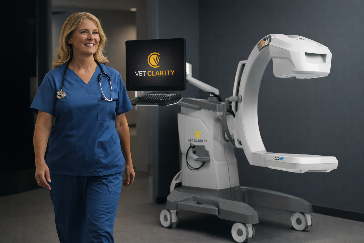
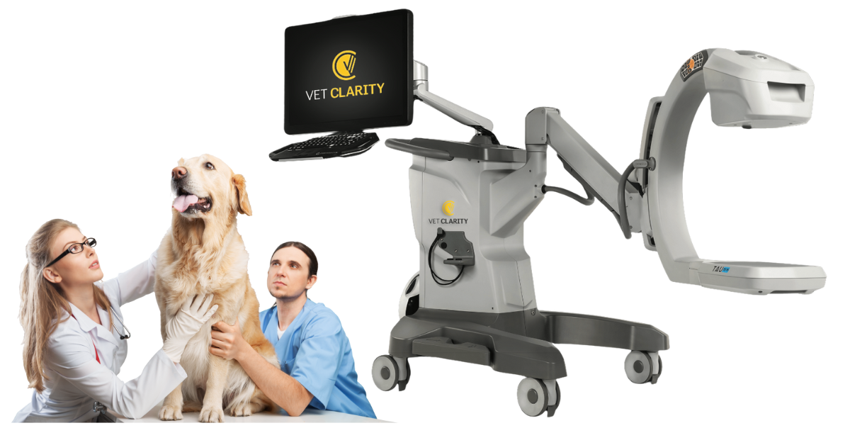
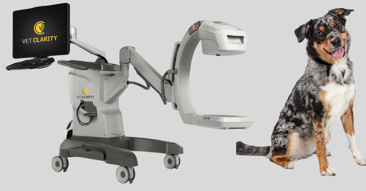
What Exactly Is a C-Arm?
A C-arm is a mobile fluoroscopy unit shaped like the letter “C,” allowing you to position the X-ray source and detector around your patient.
Unlike standard radiography, which gives you a single static image, a C-arm provides continuous, real-time imaging during procedures. That alone is a significant shift in efficiency and surgical precision!
Why C-Arms Are Becoming So Valuable in Veterinary Medicine
1. Real-Time Imaging Improves Surgical Precision
In orthopedic surgery, millimeters matter. A C-arm allows you to visualize fracture alignment, implant placement, joint spaces, and angles as you work—not after the fact.
Instead of taking a radiograph, repositioning the patient, retaking views, and hoping the alignment is correct, surgeons can adjust in the moment. The result?
More accurate repairs
Shorter anesthesia times
Fewer surprises post-op
For busy practices, this can significantly increase surgical efficiency.
2. Better Outcomes With Fewer Repeat Procedures
When you can see your hardware placement or foreign body extraction in real time, you reduce the risk of missed pathology, implant misalignment, or incomplete removal.
That translates directly to:
Decreased complication rates
Higher client satisfaction
Stronger clinical outcomes
More predictable case progression
For hospitals offering advanced procedures, this is a significant competitive advantage!
3. A Game-Changer for Complex Soft Tissue and Emergency Cases
While C-arms are often associated with orthopedics, their usefulness extends beyond bone.
They’re invaluable for:
GI foreign body retrieval (especially linear or oddly shaped objects)
Urethral obstructions and stent placement
Hepatobiliary procedures
Interventional radiology cases (coiling, embolization, biopsies, contrast studies)
Being able to visualize contrast flow or track movement in real time can significantly improve the efficiency of a case.
4. Faster Workflows and Less Guesswork
Because fluoroscopy allows for continuous guidance, you’ll spend less time repositioning patients, capturing additional radiographs, and navigating blindly.
This means:
Shorter procedure times
Reduced radiation exposure overall
Less staff fatigue
More predictable scheduling
For practices trying to increase surgical caseload while maintaining quality of care, efficiency matters.
How C-Arms Strengthen ROI for Veterinary Hospitals
Investing in a C-arm is often easier to justify when you understand where the return comes from. Practices typically recover their investment through:
1. Increased Surgical Case Volume: Offering fluoroscopy-guided procedures attracts more referrals and allows general practices to expand into higher-value services.
2. Higher Value Procedures Per Case: Fluoroscopy enables procedures that often carry higher revenue, like fracture repairs, interventional procedures, and advanced foreign body removals.
3. Improved Efficiency: Shorter anesthesia times and quicker surgeries mean you can complete more procedures safely within the same day.
4. Reduced Post-Op Complications: Fewer rechecks, fewer corrective surgeries, and fewer client issues all bolster profitability and client trust.
Even modest increases in monthly surgical volume, 1–3 additional fluoroscopy-guided cases, can rapidly close the gap on costs.
Why More Practices Are Considering a C-Arm Right Now
As more veterinary hospitals expand their orthopedic and soft-tissue capabilities, and as pet parents increasingly expect (and seek) advanced care, the demand for real-time imaging is growing.
C-arms offer:
Diagnostic clarity
Enhanced surgical confidence
The ability to perform procedures once reserved for specialty hospitals
And with modern systems becoming more cost-effective and user-friendly, they’re no longer limited to large specialty centers.
A C-arm is truly a tool that changes how surgeries are performed and how confidently your team can navigate complex cases.
Suppose your practice is looking to elevate its surgical offerings, reduce complications, or simply streamline procedures with more precision. In that case, a C-arm may be one of the best diagnostic tools to consider.