

Anesthesia & MRI: Minimizing Risk While Maximizing Image Quality
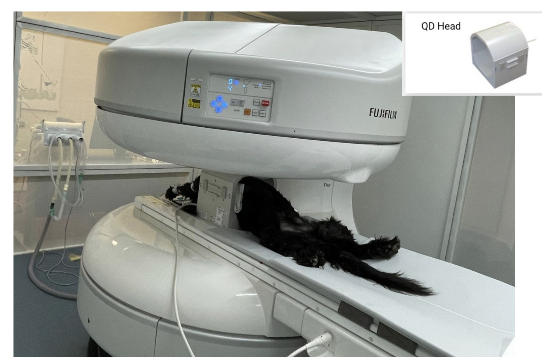
MRI is an imaging procedure, but a successful scan depends on much more than the machine.
Behind every good MRI study is a team carefully managing four important things:
Patient safety
Stable anesthesia
Efficient workflow
Clear, diagnostic images
For many veterinary teams, the anesthesia side of MRI can feel intimidating at first.
Not because it is impossible.
But because MRI is different.
The room is different.
The equipment is different.
The team setup is different.
And communication can feel different because the patient is not managed the same way as in a typical surgery room.
That is why preparation matters so much. When the team understands the MRI environment and has a clear plan before the scan begins, the entire process becomes safer, smoother, and more efficient.
Good MRI Images Start Before the Patient Enters the Scan Room
One of the easiest mistakes teams make early on is focusing only on the scan itself, but image quality starts long before imaging begins.
Things like:
Proper catheter placement
Organized line management
Patient padding
Stable anesthetic depth
Thoughtful positioning
…all directly impact the quality and efficiency of the study. A patient who’s uncomfortable, unstable, or poorly positioned often leads to:
Motion artifact
Repeat sequences
Longer anesthesia times
Increased stress for the entire team
Preparation matters more than people realize. And smooth MRI days usually come down to one thing: fewer preventable adjustments once the scan has started.
Positioning: Small Details Make a Huge Difference
Positioning is one of those things that can either save your team time or quietly steal a lot of it. Repeatable positioning helps create:
More consistent images
Faster sequence setup
Better study interpretation
Less need for rescans
Even small changes can matter, like:
Keeping the spine straight and supported
Avoiding rotation
Using consistent landmarks
Securing patients well enough to prevent subtle movement
The goal is stability without creating unnecessary pressure points or compromising ventilation. And from a workflow standpoint, consistency is everything.
When every technician positions slightly differently, studies become harder to reproduce and troubleshooting becomes more frustrating. That’s why many successful MRI teams eventually develop very standardized positioning protocols:
Same supports
Same setup sequence
Same communication process
It reduces guesswork for everyone involved.
Monitoring in MRI Environments Requires a Different Mindset
MRI anesthesia monitoring can feel uncomfortable at first because the setup naturally creates more distance between the patient and the anesthesia team.
You can’t always stand directly beside the patient the way you might during surgery. That’s why preparation and vigilance become even more important.
Some practical habits that help:
Confirm all monitoring equipment is MRI-compatible before the patient enters the room
Organize lines and cables carefully to prevent tangling or interference
Double-check connections before sequences begin
Establish clear communication between the imaging and anesthesia teams
And perhaps most importantly:
Anticipate problems early instead of reacting late
Because once imaging is underway, interruptions cost time, not just operationally, but anesthetically too.
Efficiency Between Sequences Matters More Than You Think
This is an area that often improves dramatically with experience. In the beginning, sequence transitions can feel slow:
Repositioning takes longer
Communication gets clunky
Adjustments happen reactively instead of proactively
But over time, efficient teams learn how to minimize downtime between sequences. That might look like:
Planning the study flow ahead of time
Anticipating coil changes early
Having positioning aids ready before they’re needed
Assigning clear responsibilities to each team member
The smoother those transitions become, the less time patients spend under anesthesia overall. And that benefits everyone:
Efficiency isn’t about rushing; it’s about reducing unnecessary delays.
Reducing Time Under Anesthesia Starts With Preparation
One of the best ways to shorten anesthesia time is surprisingly simple: be organized before induction ever happens. That includes:
Confirming imaging protocols ahead of time
Reviewing patient history thoroughly
Preparing supplies in advance
Ensuring the room is ready before bringing the patient in
Because once anesthesia begins, time matters. Even a few preventable delays can slowly compound:
Missing supplies
Positioning uncertainty
Communication breakdowns
Repeated setup adjustments
And those little inefficiencies add up quickly over the course of a full MRI day.
MRI Success Is Deeply Team-Dependent
One thing worth appreciating about MRI workflows is how clearly they highlight the importance of every role in the hospital. Successful MRI cases aren’t driven by one person. They rely on:
Veterinarians making thoughtful case selections
Technicians managing anesthesia and positioning
Assistants help maintain efficiency and patient safety
Imaging teams coordinating workflow and communication
When everyone understands the process, the entire experience becomes calmer and more controlled.
The Goal Isn’t Just Faster Scans
This is important. Efficiency matters, but not at the expense of patient care. The goal is not: “How fast can we get this done?”
The goal is: “How can we create consistent, high-quality studies while keeping patients as safe and stable as possible?”
When teams focus on preparation, communication, and repeatable workflow habits, both things tend to improve naturally:
Better image quality
Better anesthesia experiences
And over time, MRI shifts from feeling intimidating to feeling like just another well-coordinated part of patient care.