

Inside the Mind of a Veterinary Neurologist: What MRI Actually Reveals
When a patient presents with seizures, neck pain, weakness, ataxia, or paralysis, the neurological exam provides important clues, but it rarely tells the entire story.
It’s possible for a neurologist to localize a lesion to a specific region of the brain or spinal cord based on clinical signs alone. However, localization is only the first step. The real question remains: What is actually causing the problem?
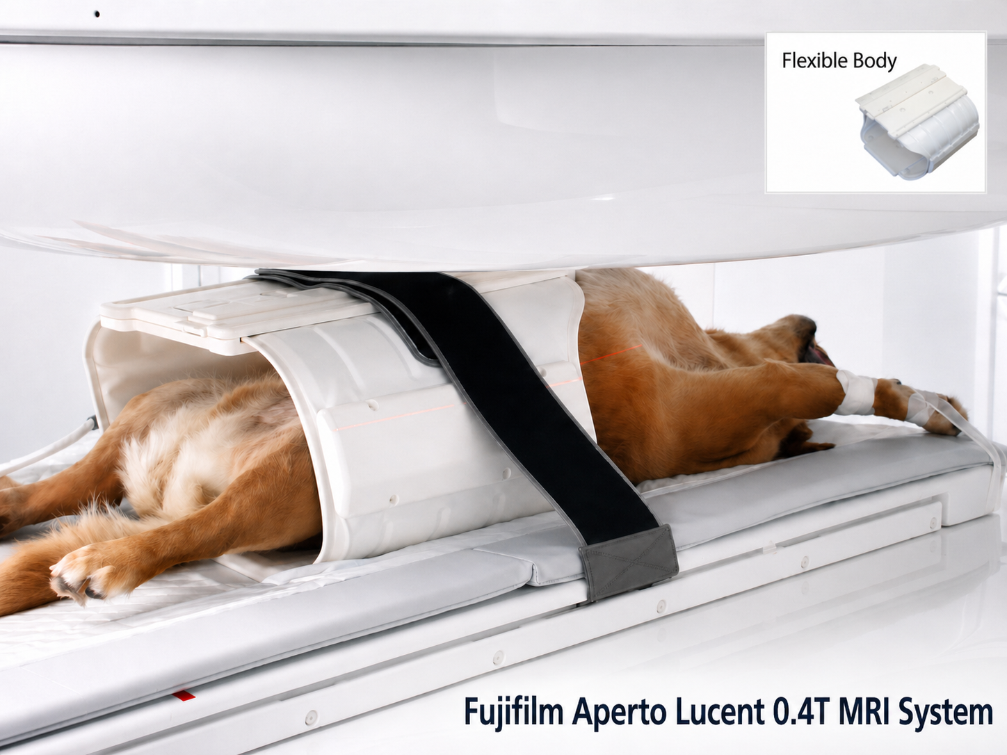
That’s where magnetic resonance imaging (MRI) becomes one of the most powerful diagnostic tools in veterinary medicine. Modern systems, such as the Fujifilm Aperto MRI System, allow clinicians to move beyond educated suspicions and visualize disease processes with remarkable detail. For veterinary neurologists, MRI is not simply about obtaining images; it is about uncovering information that directly influences diagnosis, treatment decisions, prognosis, and ultimately patient outcomes.
From Localization to Confirmation
One of the most important distinctions in neurology is between a suspected and a confirmed diagnosis.
Consider a dog presenting with acute hindlimb paralysis.
Based on history, neurological exam findings, and signalment, a neurologist may strongly suspect an intervertebral disc extrusion. However, several other conditions can produce nearly identical clinical signs, including:
Spinal neoplasia
Inflammatory disease
Hemorrhage
Vertebral abnormalities
Infectious conditions affecting the spinal cord
Without advanced imaging, treatment decisions are often based on probabilities. MRI changes that equation.
Instead of asking, “What is most likely happening?” Neurologists can ask, “What is actually happening?”
That distinction can dramatically alter a patient's treatment plan.
What Neurologists Are Actually Looking For
Many people assume neurologists simply look for obvious abnormalities on MRI images. In reality, image interpretation is far more nuanced. A veterinary neurologist evaluates dozens of variables simultaneously, including:
Tissue Characteristics
Different diseases affect tissue differently. MRI sequences help distinguish:
Normal tissue from abnormal tissue
Inflammation from neoplasia
Acute injury from chronic disease
Edema from hemorrhage
Active disease from residual structural change
These distinctions often determine whether a patient requires surgery, medication, monitoring, or referral.
Lesion Location
In neurology, location matters almost as much as the disease itself. A lesion affecting one area of the spinal cord may carry a very different prognosis than an identical lesion located elsewhere.
MRI allows neurologists to assess:
Precise anatomical location
Degree of compression
Involvement of surrounding structures
Extent of tissue damage
Even subtle differences can influence recommendations regarding surgery, rehabilitation, and long-term management.
Disease Extent
Clinical signs may underestimate, or occasionally overestimate, the true severity of the disease.
MRI frequently reveals:
Multiple lesions rather than a single lesion
More extensive inflammation than anticipated
Additional areas of compression
Previously undetected abnormalities
Understanding the full extent of disease helps clinicians build more accurate treatment plans and set realistic expectations for owners.
The Power of Subtle Findings
Some of the most valuable MRI discoveries are not dramatic. In fact, small findings often provide the information that changes everything.
Case Example: Cervical Disc Disease
A dog presents with neck pain and intermittent forelimb lameness. Radiographs may show degenerative changes, but they cannot determine whether the spinal cord is affected.
MRI may reveal:
Mild disc protrusion
Early spinal cord compression
Subtle spinal cord signal changes
These findings can help determine whether conservative management is appropriate or whether surgical intervention should be considered before neurological deficits progress.
Case Example: Seizure Patients
A young dog with seizures may have a normal neurological examination between episodes.
MRI may identify:
Congenital abnormalities
Inflammatory brain disease
Structural lesions
Previously undetected masses
Each finding points clinicians toward a completely different diagnostic and therapeutic pathway. Without imaging, those distinctions may remain hidden.
Why Image Quality Matters
Advanced MRI systems have transformed veterinary neurology by providing higher-quality images, faster acquisition times, and improved diagnostic capabilities.
TheFujifilm Aperto MRI System is designed to support the detailed visualization that neurologists rely on every day. High-resolution imaging helps clinicians identify subtle lesions that might otherwise be missed and evaluate complex neurological cases with greater confidence.
This becomes particularly important when assessing:
Early inflammatory disease
Small intracranial lesions
Mild spinal cord abnormalities
Post-surgical evaluations
Progressive neurological conditions
When image quality improves, diagnostic confidence often improves as well.
The Impact on Patient Outcomes
Ultimately, MRI is not about technology. It is about reducing uncertainty.
Every veterinary team has experienced cases where clinical signs suggest one diagnosis, only for advanced imaging to reveal something entirely different. For neurologists, MRI provides answers that physical examination alone cannot.
It confirms suspicions. It challenges assumptions. It guides treatment decisions. And most importantly, it helps clinicians deliver the best possible care for their patients.
Bringing It All Together
MRI Systems provide the detailed imaging needed to uncover subtle abnormalities, refine diagnoses, and support confident clinical decision-making. The result is more than a clearer image; it is a clearer path forward.
Because in neurology, the difference between uncertainty and certainty can mean the difference between monitoring and intervention, between symptom management and targeted treatment, and ultimately between guessing what is wrong and knowing what is truly affecting the patient.
