

What Is an Overexposed X-Ray—and How Can Veterinary Clinics Prevent It?
What Is an Overexposed X-Ray—and How Can Veterinary Clinics Prevent It?
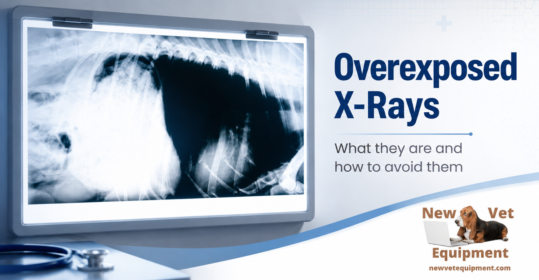
An overexposed veterinary radiograph receives more X-ray exposure than is needed to produce a diagnostic image. With traditional film, the result is usually an image that appears excessively dark. With digital radiography, however, the problem can be much harder to recognize.
Digital imaging software can automatically adjust brightness and contrast, making an overexposed image appear acceptable on the monitor. The image may look good even though the patient received more radiation than necessary.
That is why veterinary teams should not judge exposure by image brightness alone. Proper technique charts, patient measurements, positioning, collimation, exposure indicators, equipment maintenance, and staff training all play important roles in producing consistently diagnostic radiographs.
Important distinction: A radiograph that looks too dark is not necessarily overexposed, and an overexposed digital radiograph may not look too dark. In digital radiography, displayed brightness is heavily influenced by computer processing.
What Does “Overexposed” Mean?
During a radiographic exposure, X-ray photons pass through the patient and reach the image receptor. That receptor may be conventional film, a computed radiography plate, or a digital detector.
The goal is not to send as much radiation as possible through the patient. The goal is to use the lowest exposure that still produces an image with enough detail and an acceptable level of noise for diagnosis.
An exposure that is too low may create a noisy or grainy digital image, sometimes called quantum mottle. An exposure that is unnecessarily high may reduce visible noise, but it also increases radiation exposure and can eventually cause important image information to be lost.
Film and Digital Radiographs Behave Differently
Overexposure With Traditional Film
With screen-film radiography, there is a direct relationship between exposure and the darkness of the processed film. Too much exposure generally produces a film that is too dark. If the overexposure is severe, anatomical details may be obscured and the radiograph may be nondiagnostic.
A bright viewing light may occasionally help reveal information on a mildly overexposed film, but it cannot restore detail that was never properly recorded or that has been lost in a severely overexposed area.
Overexposure With Digital Radiography
Digital radiography has a much wider exposure range than film. After an image is acquired, the computer processes the data and adjusts how the image is displayed. This can make an image obtained with too much radiation appear properly exposed.
Changing the monitor brightness, window level, or window width changes how the image is displayed. It does not change the amount of radiation that was used to create it.
Severe digital overexposure can still cause detector saturation or clipping. When that happens, areas of the image may lose useful information and appear uniformly black or otherwise lack visible detail. Post-processing cannot recreate information that the detector failed to preserve.
Why Overexposure Matters
Overexposure is not simply a cosmetic image problem. It can affect patient care, radiation safety, workflow, and equipment use.
- Unnecessary patient exposure: The patient may receive more ionizing radiation than was required for the examination.
- Loss of diagnostic information: Severe exposure can saturate portions of a digital detector or obscure detail on film.
- Unnecessary retakes: A nondiagnostic image may have to be repeated, increasing radiation exposure and consuming staff time.
- More stress for the patient: Retakes prolong positioning or restraint and may require additional sedation.
- Radiation exposure to personnel: Additional exposures increase the potential occupational exposure of team members, particularly when hands-free positioning practices are not used.
- Premature equipment wear: Habitually excessive techniques create unnecessary heat loading and use of the X-ray tube.
The appropriate principle is ALARA: radiation exposure should be kept “as low as reasonably achievable” while still producing an image that answers the clinical question.
What Is Exposure Creep?
Exposure creep is a gradual increase in the techniques used for digital radiographs. It can occur because a slightly higher exposure often reduces visible image noise, while the computer prevents the image from looking obviously too dark.
Over time, staff members may begin using progressively higher settings because the images appear smoother or because higher techniques seem to reduce the risk of an underexposed image. The clinic may not recognize the increase unless it routinely reviews exposure indicators and technique-chart performance.
A cleaner-looking digital image is not automatically a better image. Once sufficient diagnostic quality has been achieved, additional exposure may provide little or no clinical benefit.
Understand kVp and mAs
The two primary exposure controls are kilovoltage peak, or kVp, and milliampere-seconds, or mAs. They affect the X-ray beam differently.
kVp
kVp primarily controls the energy and penetrating ability of the X-ray beam. Increasing kVp allows more X-rays to penetrate the patient and reach the receptor. It also affects radiographic contrast and scatter production.
Small changes in kVp can produce substantial changes in receptor exposure, so kVp should not be adjusted casually.
mAs
mAs controls the quantity of X-ray photons produced. Increasing mAs generally increases the exposure reaching the receptor. Decreasing mAs reduces the number of photons and may increase image noise if it is lowered too far.
Technique corrections should be based on the appearance of the original image, the exposure indicator, the selected examination, patient thickness, positioning, collimation, and the clinic’s validated technique chart—not on a blanket instruction to lower one setting every time an image looks dark.
How to Recognize Overexposure on a Digital System
Because displayed brightness is unreliable, veterinary teams should review the exposure feedback provided by their system.
Exposure Index
The exposure index, often abbreviated as EI, is a numerical indicator associated with the amount of radiation reaching the detector. It is not a direct measurement of the patient’s radiation dose, but it provides useful feedback about detector exposure.
Veterinary clinics should learn exactly how the index works on their equipment. Older systems and different manufacturers may use different names and scales. On some systems, a higher number means higher detector exposure. On others, especially certain manufacturer-specific scales, the relationship may be reversed.
Target Exposure Index
The target exposure index is the expected detector exposure for a particular examination when the image has acceptable diagnostic quality. Targets may differ according to the body part, projection, patient size, detector, grid use, and clinical purpose.
Deviation Index
Some systems display a deviation index, or DI, showing how far the actual detector exposure differs from the target exposure. A DI near zero generally indicates that the exposure was close to the established target.
The exposure index or deviation index should not be used as the only reason to repeat an image. The veterinary professional should first determine whether the radiograph includes the required anatomy and is diagnostic. An image should not be repeated merely to obtain a perfect number.
Common Causes of Overexposed Veterinary Radiographs
1. Incorrect Patient Measurement
Guessing the patient’s thickness is one of the fastest ways to select the wrong technique. Measurements should be taken at the thickest part of the anatomy being examined and in the position that will be used for the exposure.
For example, a thorax or abdomen may measure differently when the patient is in lateral recumbency than when the patient is standing. Measure the patient after positioning whenever practical.
2. Wrong Examination or Anatomy Selected
Many digital systems use examination-specific processing and technique presets. Selecting “abdomen” when performing a thoracic study—or choosing the wrong patient-size category—may apply inappropriate exposure factors or image-processing algorithms.
Confirm the patient, anatomical region, projection, detector, grid status, and size category before every exposure.
3. Technique Chart Errors
A technique chart may be incorrect, outdated, or based on a different generator, detector, grid, focal distance, or processing system. Technique charts should be developed and validated for the clinic’s actual equipment.
Replacing a detector or generator without reassessing the chart can create consistent exposure errors even when team members follow the chart correctly.
4. Poor Collimation
The X-ray field should be restricted to the anatomy of interest. Excessively open collimation exposes more tissue and creates additional scatter radiation, which reduces image contrast and unnecessarily increases the irradiated area.
Poor collimation can also interfere with some computed radiography systems’ ability to recognize the exposure field correctly, potentially affecting image processing and the reported exposure indicator.
5. Grid-Related Errors
Grids absorb scatter radiation but also absorb some of the primary beam. A technique intended for use with a grid will usually be higher than a nongrid technique. Accidentally using a grid technique without a grid may cause excessive detector exposure.
The team should also verify correct grid alignment, centering, orientation, and focal distance to avoid grid cutoff and inconsistent image quality.
6. Incorrect Source-to-Image Distance
Technique charts are designed around a specific source-to-image distance. Moving the tube or detector closer together without making an appropriate technique adjustment can increase the exposure reaching the receptor.
7. Detector or Plate Differences
Different detectors and computed radiography plates may respond differently to the same technique. Clinics should avoid assuming that every receptor is interchangeable without testing and calibration.
8. Equipment or Calibration Problems
Generator output, timer accuracy, detector calibration, plate-reader performance, and software configuration can all affect exposure and image quality.
An equipment problem becomes more likely when multiple team members experience the same unexplained issue across many patients, body parts, or projections.
A Practical Troubleshooting Process
When a radiograph appears abnormal or the exposure indicator is outside the clinic’s expected range, use a systematic process rather than immediately changing the technique.
| Check | What to Confirm |
|---|---|
| Diagnostic quality | Is the required anatomy included, properly positioned, sharply recorded, and visible? |
| Exposure feedback | What are the exposure index, target index, and deviation index for this examination? |
| Patient measurement | Was the correct anatomical region measured in the actual exposure position? |
| Technique selection | Was the correct chart, body part, projection, patient size, detector, and grid status selected? |
| Collimation | Was the beam restricted to the necessary anatomy without clipping the area of interest? |
| Distance and centering | Were the source-to-image distance, tube alignment, and central ray correct? |
| Pattern | Is this an isolated operator error, or is the same problem occurring repeatedly across the clinic? |
Should an Overexposed Image Always Be Repeated?
No. Repeating every image with an imperfect exposure number can create more radiation exposure without improving patient care.
Before repeating an image, ask:
- Is all required anatomy included?
- Is positioning acceptable?
- Is motion controlled?
- Can the relevant structures be evaluated confidently?
- Is there evidence of detector saturation or lost information?
- Will repeating the study reasonably change the diagnosis or treatment decision?
If the image is diagnostic, it may not need to be repeated. The exposure problem should still be documented and used to improve future technique.
How Veterinary Clinics Can Prevent Overexposure
Create Equipment-Specific Technique Charts
Develop separate techniques when necessary for different anatomical regions, patient thicknesses, projections, grids, detectors, and source-to-image distances. Do not rely exclusively on a generic chart supplied for unrelated equipment.
Measure Every Patient Consistently
Keep calipers near the X-ray table and make measurement part of the standard workflow. Guessing should be the exception, not the normal procedure.
Review Exposure Indicators
Teach every operator how the clinic’s exposure indicator works. Establish expected ranges for common examinations and review trends instead of reacting to a single image.
Track Rejected and Repeated Images
A repeat-analysis program can identify recurring problems with positioning, anatomy selection, motion, collimation, technique, or equipment. The purpose is process improvement—not punishment.
Use Appropriate Positioning and Restraint
Proper positioning reduces motion and prevents avoidable retakes. When medically appropriate, sedation and positioning aids can improve consistency while reducing the need for staff members to manually hold patients.
Collimate Carefully
Include the anatomy required for diagnosis while avoiding an unnecessarily large exposure field. Good collimation improves image quality, reduces scatter, and limits the area exposed to radiation.
Maintain and Calibrate the Equipment
Follow the manufacturer’s recommended preventive-maintenance schedule. Detector calibration, plate care, generator testing, monitor quality, and software configuration should all be part of the clinic’s quality-assurance program.
Train the Entire Team
Every person who performs radiography should understand basic exposure principles, the clinic’s technique chart, the equipment’s exposure-index scale, positioning standards, collimation, repeat criteria, and radiation-safety procedures.
When to Request Equipment Service
Contact a qualified service provider when:
- Exposure problems occur across multiple patients and examinations.
- Identical techniques produce inconsistent results.
- Exposure indicators shift unexpectedly.
- One detector or computed radiography plate behaves differently from the others.
- Images show recurring lines, dead pixels, uneven density, artifacts, or unusual processing.
- The generator, detector, plate reader, workstation, or software was recently repaired or replaced.
- The technique chart no longer produces consistent diagnostic results.
A persistent problem should not be “fixed” by continuing to lower or raise exposure settings without determining the underlying cause. Doing so may hide an equipment fault and create new image-quality problems.
A Simple Pre-Exposure Checklist
Before making the exposure, confirm:
☐ Correct patient and examination
☐ Correct anatomical region and projection
☐ Patient measured in the exposure position
☐ Correct technique-chart setting selected
☐ Grid or nongrid technique confirmed
☐ Detector and source-to-image distance confirmed
☐ Patient positioned and immobilized
☐ Beam centered and collimated
☐ Personnel protected and positioned according to clinic policy
Frequently Asked Questions
Is an Overexposed X-Ray Always Black?
No. An overexposed film radiograph normally appears too dark. An overexposed digital radiograph may appear normal because the computer adjusts its displayed brightness and contrast.
Can Image-Processing Software Correct Overexposure?
Software can change how digital image data are displayed, but it cannot reduce the radiation already delivered to the patient. It also cannot recover data lost through detector saturation or clipping.
Should We Lower kVp or mAs When an Image Is Overexposed?
Not automatically. First confirm that the measurement, examination selection, grid status, distance, collimation, detector, processing, and exposure indicator are correct. Then adjust the appropriate factor according to the clinic’s validated technique chart and equipment instructions.
Does the Exposure Index Equal Patient Dose?
No. The exposure index primarily reflects exposure at the detector. Patient dose is also affected by patient size, beam quality, filtration, collimation, projection, distance, and other technical factors.
Why Do Exposure-Index Values Vary Between Machines?
Different manufacturers and older systems may use different calculations, names, and numerical scales. Veterinary teams should use the documentation and target ranges supplied for their specific equipment.
The Bottom Line
Preventing overexposed radiographs requires more than looking for an image that appears too dark. That approach may work with traditional film, but it is unreliable with modern digital systems.
Veterinary teams should evaluate diagnostic quality, exposure indicators, patient measurements, technique-chart accuracy, positioning, collimation, grid use, equipment performance, and repeat trends together.
A consistent quality-assurance program helps clinics obtain diagnostic images the first time, reduce unnecessary retakes, protect patients and staff, and make better use of their imaging equipment.
Need Help Choosing Veterinary X-Ray Equipment?
We help veterinary clinics compare digital X-ray systems, understand equipment options, and make a confident purchasing decision without paying more than necessary.
Call 877-545-1202 Is It Time to Upgrade Your X-Ray? Click HereThis article provides general educational information and is not a substitute for the instructions supplied by your equipment manufacturer, applicable radiation-safety regulations, or advice from a qualified veterinary radiologist, medical physicist, radiation-safety professional, or service engineer.